Review
. 2016 Oct 3;8(10):270.
doi: 10.3390/v8100270.

Zoonotic Hepatitis E Virus: Classification, Animal Reservoirs and Transmission Routes

Affiliations
Review

Zoonotic Hepatitis E Virus: Classification, Animal Reservoirs and Transmission Routes

Virginie Doceul et al. Viruses. .

Abstract

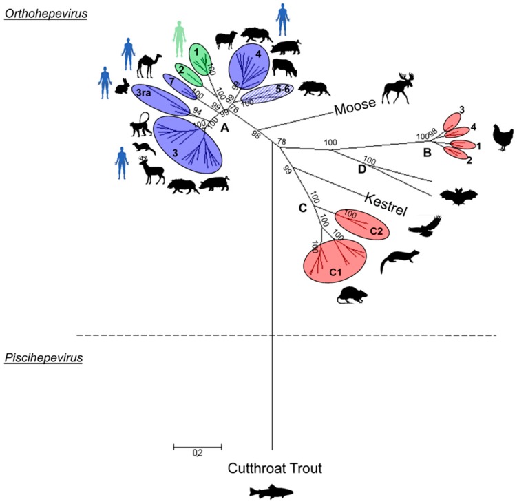
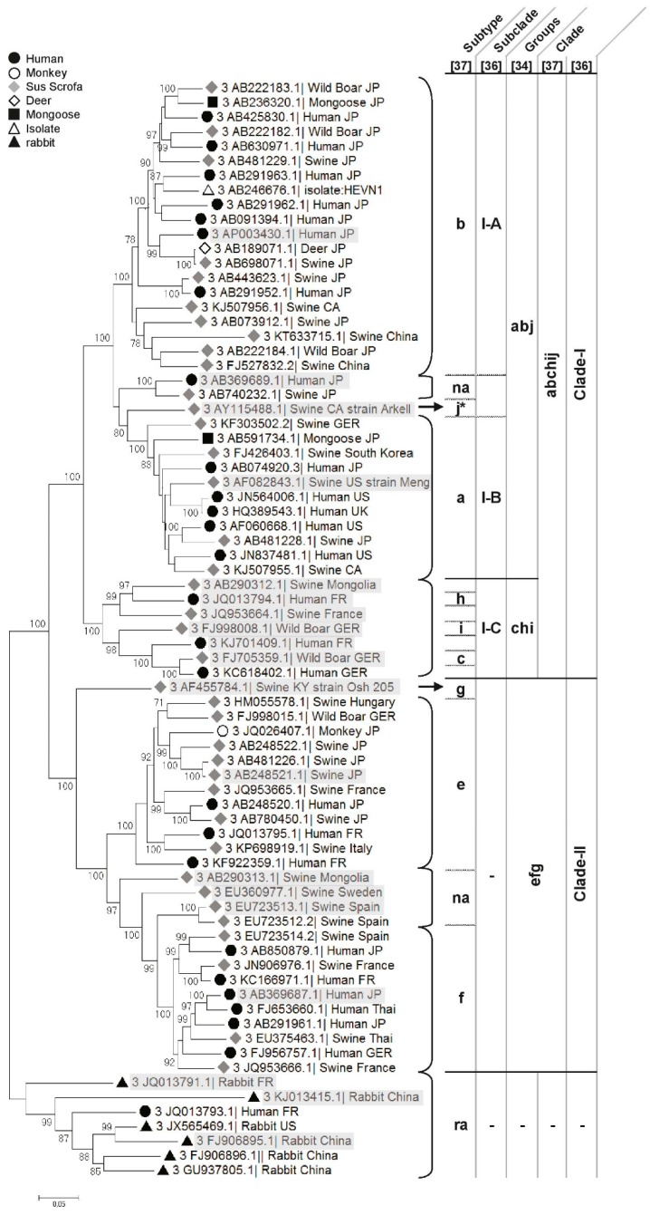
During the past ten years, several new hepatitis E viruses (HEVs) have been identified in various animal species. In parallel, the number of reports of autochthonous hepatitis E in Western countries has increased as well, raising the question of what role these possible animal reservoirs play in human infections. The aim of this review is to present the recent discoveries of animal HEVs and their classification within the Hepeviridae family, their zoonotic and species barrier crossing potential, and possible use as models to study hepatitis E pathogenesis. Lastly, this review describes the transmission pathways identified from animal sources.

Keywords: animals; foodborne transmission; hepatitis E virus (HEV); zoonotic reservoir.


Conflict of interest statement

The authors declare no conflict of interest.

Figures

Figure 1
Figure 1
Phylogenetic tree of representative members of the Hepeviridae family. The tree was inferred using the Maximum Likelihood method based on the Tamura–Nei model. The analysis was performed with 67 hepatitis E virus (HEV) complete genomes or complete coding sequences available in the GenBank database and representative of each genotype. The sequence size varies between 6543 and 7318 nt in length, and they were aligned using Clustal W. The bootstraps were obtained from 1000 replicates and values over 70% are indicated at the genotype level. The initial tree was obtained by applying the Neighbour-Joining method to a matrix of pairwise distances estimated using the Maximum Composite Likelihood (MCL) approach. The tree is drawn to scale, with branch lengths proportional to the number of substitutions per site. Evolutionary analyses were conducted using Molecular Evolutionary Genetics Analysis (Version 6.0). The Orthohepevirus species taxon name is added at the junction of the last common ancestor for each species. Genotypes of non-zoonotic HEV species (red), genotypes including HEV strains isolated from animals and human (blue), genotypes infecting human only (green) and genotypes infecting wild boar that are not linked to human infections (striped blue) are shown.
Figure 2
Figure 2
Phylogenetic tree of HEV-3. The tree was inferred using the Maximum Likelihood method based on the Tamura–Nei model. The analysis involved the 75 most representative HEV-3 complete sequences/cds available on the GenBank database and aligned using the clustal W method. The bootstraps were obtained from 1000 replicates and values >70% are indicated. The initial tree was obtained by applying the Neighbour-Joining method to a matrix of pairwise distances estimated using the MCL approach. The tree is drawn to scale, with branch lengths proportional to the number of substitutions per site. Evolutionary analyses were conducted using Molecular Evolutionary Genetics Analysis (Version 6.0). Cluster names from the classification proposed by Lu et al., Vina-Rodrigues et al. and Smith et al. (letters a to j and ra cluster) are indicated in the table on the right side of the tree [35,38,39] and by Mirazo et al. (Clade-I and -II and subclades I-A to I-C) [37]. Reference sequences of the different HEV subtypes used by Smith et al. [38] are highlighted in grey. The symbols to the left of the different HEV strains indicate the host of origin. na = non assigned.
Figure 3
Figure 3
Experimental inter-species transmissions of HEV. Inter-species transmission of different HEV strains determined by the experimental infection of animal reservoirs (black silhouette) or animal models (white silhouette). Details and references of the different experiments presented are given in the text.

References

    1. Kamar N., Marion O., Abravanel F., Izopet J., Dalton H.R. Extrahepatic manifestations of hepatitis E virus. Liver Int. 2016;36:467–472. doi: 10.1111/liv.13037. - DOI - PubMed
    1. Sayed I.M., Vercouter A.-S., Abdelwahab S.F., Vercauteren K., Meuleman P. Is hepatitis E virus an emerging problem in industrialized countries? Hepatology. 2015;62:1883–1892. doi: 10.1002/hep.27990. - DOI - PubMed
    1. Murali A.R., Kotwal V., Chawla S. Chronic hepatitis E: A brief review. World J. Hepatol. 2015;7:2194–2201. doi: 10.4254/wjh.v7.i19.2194. - DOI - PMC - PubMed
    1. Khuroo M.S. Study of an epidemic of non-A, non-B hepatitis. Possibility of another human hepatitis virus distinct from post-transfusion non-A, non-B type. Am. J. Med. 1980;68:818–824. doi: 10.1016/0002-9343(80)90200-4. - DOI - PubMed
    1. Balayan M.S., Andjaparidze A.G., Savinskaya S.S., Ketiladze E.S., Braginsky D.M., Savinov A.P., Poleschuk V.F. Evidence for a virus in non-A, non-B hepatitis transmitted via the fecal-oral route. Intervirology. 1983;20:23–31. - PubMed

Publication types

LinkOut - more resources