. 2016 Oct 5;11(10):e0162578.
doi: 10.1371/journal.pone.0162578. eCollection 2016.

Mechanism of Rifampicin Inactivation in Nocardia farcinica

Affiliations

Mechanism of Rifampicin Inactivation in Nocardia farcinica

Heba Abdelwahab et al. PLoS One. .

Abstract

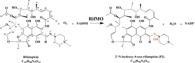
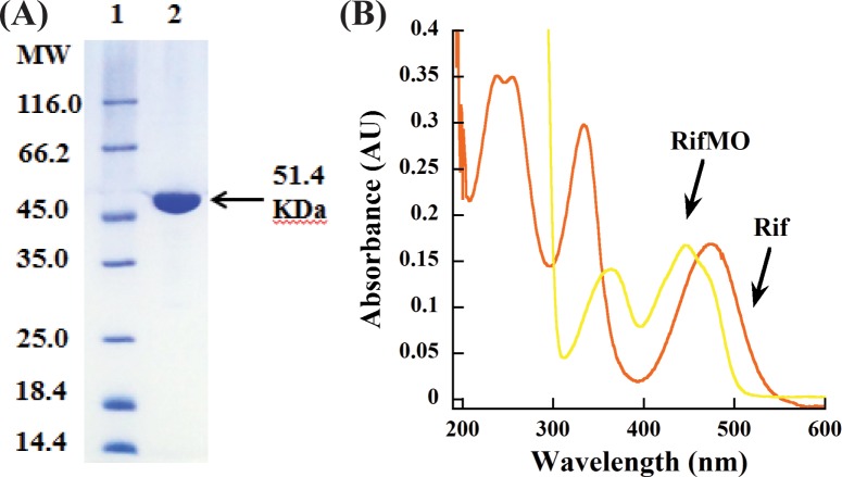
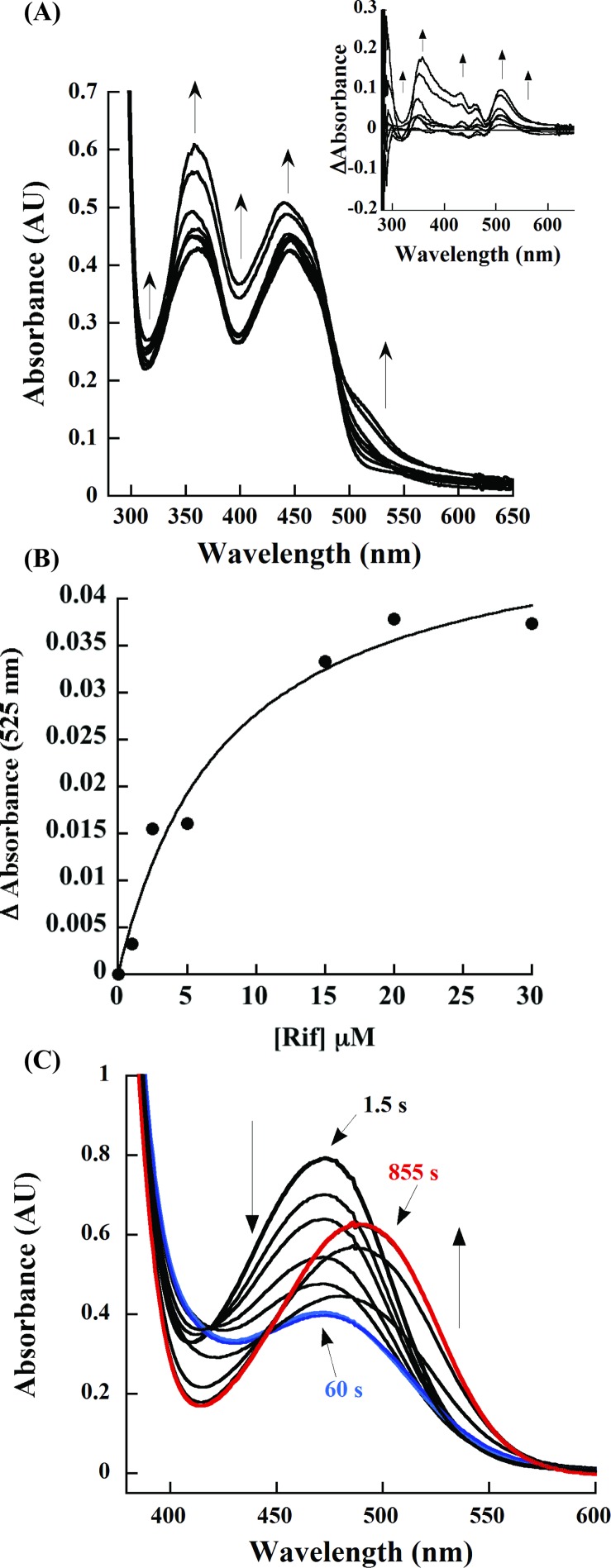
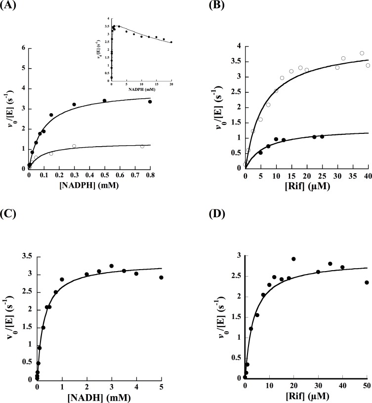
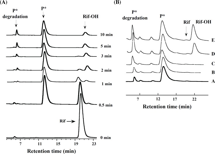
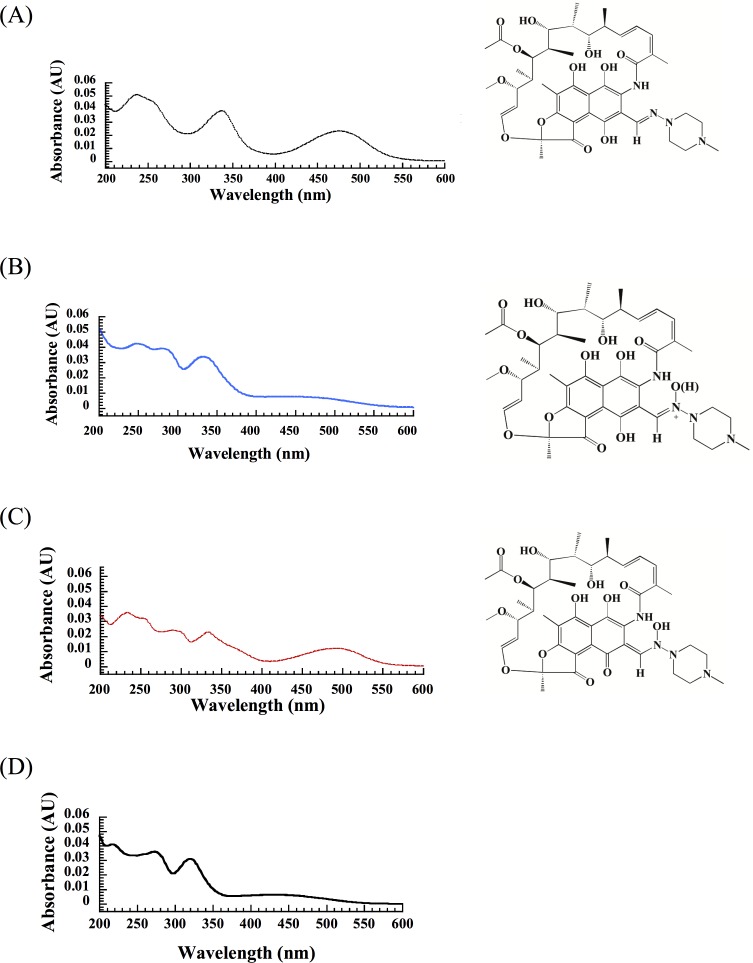
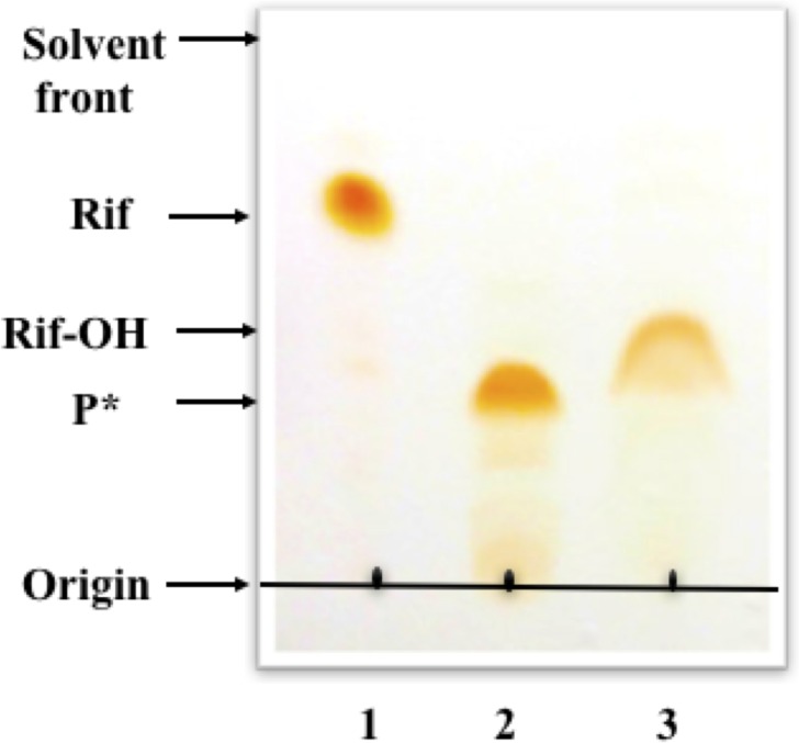
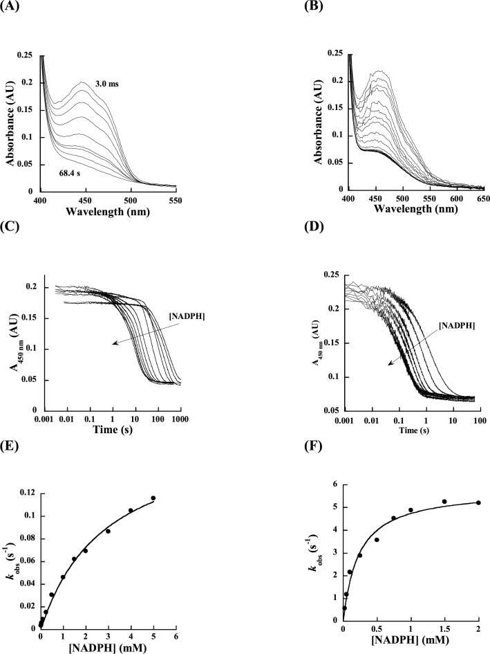
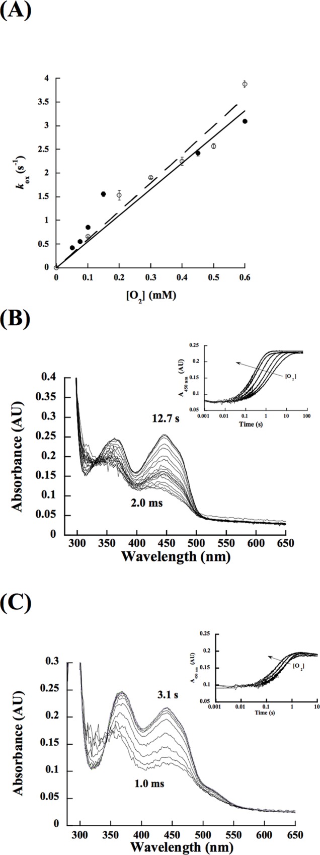
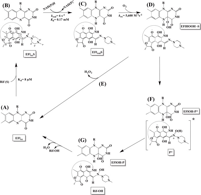
A novel mechanism of rifampicin (Rif) resistance has recently been reported in Nocardia farcinica. This new mechanism involves the activity of rifampicin monooxygenase (RifMO), a flavin-dependent monooxygenase that catalyzes the hydroxylation of Rif, which is the first step in the degradation pathway. Recombinant RifMO was overexpressed and purified for biochemical analysis. Kinetic characterization revealed that Rif binding is necessary for effective FAD reduction. RifMO exhibits only a 3-fold coenzyme preference for NADPH over NADH. RifMO catalyzes the incorporation of a single oxygen atom forming an unstable intermediate that eventually is converted to 2'-N-hydroxy-4-oxo-Rif. Stable C4a-hydroperoxyflavin was not detected by rapid kinetics methods, which is consistent with only 30% of the activated oxygen leading to product formation. These findings represent the first reported detailed biochemical characterization of a flavin-monooxygenase involved in antibiotic resistance.


Conflict of interest statement

The authors have declared that no competing interests exist.

Figures

Fig 1
Fig 1. Reaction catalyzed by RifMO.
Fig 2
Fig 2
(A) SDS-PAGE of purified RifMO. Lane 1, Molecular weight markers; lane 2, final RifMO sample. (B) UV-visible spectrum of 15 μM Rif and RifMO. The flavin spectrum shows absorbance maxima at 366 nm and 449 nm.
Fig 3
Fig 3. Spectral changes during Rif binding and turnover.
(A) Flavin spectral changes as a function of increasing concentration of Rif (0–40 μM). The spectral changes show increases in absorbance at (320, 360, 400, 443, and 525 nm) and the isobestic point at ~ 480 nm. The inset shows spectral differences after subtracting the spectrum of RifMO with 0 μM Rif. (B) Determination of the KD value of Rif. The change in absorbance of the RifMO-Rif complex at 525 nm was plotted as a function of Rif to determine a KD value. (C) UV-Vis spectrophotometric monitoring of RifMO product formation representing the decline of the Rif peak at 475 nm (blue), followed by red shifting and an absorbance increase at 493 nm (red).
Fig 4
Fig 4. Steady-state kinetics of oxygen consumption compared to HPLC analysis.
(A) Reaction rates as a function of NADPH using oxygraph (solid circles) and HPLC (open circles). The inset shows the oxygen consumption activity at higher NADPH concentration. (B) Reaction rates as a function of Rif using oxygraph (open circles) and HPLC (closed circles) in the presence of 1 mM NADPH as the electron donor. Oxygen consumption assays were done in 1 mL of 100 mM sodium phosphate, pH 7.5, at 25°C. (C) Oxygen consumption as a function of NADH. (D) Oxygen consumption as a function of Rif in the presence of 2 mM NADH as the electron donor.
Fig 5
Fig 5. Time-dependent HPLC analysis of RifMO reactions.
(A) Stacked chromatograms showing time traces for the elution of the Rif peak (21.2 min), P* (13.4 min), Rif-OH (22.1 min), and the P* degradation compound (6.7 min). (B) Stacked chromatograms show P* (A) extracted in 100 mM sodium phosphate buffer, pH 7.5, incubated with: (B) NADPH, (C) RifMO, (D, E) NADPH and RifMO, for 5, and 20 min., respectively.
Fig 6
Fig 6. Individual UV-spectra extracted for pure peaks during HPLC analysis, representing all species involved in the RifMO reaction.
(A) Rif, (B)The first product, (P*), (C) The final product, Rif-OH, and (D) Rif degradation compound of the first product. Rif-OH structure was elucidated from the NMR analysis.
Fig 7
Fig 7. TLC monitoring of RifMO activity with Rif.
Lane 1, Rif; lane 2, P*-generating reaction; lane 3, Rif-OH-generating reaction.
Fig 8
Fig 8. Flavin reduction with NADPH.
Spectra changes for the substrate-free RifMO with 2 mM NADPH. (B) Change in the flavin absorbance at 450 nm for substrate-free RifMO at various concentrations of NADPH (0.025–2 mM). (C) Dependence of the kobs values as a function of NADPH in the presence of 15 μM Rif. Data was fit to a single exponential decay equation. (D) Spectra changes for the Rif-RifMO complex with 2 mM NADPH. (E) Change in the flavin absorbance at 450 nm for substrate-complexed RifMO at various concentrations of NADPH (0.025–5 mM). (F) Dependence of the kobs values as a function of NADPH in the absence of Rif. Data was fit to a single exponential decay equation.
Fig 9
Fig 9. Flavin oxidation.
(A) kobs values as a function of [O2] without Rif (closed circles) and with 15 μM Rif (opened circles). (B) Spectra changes during the oxidation of free reduced RifMO with 250 μM O2. The inset shows the changes in absorbance at 450 nm as a function of oxygen concentration. (C) Spectra changes during the oxidation of free, reduced RifMO with 200 μM O2. The inset shows changes in absorbance at 450 nm as a function of oxygen concentration. Both data were fit to a single phase exponential equation.
Fig 10
Fig 10. Catalytic cycle of RifMO.
The reaction starts by the binding of Rif to oxidized RifMO (A), which primes the enzyme for the next step by inducing conformational changes in the flavin (B). NAPDH binds and reduces the flavin (C), and the reduced Rif-MO/Rif complex reacts with molecular oxygen to form the C4a-hydroperoxyflavin (D). This intermediate is not very stable and only ~30% hydroxylates Rif (F). The rest decays to hydrogen peroxide (E). The first product is the quinol (P*) (F), which is then converted to the final quinone product, Rif-OH (G). Release of Rif-OH and dehydration of the flavin are the final steps in the reaction.

Similar articles

Cited by

References

    1. Lerner PI. Nocardiosis. Clin Infect Dis 1996:891–903. - PubMed
    1. McMurray DN. Mycobacteria and Nocardia. In: Baron S, editor. Medical Microbiology. 4th ed. Galveston (TX)1996. - PubMed
    1. Hartmann G, Honikel KO, Knusel F, Nuesch J. The specific inhibition of the DNA-directed RNA synthesis by rifamycin. Biochim Biophys Acta. 1967;145(3):843–844. - PubMed
    1. Kim TK, Hewavitharana AK, Shaw PN, Fuerst JA. Discovery of a new source of rifamycin antibiotics in marine sponge actinobacteria by phylogenetic prediction. Appl Environ Microbiol. 2006;72(3):2118–2125. - PMC - PubMed
    1. Aristoff PA, Garcia GA, Kirchhoff PD, Showalter HH. Rifamycins–Obstacles and opportunities. Tuberculosis. 2010;90(2):94–118. 10.1016/j.tube.2010.02.001 - DOI - PubMed

MeSH terms

LinkOut - more resources