. 2016 Oct 5;11(10):e0164139.
doi: 10.1371/journal.pone.0164139. eCollection 2016.

Protective Effects of Acetylation on the Pathological Reactions of the Lens Crystallins with Homocysteine Thiolactone

Affiliations

Protective Effects of Acetylation on the Pathological Reactions of the Lens Crystallins with Homocysteine Thiolactone

Zeinab Moafian et al. PLoS One. .

Abstract

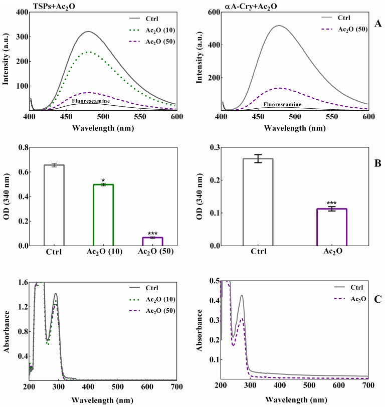
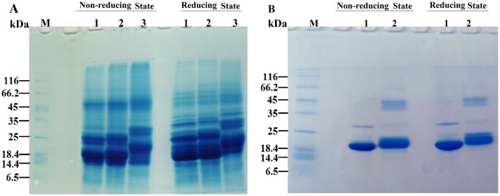
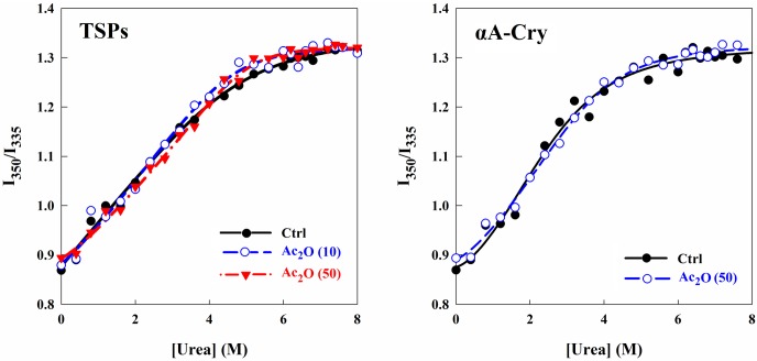
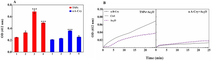
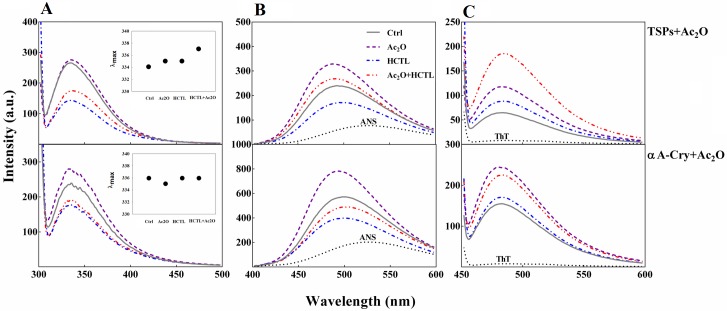
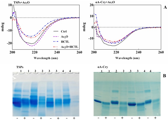
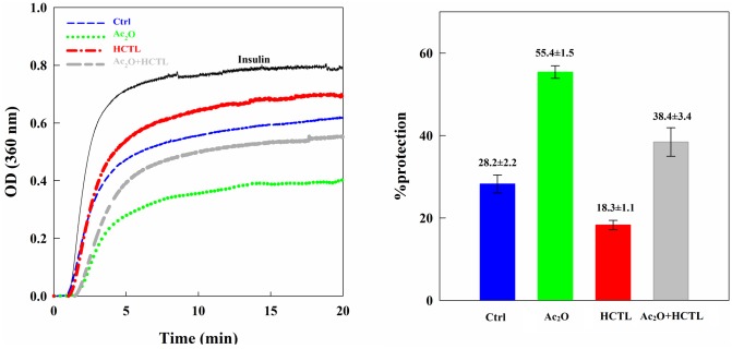
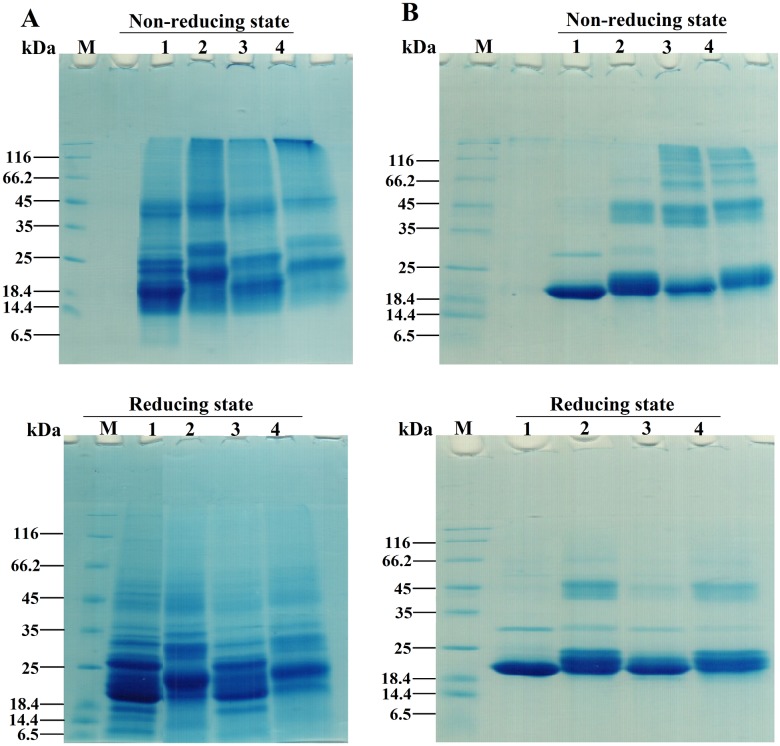
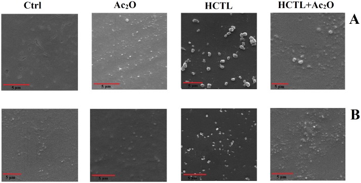
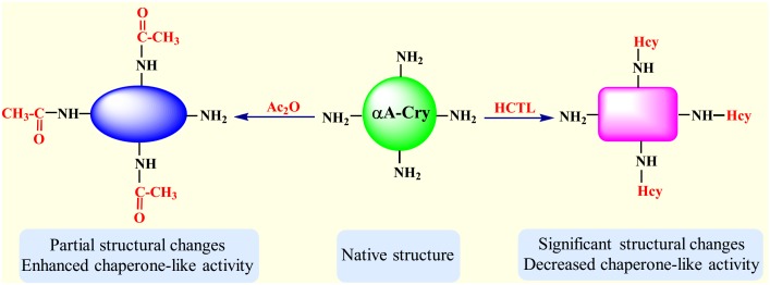
Various post-translational lens crystallins modifications result in structural and functional insults, contributing to the development of lens opacity and cataract disorders. Lens crystallins are potential targets of homocysteinylation, particularly under hyperhomocysteinemia which has been indicated in various eye diseases. Since both homocysteinylation and acetylation primarily occur on protein free amino groups, we applied different spectroscopic methods and gel mobility shift analysis to examine the possible preventive role of acetylation against homocysteinylation. Lens crystallins were extensively acetylated in the presence of acetic anhydride and then subjected to homocysteinylation in the presence of homocysteine thiolactone (HCTL). Extensive acetylation of the lens crystallins results in partial structural alteration and enhancement of their stability, as well as improvement of α-crystallin chaperone-like activity. In addition, acetylation partially prevents HCTL-induced structural alteration and aggregation of lens crystallins. Also, acetylation protects against HCTL-induced loss of α-crystallin chaperone activity. Additionally, subsequent acetylation and homocysteinylation cause significant proteolytic degradation of crystallins. Therefore, further experimentation is required in order to judge effectively the preventative role of acetylation on the structural and functional insults induced by homocysteinylation of lens crystallins.


Conflict of interest statement

The authors have declared that no competing interests exist.

Figures

Fig 1
Fig 1. The in vitro acetylation of TSPs and recombinant αA-Cry with Ac2O.
TSPs (5 mg mL-1) and αA-Cry (3 mg mL-1) were incubated respectively with different molar ratios of Ac2O at room temperature over 1 h. Panel (A) Fluorescamine assay was performed at excitation/emission wavelengths of 390/490 nm as described in the experimental section. The excitation/emission slit widths were set at 10 nm for all protein samples. Panel (B) The spectroscopic evaluation of protein acetylation was performed via an OPA assay. Native and acetylated TSPs (4 mg mL-1) and αA-Cry (3 mg mL-1) were added to OPA solution and incubated for 2 min at room temperature. Then, the absorbance was measured at 340 nm. All results are reported as the average of triplicate experiments. The bars represent the means ± standard deviation (SD) of three independent experiments. The values of *p < 0.05 were considered significant. Panel (C) UV-Vis spectroscopic analysis of acetylated TSPs and αA-Cry with Ac2O. All protein samples were diluted to 0.5 mg mL-1 prior to collect the absorption spectra between 200 and 700 nm. Ac2O (10) and Ac2O (50) indicate protein to Ac2O molar ratios of 1:10 and 1:50, respectively.
Fig 2
Fig 2. SDS–PAGE analysis of acetylated TSPs and αA-Cry with Ac2O and ASA.
TSPs (4 mg mL-1) and αA-Cry (3 mg mL-1) were acetylated with Ac2O at room temperature over 1h. Then, a 15 μg of each protein sample was subjected to the reducing and non-reducing SDS-PAGE analysis (12% gel). (A) The SDS-PAGE analysis of TSPs acetylation. Lanes 1, 2 and 3 indicate native, acetylated TSPs which incubated with 1:10 and 1:50 molar ratios of TSPs/Ac2O, respectively. (B) The SDS-PAGE analysis of αA-Cry acetylation. Lanes 1 and 2 stand for native and acetylated αA-Cry with 1:50 molar ratio of αA-Cry/Ac2O. The staining of protein bands was done with an appropriate Coomassie Brilliant Blue (CBB) staining method. Ctrl and M indicate control sample and molecular mass markers, respectively.
Fig 3
Fig 3. Estimation of the structural stability of lens proteins upon acetylation.
The protein samples were incubated in the presence of increasing concentrations of urea (0–8 M) for 12 h in NaPi buffer (50 mM, pH 7.4). At the end of incubation, Trp fluorescence emission spectra were acquired between 300–500 nm. The results are presented as the ratio of fluorescence intensities at 350 and 335 nm for different protein samples against urea concentrations.
Fig 4
Fig 4. The assessment of sulfhydryl groups in lens proteins.
(A) Evaluation of free sulfhydryl groups via DTNB assay. Native and modified TSPs (1 mg mL-1) and αA-Cry (1 mg mL-1) were solubilized with urea (8M) and then incubated with DTT (5 mM). Subsequently, the protein samples were dialyzed against buffer C. Evaluation of free thiols was determined with DTNB at a 7-fold molar excess of the protein. The absorbance was measured at 412 nm. All results are reported as the average of triplicate experiments. αB-crystallin was used as negative control because this protein has no Cys residue. The bars represent the means ± standard deviation (SD) of three independent experiments. The values of *p < 0.05 and ***p < 0.001 were considered significant. Also, 1, 2, 3 and 4 stand for control, acetylated, homocysteinylated and double-modified protein samples, respectively. (B) Kinetic analysis of the reaction of DNTB with thiol groups of lens proteins. The kinetic profiles of DTNB reaction with native and acetylated proteins (0.1 mg mL-1) were measured over 25 min in NaPi buffer (50 mM pH 7.4), containing EDTA (1 mM) at 25°C. Protein/DTNB was added at a molar ratio of 1:7 and the absorbance was measured at 412 nm.
Fig 5
Fig 5. Aggregation propensity of acetylated TSPs and αA-Cry in the presence of HCTL.
The native and acetylated TSPs (4 mg mL-1) were incubated with HCTL (10 mM) in buffer C at 37°C for 3 days. Then, the protein samples were diluted to 0.5 mg mL -1 in the same buffer and the absorption spectra were collected between 200 and 700 nm. Also, the protein samples were centrifuged at 13,000 rpm for 30 min. The absorption spectra of supernatant were obtained over the same wavelength range. The symbols used in this figure are thick blue dotted line: HCTL-modified proteins; thin red dotted line: double-modified protein; thick blue solid line: supernatant of HCTL-modified protein; thin red solid line: supernatant of double-modified proteins.
Fig 6
Fig 6. Fluorescence analysis of lens proteins upon modification with Ac2O and HCTL.
(A) Trp fluorescence assessment of lens proteins. Protein samples including native and acetylated proteins incubated without and with HCTL diluted to 0.15 mg mL-1 in buffer C. The excitation wavelength was 295 nm and emission spectra were collected between 300 and 500 nm. The excitation/emission slit widths were 5/10 nm for TSPs and 10/10 nm for αA-Cry samples. The inset figures display λmax for control (334 nm), acetylated (335 nm), homocysteinylated (335 nm) and doubled modified (337 nm) TSPs. Also, this value for the control sample, homocysteinylated and doubled modified αA-Cry is 336 nm and for acetylated αA-Cry is 335 nm. (B) ANS fluorescence analysis of the lens proteins. Protein samples (0.15 mg mL-1 diluted in buffer C) were incubated with ANS (100 μM) for 30 min. The ANS fluorescence emission spectra were collected between 400 and 600 nm, with an excitation wavelength of 365 nm. The excitation/emission slit widths were set at 10/10 nm for TSPs and at 10/20 for αA-Cry. (C) The fluorescence experiment was performed with incubation of the protein samples (0.15 mg mL -1 diluted in buffer C) in the presence of ThT (20 μM) for 10 min. The protein samples were excited at 440 nm and the slit widths for excitation/emission were fixed at 10/10 nm.
Fig 7
Fig 7. Estimation of the secondary structural contents and evaluation of α-chymotrypsin digestion of lens crystallins upon different modifications.
(A) CD spectroscopic assessment of lens proteins after modification with Ac2O and HCTL. Far UV-CD spectra of TSPs and αA-Cry upon modification with Ac2O and HCTL. Proteins were diluted to 0.3 mg mL-1 in buffer C. The measurements were undertaken using a cuvette with 0.1 cm path length at 25°C. (B) SDS-PAGE assessment of chymotryptic digestion of modified lens proteins. The lens proteins, after modification with Ac2O and HTCL, were subjected to reducing SDS-PAGE (12% gel) for the assessment of their proteolytic susceptibility with α-chymotrypsin. The lens proteins (2 mg mL-1) were incubated with α-chymotrypsin with a 1:50 (w/w) ratio of enzyme/substrate at 37°C for 6 h. Incubation was done in 100 mM NaPi buffer containing 0.01% NaN3 at pH 7.8. At the end of incubation, a 15 μg of each protein sample was loaded to SDS-PAGE gel. 1–4, respectively stand for the control sample, acetylated proteins, HCTL-modified proteins and double modified proteins. Also (-) and (+) indicate the absence and presence of α-chymotrypsin, respectively. The protein bands were visualized by a CBB staining protocol.
Fig 8
Fig 8. DLS analysis of lens proteins upon modification with Ac2O and HCTL.
Lens proteins were assessed for their hydrodynamic size distribution, upon modification with Ac2O and HCTL, by DLS. Prior to the measurements, the protein samples were diluted to 3 mg mL -1 in 100 mM phosphate buffer, pH 7.4. The size distributions were reported to their relative volumes. The insets indicate their relative scattering intensity. A, B, C and D, respectively indicate native proteins, acetylated proteins, HCTL-modified proteins and double modified proteins.
Fig 9
Fig 9. The chaperone-like activity of αA-Cry upon modification with Ac2O and HCTL.
(A) The chaperone-like activity of different modified forms of αA-Cry was assessed in a chemical-induction aggregation system. Native and modified αA-Cry samples (0.15 mg mL-1) were assessed to protect the aggregation of bovine pancreatic insulin (0.3 mg mL-1) with DTT (20 mM) in NaPi buffer (100 mM) pH 7.2 at 40°C. The aggregation progress was monitored at 360 nm for 20 min. (B) The chaperone-like activity was quantified, based on Eq 1, in terms of the percentage of protection.
Fig 10
Fig 10. Gel mobility shift analysis of the acetylated lens proteins incubated with HCTL.
Native and acetylated TSPs were incubated with HCTL (10 mM) in buffer C at 37°C for 3 days. At the end of incubation, 15 μg of each protein was loaded into wells of electrophoresis gel (12% gel) under both non-reducing and reducing conditions. (A) SDS-PAGE analysis of acetylated and double modified TSPs. M indicates the molecular mass markers. Lanes 1–5 respectively are the fresh proteins, control proteins, acetylated proteins, HCTL-modified proteins and HCTL/Ac2O double modified proteins. (B) SDS-PAGE analysis of acetylated αA-Cry and double modified αA-Cry. Lanes 1–4, respectively are the control αA-Cry, acetylated αA-Cry, HCTL-modified and double modified αA-Cry. The protein bands were visualized by a proper CBB staining method.
Fig 11
Fig 11. Scanning electron microscope (SEM) images of modified lens crystallins.
The symbols are as following: Ctrl: control TSPs; Ac2O: acetylated TSPs; HCTL: homocysteineteinylated TSPs and Ac2O+HCTL: double-modified TSPs. Also, A and B stand for TSPs and αA-Cry, respectively.
Fig 12
Fig 12. Structural properties and chaperone-like activity of αA-Cry upon modification with Ac2O and HCTL.
Homocysteinylation of free amino groups results in significant structural changes and attenuates the chaperone-like activity of αA-Cry while acetylation leads to partial structural alteration and enhanced efficiency in suppressing aggregation of a client protein.

Similar articles

Cited by

References

    1. Sharma KK, Santhoshkumar P. Lens aging: effects of crystallins. Biochim Biophys Acta 2009; 1790: 1095–1108. 10.1016/j.bbagen.2009.05.008 - DOI - PMC - PubMed
    1. Wistow GJ, Piatigorsky J. Lens crystallins: the evolution and expression of proteins for highly specialized tissue. Annu Rev Biochem. 1988; 57: 479–504. 10.1146/annurev.bi.57.070188.002403 - DOI - PubMed
    1. Lubsen NH, Aarts HJ, Schoenmakers JG. The evolution of lenticular proteins: the beta- and gamma-crystallin super gene family. Prog Biophys Mol Biol. 1988; 51: 47–76. 10.1016/0079-6107(88)90010-7 - DOI - PubMed
    1. Bhat SP, Nagineni CN. Alpha B subunit of lens-specific protein alpha crystallin is present in other ocular and non-ocular tissues. Biochem Biophys Res Commun. 1989; 158: 319–325. - PubMed
    1. Horwitz J. The function of alpha-crystallin in vision. Semin Cell Dev Biol. 2000; 11: 53–60. 10.1006/scdb.1999.0351 - DOI - PubMed

LinkOut - more resources