. 2017 Jan;26(1):122-129.
doi: 10.1002/pro.3060. Epub 2016 Oct 15.

On the interpretation of electron microscopic maps of biological macromolecules

Affiliations

On the interpretation of electron microscopic maps of biological macromolecules

Jimin Wang et al. Protein Sci. 2017 Jan.

Abstract

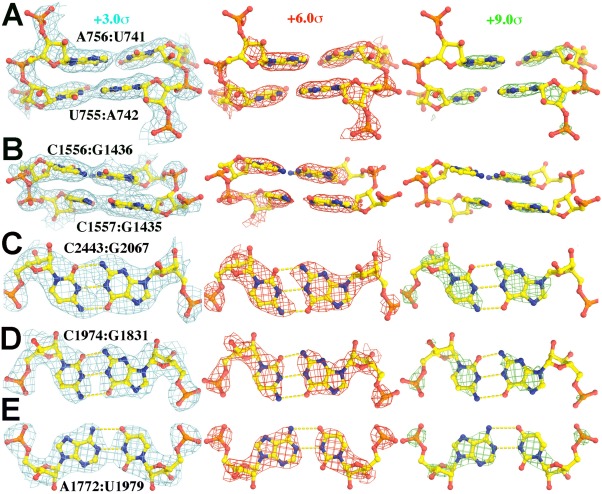
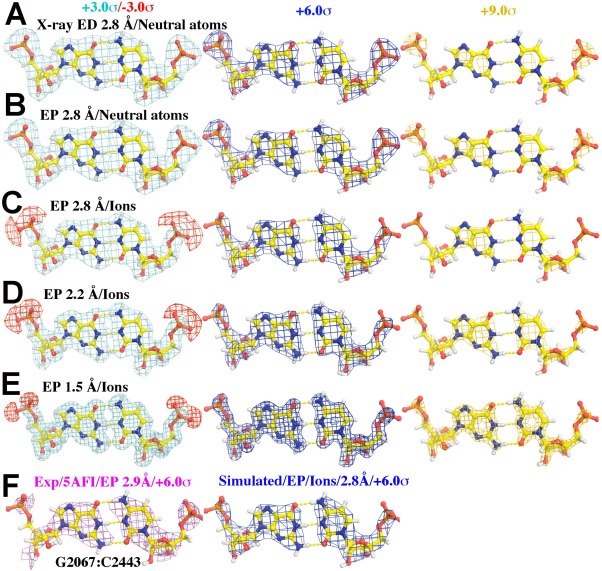
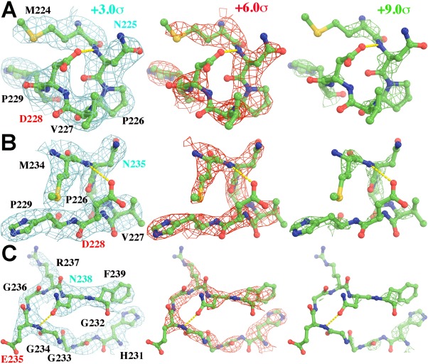
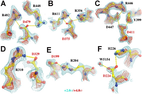
The images of flash-frozen biological macromolecules produced by cryo-electron microscopy (EM) can be used to generate accurate, three-dimensional, electric potential maps for these molecules that resemble X-ray-derived electron density maps. However, unlike electron density maps, electric potential maps can include negative features that might for example represent the negatively charged, backbone phosphate groups of nucleic acids or protein carboxylate side chains, which can complicate their interpretation. This study examines the images of groups that include charged atoms that appear in recently-published, high-resolution EM potential maps of the ribosome and β-galactosidase. Comparisons of simulated maps of these same groups with their experimental counterparts highlight the impact that charge has on the appearance of electric potential maps.

Keywords: X-ray scattering; electric potential; electron atomic scattering factor; electron density; electron microscopy; electron scattering.


Figures

Figure 1
Figure 1
Experimental EP map of portions of the nucleic acid part of the E. coli ribosome‐EF‐Tu complex (5AFI5), contoured at +3.0σ (cyan, left), +6.0σ (red, middle), and +9.0σ (green, right). (A) and (B) show a side‐on view of two base pairs in a helix found in a highly ordered part of the structure (A), and a less ordered part (B) of the structure. (C) and (D) are face on views of selected Watson‐Crick G:C base pairs) and (E) is a face‐on view of an A:U base pair. Original EM‐derived atomic models (PDB accession number in parenthesis along with citation reference) were used in all figures in this paper with the exception of simulations in which H atoms were added. An expanded view of duplexes for (A) and (B) can be found in Supporting Information Figure S1.
Figure 2
Figure 2
Simulated and experimental maps for GC base pairs contoured at +3.0σ (cyan, left)/‐3.0σ (red, left), +6.0σ (blue, middle), and +9.0σ (gold, right) for (A) through (E). (A) X‐ray ED maps simulated using independent, neutral atoms X‐ray scattering factors at 2.80‐Å resolution. (B) EP maps simulated using neutral atom electron scattering factors at 2.80‐Å resolution. (C‐E) EP maps using partially ionized electron scattering factors for OP1 and OP2 at 2.80‐Å (C), 2.20‐Å (D), and 1.50‐Å (E) resolution. (F) An experimental EP map of a G:C pair determined at 2.90‐Å resolution and contoured at +6.0σ (cyan, left) compared to a simulated EP map at 2.80‐Å resolution similar to (C) contoured at +6.0σ (blue, right).
Figure 3
Figure 3
Portions of the experimental EP map reported for segments of protein in the ribosome (5AFI5) are shown that include Asp, Glu, and Asn side chains (Asn residues, cyan labels; and carboxylate residues, red labels), contoured at +3.0σ (cyan, left), +6.0σ (red, middle), and +9.0σ (green, right). (A, B) Two views of residues M224‐P229 in chain C. (C) Residues H231‐F239 in chain C.
Figure 4
Figure 4
Portions of the experimental EP map reported for the segments in β‐galactosidase (5A1A6) contoured at +2.0σ (cyan) and +4.0σ (red). Asp residues are labeled in red. (A) D479. (B) D375, (C) D411. It is noted that both Oδ1 and Oδ2 atoms of this Asp residue are inside the iso‐potential envelope at the +4.0σ level. (D) D329. (E) D199. (F) D224. Additional views of Gln and Glu residues can be found in Supporting Information Figure S2.

References

    1. Henderson R (2015) Overview and future of single particle electron cryomicroscopy. Arch Biochem Biophys 581:19–24. - PubMed
    1. Subramaniam S, Kuhlbrandt W, Henderson R (2016) CryoEM at IUCrJ: a new era. IUCr J 3:3–7. - PMC - PubMed
    1. Subramaniam S, Earl LA, Falconieri V, Milne JL, Egelman EH (2016) Resolution advances in cryo‐EM enable application to drug discovery. Curr Opin Struct Biol 41:194–202. - PMC - PubMed
    1. Shalev‐Benami M, Zhang Y, Matzov D, Halfon Y, Zackay A, Rozenberg H, Zimmerman E, Bashan A, Jaffe CL, Yonath A, Skiniotis G (2016) 2.8‐A cryo‐EM structure of the large ribosomal subunit from the eukaryotic parasite Leishmania. Cell Rep 16:288–294. - PMC - PubMed
    1. Fischer N, Neumann P, Konevega AL, Bock LV, Ficner R, Rodnina MV, Stark H (2015) Structure of the E. coli ribosome‐EF‐Tu complex at <3 A resolution by Cs‐corrected cryo‐EM. Nature 520:567–570. - PubMed

Publication types

MeSH terms

LinkOut - more resources