. 2016 Nov 10;128(19):2338-2342.
doi: 10.1182/blood-2016-08-736249. Epub 2016 Oct 5.

Strict in vivo specificity of the Bcl11a erythroid enhancer

Affiliations

Strict in vivo specificity of the Bcl11a erythroid enhancer

Elenoe C Smith et al. Blood. .

Abstract

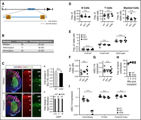
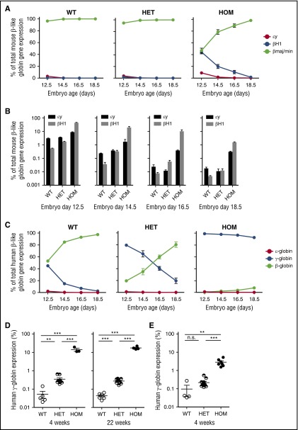
BCL11A, a repressor of human fetal (γ-)globin expression, is required for immune and hematopoietic stem cell functions and brain development. Regulatory sequences within the gene, which are subject to genetic variation affecting fetal globin expression, display hallmarks of an erythroid enhancer in cell lines and transgenic mice. As such, this enhancer is a novel, attractive target for therapeutic gene editing. To explore the roles of such sequences in vivo, we generated mice in which the orthologous 10-kb intronic sequences were removed. Bcl11a enhancer-deleted mice, Bcl11a(Δenh), phenocopy the BCL11A-null state with respect to alterations of globin expression, yet are viable and exhibit no observable blood, brain, or other abnormalities. These preclinical findings provide strong in vivo support for genetic modification of the enhancer for therapy of hemoglobin disorders.


Figures

Figure 1
Figure 1
Loss of the Bcl11a erythroid enhancer has no effect outside of the erythroid compartment. (A) Diagram of the murine Bcl11a locus showing the location of the intronic 10-kb element and the 2 orthologous DHSs. Dashed lines indicate position of the deletions made in the Bcl11a(Δenh) and single +62DHS deletion mouse lines. (B) Bcl11a(Δenh) mice are viable. Heterozygous enhancer deleted female and male mice were bred together. Numbers of combined progeny and their genotypes from these breedings were tallied. Expected percentages based on normal Mendelian genetics are indicated in parentheses. (C) The integrity of the cortical and subcortical organization of Bcl11a(Δenh) mice is maintained. At postnatal day 4 (P4), mice were perfused with 4% paraformaldehyde for preparation of brain specimens. (i and ii) After sectioning, tissue was stained for nuclei (DAPI-blue), BCL11A-red (mouse anti-BCL11A clone 14B5 [Abcam]), and BCL11B-green (rat anti-BCL11B [Abcam]). (i’ and ii’) BCL11A expression in Bcl11a(Δenh) mice is comparable to wild-type levels. (i’’ and ii”) The intensity of BCL11B staining establishes the boundary between neocortical layers V and VI. (iii) The number of BCL11B positive neurons is unaffected by the loss of the Bcl11a enhancer. (iv) Quantification of the thickness of the BCL11B defined cortical layers indicates no significant difference in the depth of each layer. Cells positive for indicated marker(s) were counted in a box of predefined size and applied to both wild-type and Bcl11a(Δenh) single confocal slices. Each layer was defined as a percentage of the total cortical thickness. Sections were mounted using DAPI-Fluoromount G (Southern Biotech) and images obtained at room temperature by a Zeiss AxioCam MRm camera attached to a Zeiss LSM 700 microscope using ZEN Black 2011 acquisition software (Zeiss). Images were processed for brightness and cropped using Photoshop. All transformations were applied evenly across all images. (D) Mature blood cell production and HSCs were unaffected by the loss of the 10-kb Bcl11a enhancer. Peripheral blood was harvested from the tail veins of 4-month-old animals and analyzed by flow cytometry for B cells (B220+/CD19+), T cells (CD3e+/NK1-1), and myeloid cells (Gr-1+/Mac1+). (E) Bcl11a(Δenh) mice had normal numbers of B-cell progenitors (PreProB: B220+, CD43+, IgM, AA4-1+, CD19; ProB+PreB: B220+, CD43+, IgM, AA4-1+, CD19+) and differentiated B cells (B220+, IgM+). (F) Sixteen-week Bcl11a(Δenh) mice have normal BM HSC frequencies as defined by Lineage, Sca-1+, c-kit+ CD48, Flt3, and CD150+. (G) Bcl11a expression in HSCs does not change in Bcl11a(Δenh) mice. Greater than 5000 HSCs (Lineage, Sca-1+, c-kit+ CD48, and CD150high) were sorted from 32- to 35-week-old mouse BM and RNA isolated. Quantitative polymerase chain reaction (qPCR) shows no difference in Bcl11a expression between the genotypes when normalized to GAPDH. (H) Bcl11a(Δenh) HSCs are functional. Whole BM was isolated from either wild-type (CD45.2) or Bcl11a(Δenh) (CD45.2) mice and transplanted into lethally irradiated recipients (CD45.1/.2). Engraftment was measured as the CD45.2+ percentage of total viable cells. (I) The 10-kb enhancer only affects erythroid Bcl11a transcription. Date of vaginal plug detection was designated E0.5. RNA analysis of Bcl11a expression in embryonic day 16.5 (E16.5) tissues shows decreased Bcl11a expression in the erythroid compartment of homozygous Bcl11a(Δenh) mice, but not in B cells or brain tissue. (Data are represented as mean ± standard error of the mean; ****P < .0001; n.s., not significant; all conditions represent n ≥ 4). DAPI, 4′,6-diamidino-2-phenylindole.
Figure 2
Figure 2
Deletion of erythroid specific enhancer sequences delays embryonic and fetal globin gene silencing. (A) Fetal liver was dissected from indicated days of embryonic development. qPCR was done on RNA from the dissected tissue. Globin genes were normalized to GAPDH expression. Delayed hemoglobin switching of the mouse embryonic globins εy and βH1 was observed in Bcl11a(Δenh) mice. (B) Derepression of mouse embryonic globins (εy and βH1) in developmental erythropoiesis as assessed by qPCR. (C) Bcl11a(Δenh) mice were mated with β-YAC mice in order to evaluate human globin gene expression. Bcl11a(Δenh) mice failed to silence γ-globin gene expression during embryonic development. (D) qPCR analysis of RNA from 4-week and 22-week adult mice peripheral blood showed incomplete silencing of γ-globin. (E) Deletion of the in vitro defined core enhancer (+62DHS) region also fails to completely silence γ-globin; however, derepression is not to the level seen in Bcl11a(Δenh) mice. (Data are represented as mean ± standard error of the mean; ***P < .001; **P < .01; n.s., not significant; all conditions represent n ≥ 4.)

References

    1. Lettre G, Sankaran VG, Bezerra MA, et al. DNA polymorphisms at the BCL11A, HBS1L-MYB, and beta-globin loci associate with fetal hemoglobin levels and pain crises in sickle cell disease. Proc Natl Acad Sci USA. 2008;105(33):11869–11874. - PMC - PubMed
    1. Menzel S, Garner C, Gut I, et al. A QTL influencing F cell production maps to a gene encoding a zinc-finger protein on chromosome 2p15. Nat Genet. 2007;39(10):1197–1199. - PubMed
    1. Uda M, Galanello R, Sanna S, et al. Genome-wide association study shows BCL11A associated with persistent fetal hemoglobin and amelioration of the phenotype of beta-thalassemia. Proc Natl Acad Sci USA. 2008;105(5):1620–1625. - PMC - PubMed
    1. Sankaran VG, Menne TF, Xu J, et al. Human fetal hemoglobin expression is regulated by the developmental stage-specific repressor BCL11A. Science. 2008;322(5909):1839–1842. - PubMed
    1. Sankaran VG, Xu J, Ragoczy T, et al. Developmental and species-divergent globin switching are driven by BCL11A. Nature. 2009;460(7259):1093–1097. - PMC - PubMed