. 2016 Nov 28;90(24):11075-11086.
doi: 10.1128/JVI.01563-16. Print 2016 Dec 15.

The Tetherin Antagonism of the Ebola Virus Glycoprotein Requires an Intact Receptor-Binding Domain and Can Be Blocked by GP1-Specific Antibodies

Affiliations

The Tetherin Antagonism of the Ebola Virus Glycoprotein Requires an Intact Receptor-Binding Domain and Can Be Blocked by GP1-Specific Antibodies

Constantin Brinkmann et al. J Virol. .

Abstract

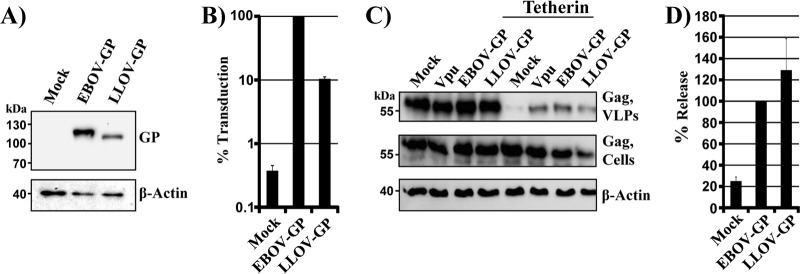
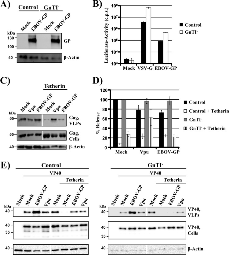
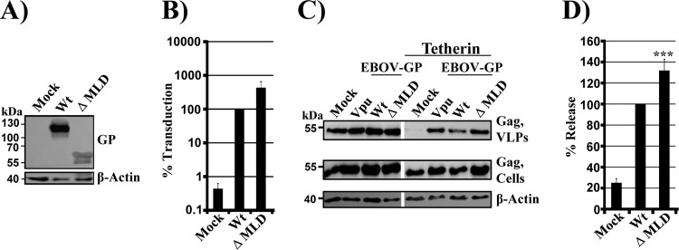
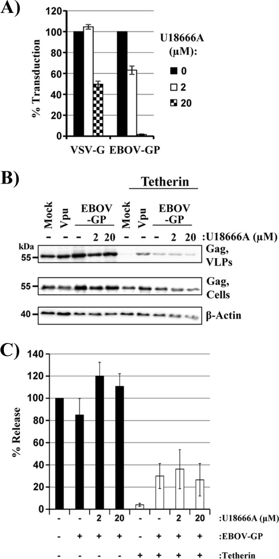
The glycoprotein of Ebola virus (EBOV GP), a member of the family Filoviridae, facilitates viral entry into target cells. In addition, EBOV GP antagonizes the antiviral activity of the host cell protein tetherin, which may otherwise restrict EBOV release from infected cells. However, it is unclear how EBOV GP antagonizes tetherin, and it is unknown whether the GP of Lloviu virus (LLOV), a filovirus found in dead bats in Northern Spain, also counteracts tetherin. Here, we show that LLOV GP antagonizes tetherin, indicating that tetherin may not impede LLOV spread in human cells. Moreover, we demonstrate that appropriate processing of N-glycans in tetherin/GP-coexpressing cells is required for tetherin counteraction by EBOV GP. Furthermore, we show that an intact receptor-binding domain (RBD) in the GP1 subunit of EBOV GP is a prerequisite for tetherin counteraction. In contrast, blockade of Niemann-Pick disease type C1 (NPC1), a cellular binding partner of the RBD, did not interfere with tetherin antagonism. Finally, we provide evidence that an antibody directed against GP1, which protects mice from a lethal EBOV challenge, may block GP-dependent tetherin antagonism. Our data, in conjunction with previous reports, indicate that tetherin antagonism is conserved among the GPs of all known filoviruses and demonstrate that the GP1 subunit of EBOV GP plays a central role in tetherin antagonism.

Importance: Filoviruses are reemerging pathogens that constitute a public health threat. Understanding how Ebola virus (EBOV), a highly pathogenic filovirus responsible for the 2013-2016 Ebola virus disease epidemic in western Africa, counteracts antiviral effectors of the innate immune system might help to define novel targets for antiviral intervention. Similarly, determining whether Lloviu virus (LLOV), a filovirus detected in bats in northern Spain, is inhibited by innate antiviral effectors in human cells might help to determine whether the virus constitutes a threat to humans. The present study shows that LLOV, like EBOV, counteracts the antiviral effector protein tetherin via its glycoprotein (GP), suggesting that tetherin does not pose a defense against LLOV spread in humans. Moreover, our work identifies the GP1 subunit of EBOV GP, in particular an intact receptor-binding domain, as critical for tetherin counteraction and provides evidence that antibodies directed against GP1 can interfere with tetherin counteraction.


Figures

FIG 1
FIG 1
LLOV GP is a tetherin antagonist. (A) Plasmids encoding V5-tagged versions of the indicated glycoproteins were transiently transfected into 293T cells. Transfection of empty plasmid (Mock) served as a negative control. Glycoprotein expression in cell lysates was detected by Western blotting, using anti-V5 antibody. Detection of β-actin served as a loading control. The results were confirmed in two separate experiments. (B) MLV vectors bearing the indicated glycoproteins were used to transduce 293T cells, and luciferase activities in cell lysates were measured at 72 h postransduction. Transduction mediated by EBOV GP wt was set as 100%. The averages and standard errors of the mean (SEM) of five independent experiments are shown. (C) 293T cells were transiently transfected with plasmids encoding HIV Gag, tetherin, and the indicated viral glycoproteins or with empty plasmid as a negative control (Mock). HIV-1 Vpu served as a positive control for tetherin antagonism. The presence of Gag in supernatants and cell lysates was determined by Western blotting using an anti-Gag antibody. Detection of β-actin in cell lysates served as a loading control. (D) Averages of four independent experiments conducted as described for panel C and quantified via the ImageJ program. Release of Gag from cells coexpressing EBOV GP and tetherin was set as 100%.
FIG 2
FIG 2
Processing of N-glycans is required for tetherin antagonism by EBOV GP, but not Vpu. (A) A plasmid encoding EBOV GP was transiently transfected into control 293T or GnTI cells. Transfection of empty plasmid (Mock) served as a negative control. Glycoprotein expression in cell lysates was detected by Western blotting, using serum raised against the GP1 subunit of EBOV GP. Detection of β-actin served as a loading control. Three separate experiments yielded similar results. (B) Equal volumes of MLV vectors produced in control or GnTI cells and bearing the indicated viral glycoproteins were used to transduce 293T cells. At 72 h postransduction, luciferase activities were measured in cell lysates. The results of a single representative experiment carried out with triplicate samples are shown. The error bars indicate standard deviations (SD). Similar results were obtained in three separate experiments. c.p.s., counts per second. (C) GnTI cells were transfected with plasmids encoding HIV Gag, the indicated viral glycoproteins, and tetherin or with empty plasmid (Mock). HIV-1 Vpu served as a positive control for tetherin antagonism. The presence of Gag protein in culture supernatants and cell lysates was determined by Western blotting. Detection of β-actin in cell lysates served as a loading control. (D) Averages of five independent experiments with control and GnTI cells conducted as described for panel C and quantified via the ImageJ program. The release of Gag from cells expressing only Gag without any antagonist and without tetherin was set as 100%; the error bars indicate SEM. (E) Control and GnTI cells were transfected with plasmids encoding VP40 harboring a myc tag, the indicated viral glycoproteins, and tetherin or with empty plasmid (Mock). HIV-1 Vpu served as a positive control for tetherin antagonism. The presence of VP40 in culture supernatants and cell lysates was determined by Western blotting using an anti-myc antibody. The results of single blots are shown, from which irrelevant lanes were cut out. Detection of β-actin in cell lysates served as a loading control. Similar results were obtained in three separate experiments.
FIG 3
FIG 3
The MLD in EBOV GP is dispensable for tetherin antagonism. (A) Plasmids encoding the indicated viral glycoproteins were transiently transfected into 293T cells. Transfection of empty plasmid (Mock) served as a negative control. Glycoprotein expression in cell lysates was detected by Western blotting using serum raised against the GP1 subunit of EBOV GP. Detection of β-actin served as a loading control. Three separate experiments yielded similar results. (B) Equal volumes of MLV vectors bearing the indicated glycoproteins were used for transduction of 293T target cells. Luciferase activities in cell lysates were measured at 72 h postransduction. Transduction mediated by EBOV GP wt was set as 100%. The averages and SEM of five independent experiments are shown. (C) 293T cells were transiently transfected with plasmids encoding HIV Gag, the indicated viral glycoproteins, and tetherin or with empty plasmid (Mock). HIV-1 Vpu served as a positive control for tetherin antagonism. The presence of Gag protein in supernatants and cell lysates was determined by Western blotting using an anti-Gag antibody. Detection of β-actin in cell lysates served as a loading control. (D) Averages of at least five independent experiments conducted as described for panel C and quantified via the ImageJ program. The release of Gag from cells expressing GP and tetherin was set as 100%; the error bars indicate SEM. ***, P < 0.0001.
FIG 4
FIG 4
EBOV GP requires an intact receptor-binding domain for tetherin antagonism. (A) Amino acid sequence alignment of portions (residues 85 to 125 in EBOV GP) of filovirus RBDs that harbor the amino acid residues investigated for tetherin antagonism (green; numbering according to EBOV GP). *, positions which have a single, fully conserved residue; :, conservation between groups of strongly similar properties (scoring >0.5 in the Gonnet PAM 250 matrix); ., conservation between groups of weakly similar properties (scoring ≤0.5 in the Gonnet PAM 250 matrix). (B) Plasmids encoding the indicated viral glycoproteins were transiently transfected into 293T cells. Transfection of empty plasmid (Mock) served as a negative control. Glycoprotein expression in cell lysates was detected by Western blotting, using serum raised against GP1 of EBOV GP. Detection of β-actin served as a loading control. Four independent experiments yielded highly comparable results. (C) Equal volumes of MLV vectors bearing the indicated viral glycoproteins were used to transduce 293T cells. Luciferase activity in cell lysates was measured at 72 h postransduction. Transduction mediated by EBOV GP wt was set as 100%. The averages and SEM of five independent experiments are shown. (D) 293T cells were transfected with plasmids encoding HIV Gag, tetherin, and the indicated viral glycoproteins or empty plasmid (Mock). HIV-1 Vpu served as a positive control for tetherin antagonism. The presence of HIV Gag in culture supernatants and cell lysates was determined by Western blotting. Detection of β-actin served as a loading control. (E) Averages of at least five independent experiments conducted as described for panel C and quantified via the ImageJ program. The release of Gag from cells coexpressing GP and tetherin was set as 100%; the error bars indicate SEM. (F) 293T cells were transiently transfected with plasmids encoding VP40 harboring a myc tag, tetherin, and the indicated viral glycoproteins or with empty plasmid (Mock). HIV-1 Vpu served as a positive control for tetherin antagonism. The presence of VP40 in culture supernatants and cell lysates was determined by Western blotting. Detection of β-actin in cell lysates served as a loading control. *, P < 0.05; ***, P < 0.0001.
FIG 5
FIG 5
U18666A does not block tetherin antagonism by the Ebola virus glycoprotein. 293T cells were treated with the indicated concentrations of compound, incubated with equal volumes of VSV pseudotypes bearing VSV G or EBOV GP, and luciferase activities in the cell lysates were determined at 16 h postransduction. The averages of two independent experiments performed with triplicate samples are shown; the error bars indicate SEM. Transduction in the absence of inhibitor was set as 100%. (B) 293T cells were cotransfected with plasmids encoding HIV Gag, tetherin, and EBOV GP or with empty plasmid (Mock). HIV-1 Vpu served as a positive control for tetherin antagonism. At 12 h posttransfection, the indicated concentrations of U18666A were added to cultures expressing EBOV GP. The presence of HIV Gag in culture supernatants and cell lysates was determined by Western blotting. Detection of β-actin served as a loading control. (C) Averages of three independent experiments conducted as described for panel B and quantified via the ImageJ program. The release of Gag from untreated, tetherin-negative control cells was set as 100%; the error bars indicate SEM.
FIG 6
FIG 6
Mutations in the receptor-binding domain of the Ebola virus glycoprotein that interfere with tetherin antagonism are compatible with tetherin binding. (A) 293T cells were cotransfected with plasmids encoding tetherin with an N-terminal AU1 tag and EBOV GP wt or the indicated EBOV GP mutants. Coimmunoprecipitation was performed with anti-AU1 antibody coupled to agarose beads, and proteins in cell lysates and in precipitates were detected by Western blotting, employing rabbit serum raised against the GP1 subunit of EBOV GP and a rabbit monoclonal antibody directed against tetherin. The results of a single representative experiment, confirmed in a separate experiment, are shown. (B and C) HeLa cells were transfected with plasmids encoding EBOV GP or the indicated EBOV GP mutants or with empty plasmid as a control. For the PLA, the cells were stained with anti-tetherin and anti-EBOV GP primary antibodies. The images were analyzed by automatically counting the red spots of 20 transfected cells per sample using Volocity software (version 6.3). (B) Representative microscopy images. (C) Mean values and standard deviations of the relative numbers of PLA spots per cell (n = 20). The PLA spot count for cells transfected with EBOV GP wt was set as 100%. DAPI, 4′,6-diamidino-2-phenylindole.
FIG 7
FIG 7
Evidence that an antibody directed against the GP1 subunit of EBOV GP can block tetherin counteraction. (A) 293T cells were transiently transfected with plasmids encoding HIV Gag and EBOV GP or with empty plasmid (Mock). HIV-1 Vpu served as a positive control for tetherin antagonism. At 16 h posttransfection, the medium was replaced with fresh culture medium supplemented with the indicated antibodies at a final concentration of 20 μg/ml or with PBS. The presence of Gag in culture supernatants and cell lysates was determined by Western blotting. Detection of β-actin served as a loading control. mAb, monoclonal antibody. (B) Averages of two to five independent experiments conducted as described for panel A and quantified via the ImageJ program. The release of Gag from cells expressing GP and treated with PBS was set as 100%; the error bars indicate SEM. (C) The experiment was conducted as described for panel A but cells coexpressing tetherin were examined. (D) Averages of three independent experiments conducted as described for panel A and quantified via the ImageJ program. The release of Gag from cells expressing GP and tetherin and treated with PBS was set as 100%; the error bars indicate SEM. The dashed line indicates the assay background, which was defined by VLP release from tetherin-positive cells, which do not express a tetherin antagonist. *, P < 0.05; ***, P < 0.0001.

Similar articles

Cited by

References

    1. Ebola Response Team WHO. 2016. Ebola virus disease among male and female persons in West Africa. N Engl J Med 374:96–98. doi:10.1056/NEJMc1510305. - DOI - PMC - PubMed
    1. WHO. 17 February 2016. Ebola situation report-17 February 2016. http://apps.who.int/ebola/current-situation/ebola-situation-report-17-fe....
    1. Basler CF. 2015. Innate immune evasion by filoviruses. Virology 479-480:122–130. doi:10.1016/j.virol.2015.03.030. - DOI - PMC - PubMed
    1. Misasi J, Sullivan NJ. 2014. Camouflage and misdirection: the full-on assault of ebola virus disease. Cell 159:477–486. doi:10.1016/j.cell.2014.10.006. - DOI - PMC - PubMed
    1. Schoggins JW, Wilson SJ, Panis M, Murphy MY, Jones CT, Bieniasz P, Rice CM. 2011. A diverse range of gene products are effectors of the type I interferon antiviral response. Nature 472:481–485. doi:10.1038/nature09907. - DOI - PMC - PubMed

MeSH terms

LinkOut - more resources