. 2016 Sep 30;353(6307):aah3374.
doi: 10.1126/science.aah3374.

Pathological α-synuclein transmission initiated by binding lymphocyte-activation gene 3

Affiliations

Pathological α-synuclein transmission initiated by binding lymphocyte-activation gene 3

Xiaobo Mao et al. Science. .

Abstract

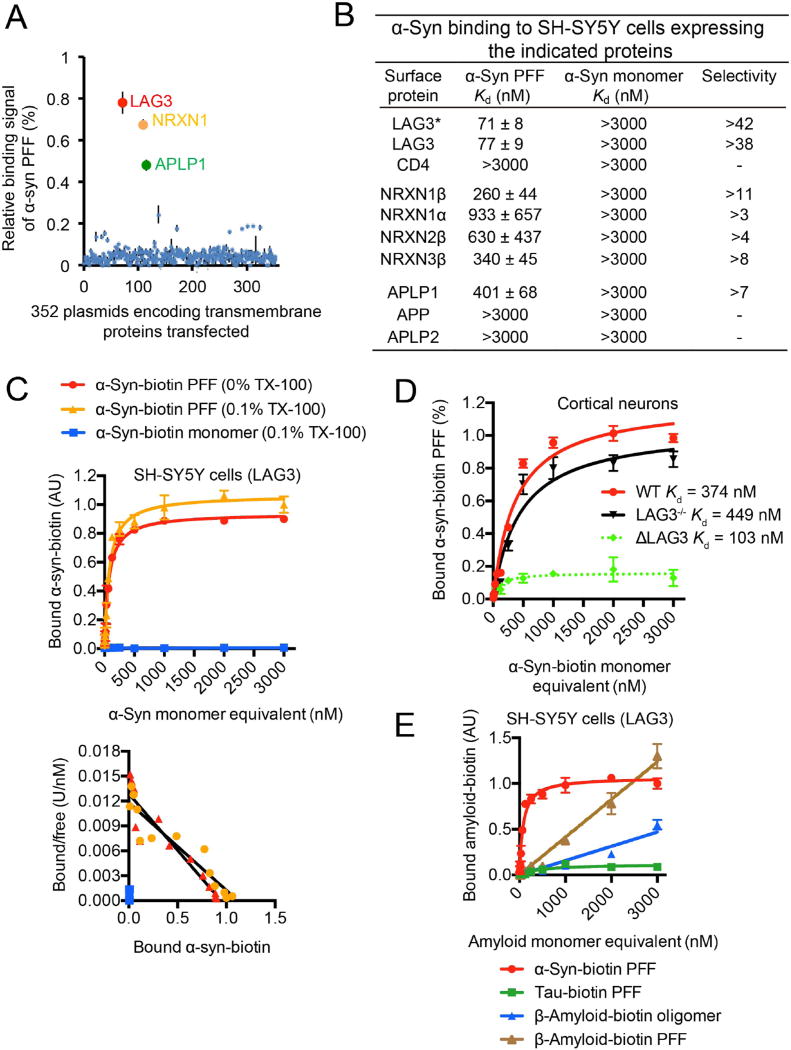
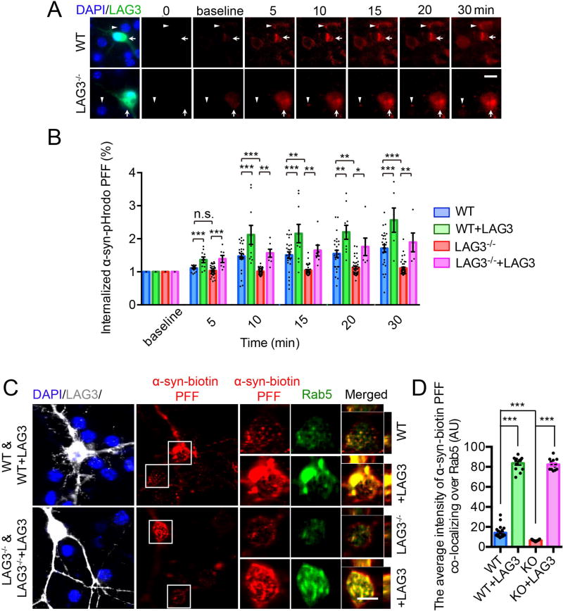
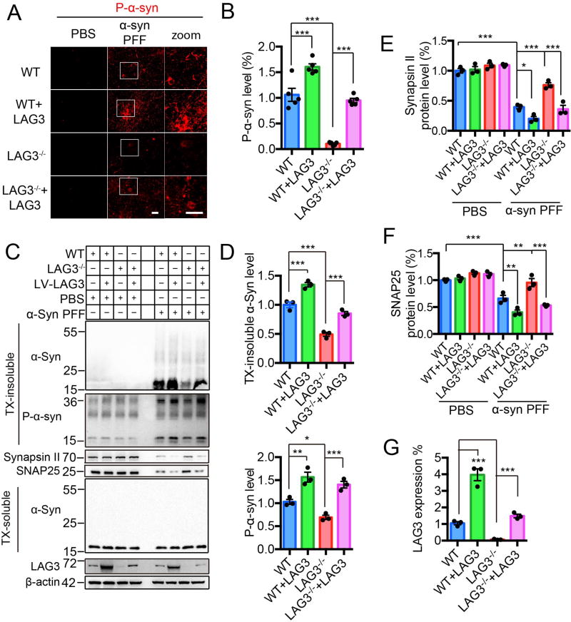
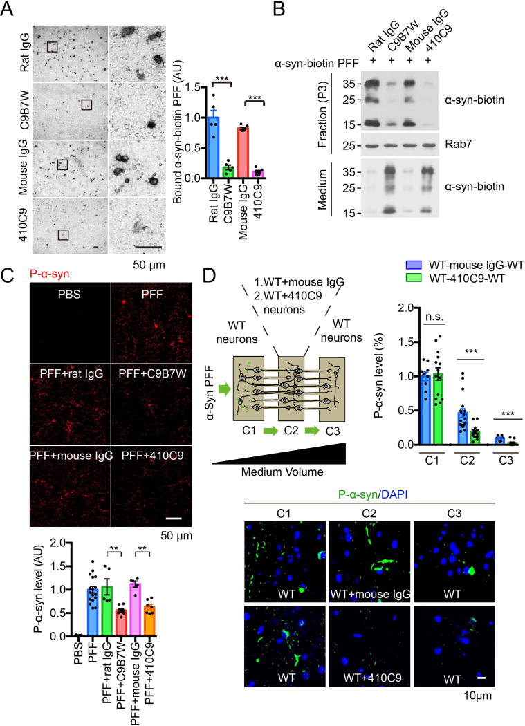
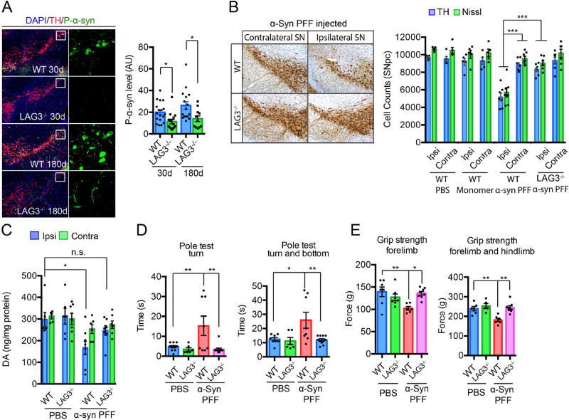
Emerging evidence indicates that the pathogenesis of Parkinson's disease (PD) may be due to cell-to-cell transmission of misfolded preformed fibrils (PFF) of α-synuclein (α-syn). The mechanism by which α-syn PFF spreads from neuron to neuron is not known. Here, we show that LAG3 (lymphocyte-activation gene 3) binds α-syn PFF with high affinity (dissociation constant = 77 nanomolar), whereas the α-syn monomer exhibited minimal binding. α-Syn-biotin PFF binding to LAG3 initiated α-syn PFF endocytosis, transmission, and toxicity. Lack of LAG3 substantially delayed α-syn PFF-induced loss of dopamine neurons, as well as biochemical and behavioral deficits in vivo. The identification of LAG3 as a receptor that binds α-syn PFF provides a target for developing therapeutics designed to slow the progression of PD and related α-synucleinopathies.


Figures

Fig. 1
Fig. 1. α-Syn PFF binds to LAG3
(A) Individual clones from a library consisting of 352 individual cDNAs encoding transmembrane proteins (GFC-transfection array panel, Origene) were transfected into SH-SY5Y cells, and the relative binding signals of human α-syn PFF to individual transmembrane proteins are shown. Positive candidates are LAG3 (NM_002286), NRNX1 (NM_138735) and APLP1 (NM_005166). (B) Mouse α-syn-biotin monomer and α-syn-biotin PFF binding affinity to SH-SY5Y cells expressing the indicated proteins. LAG3* Kd assessment was performed without Triton X-100. All other experiments were performed with 0.1% Triton X-100. Transmembrane proteins similar to the candidates were also tested. Quantification of bound α-syn-biotin PFF to the candidates was performed with ImageJ. Kd values are means ± SEM and are based on monomer equivalent concentrations. Selectivity was calculated by dividing Kd (monomer) by Kd (PFF). Binding of α-syn-biotin monomer was detected at a concentration of 3000 nM, but binding was not saturable. (C) α-Syn-biotin monomer or α-syn-biotin PFF binding to LAG3-overexpressing SH-SY5Y cells as a function of total α-syn concentration in 0% Triton X-100 (TX-100) or 0.1% TX-100 conditions (monomer equivalent for PFF preparations, top panel). Scatchard analysis (bottom panel). Kd= 71 nM (0% TX-100) and 77 nM (0.1% TX-100), data are the means ± SEM, n = 3. (D) Binding of α-syn-biotin PFF to cultured cortical neurons (21 days in vitro (DIV)) is reduced by LAG3 knockout (LAG3−/−), as assessed by alkaline phosphatase assay. α-Syn-biotin PFF WT-Kd = 374 nM, LAG3−/−-Kd = 449 nM, estimated Kd for neuronal LAG3 [dashed line: ΔLAG3 = wild-type (WT) minus LAG3−/−] is 103 nM. Data are the means ± SEM, n = 3. * P < 0.05, Student’s t-test. Power (1-β err prob) = 1. (E) Specificity of LAG3 binding with α-syn-biotin PFF (Fig. S4). Tau-biotin PFF (Fig. S8), β-amyloid-biotin oligomer and β-amyloid-biotin PFF (Fig. S9) are negative controls.
Fig. 2
Fig. 2. Endocytosis of α-syn PFF is dependent on LAG3
(A) Live image analysis of the endocytosis of α-syn-pHrodo PFF. α-Syn PFF was conjugated with a pH dependent dye (pHrodo red), in which fluorescence increases as pH decreases from neutral to acidic environments. White triangles indicate non-transfected wild-type (WT) or LAG3−/− neurons and white arrows indicate LAG3 transfected neurons. Scale bar, 10 µm. (B) Quantification of panel A, cell number (–46) from n = 3. (C) Internalized α-syn-biotin PFF co-localizes with Rab5. Co-localization of internalized α-syn-biotin PFF and Rab5 was assessed by confocal microscopy, scale bar, 10 µm. (D) Quantification of panel C, cell number (–32) from n = 4. One-way ANOVA with Tukey’s correction. Data in B and D are as means ± SEM. *P < 0.05, **P < 0.01, ***P < 0.001. Power (1-β err prob) = 1.
Fig. 3
Fig. 3. α-Syn PFF induced pathology is reduced by deletion of LAG3 in vitro
(A) WT and LAG3−/− primary cortical neurons at 7 DIV were treated with α-syn PFF or PBS. LAG3 was overexpressed via Lenti-virus (LV) transduction in WT or LAG3−/− neurons at 4 DIV. 3 days after transduction, 7 DIV cultures were treated with α-syn PFF or PBS. All the cultures were fixed 10-day post-treatment in 4% PFA. Neurons were stained with rabbit mAb MJF-R13 (8-8) for P-α-syn. Scale bar, 40 µm. (B) Quantification of panel A, n = 5 independent experiments, each performed in duplicate. Values are given as the means ± SEM. Statistical significance was determined using one-way ANOVA followed with Tukey’s correction, ***P < 0.001. Power (1-β err prob) = 1. (C) Immunoblots in WT and LAG3−/− neuron lysates of misfolded α-syn, P-α-syn, synapsin II, SNAP25 and LAG3. β-actin served as a loading control. WT and LAG3−/− neuron lysates were sequentially extracted in 1% TX-100 (TX-soluble) followed by 2% SDS (TX-insoluble) 14 days after α-syn PFF treatment. α-Syn PFF recruited endogenous α-Syn into TX-insoluble and hyperphosphorylated aggregates, which was ameliorated by deletion of LAG3. α-Syn PFF caused a reduction in levels of SNAP25 and synapsin II compared to PBS 14 days post-treatment. Deletion of LAG3 prevented PFF-induced synaptic protein loss. (D–G) Quantification of panel C. Values are given as means ± SEM, n = 3 independent experiments. Statistical significance was determine using one-way ANOVA followed by Tukey’s correction, *P < 0.05, **P < 0.01, ***P < 0.001.
Fig. 4
Fig. 4. α-Syn PFF transmission is reduced by deletion of LAG3 in vitro
(A) Schematic representation of the 3 chambers in which neurons were cultured in chamber 1 (C1) chamber 2 (C2) and chamber 3 (C3) (top) or C1 and C3 (bottom). (B) α-Syn PFF was added to C1 of the microfluidic device. On day 14, α-syn-biotin PFF was detected via P-α-syn in C2 and C3 when neurons were present in all three chambers. Transmission to C3 is not detectable when neurons are not present in C2. (C) Quantification of immunofluorescent images in B. Data are the means ± SEM, n = 3. one-way ANOVA followed by Sidak’s correction. ***P < 0.001 versus C1. Power (1-β err prob) = 1. Scale bars, 100 µm. (D) Schematic of microfluidic neuron device with three chambers to separate neurons seeded in three chambers. (E)Transmission of pathologic P-α-syn from chamber 1 (C1) to chamber 2 (C2) to chamber 3 (C3) 14 days post-addition of α-syn PFF in C1. The different combinations of neurons tested in C2, listed as C1-(C2)-C3, are: WT-(WT)-WT, WT-(WT+LAG3)-WT, WT-(LAG3−/−)-WT, WT-(LAG3−/−+LAG3)-WT. Scale bar, 10 µm. (F) Quantification of panel E. Values are given as means ± SEM, n = 3. Statistical significance was determine using one-way ANOVA followed by Tukey’s correction, *P < 0.05, **P < 0.01, ***P < 0.001. Power (1-β err prob) = 1.
Figure 5
Figure 5. Anti-LAG3 antibodies block α-syn PFF binding, endocytosis, pathology and transmission
(A) Anti-LAG3 antibodies C9B7W and 410C9 (both 50 µg/mL) block the binding of α-syn-biotin PFF (500 nM) on SH-SY5Y cells expressing LAG3, Scale bar, 50 µm. Right panel, quantification of images in Panel A. Data are the means ± SEM, n =3, Student’s t-test. ***P < 0.001. (B) Anti-LAG3 antibodies C9B7W and 410C9 (both 50 µg/mL) reduced the endocytosis of α-syn-biotin PFF (1 µM) in 12 DIV primary cortical neurons. Rab7 was used to confirm the isolation of endosomes. (C) Phosphorylated α-syn (P-α-syn) as detected by rabbit mAb MJF-R13 (8-8) was reduced by anti-LAG3 antibodies in primary cortical neurons. Scale bar, 50 µm. Right panel, quantification of images in Panel C. Data are the means ± SEM, n = 3, one-way ANOVA followed by Tukey’s correction. **P < 0.01. (D) Anti-LAG3 410C9 antibody delays α-syn PFF transmission in neurons. Left panel is a schematic representation of the 3 microchambers in which neurons were cultured in C1, C2 and C3. α-syn PFF was added to C1 of the microfluidic device on day 7. Mouse IgG or anti-LAG3 410C9 antibody (both 50 µg/mL) was added to C2 on day 7. α-syn PFF transmission was detected via P-α-syn immunostaining in C2 and C3 on day 21. Scale bar, 10 µm. Right panel, quantification of images in bottom panel. Data are the means ± SEM, N =3, one-way ANOVA followed by Tukey’s correction. ***P < 0.001, n.s., non-significant.
Fig. 6
Fig. 6. α-Syn PFF induced pathology is reduced by deletion of LAG3 in vivo
(A) Representative P-α-syn immunostaining and quantification in the substantia nigra par compacta (SNpc) of WT and LAG3−/− mice sacrificed at 30 and 180 days after intrastriatal α-syn PFF injection. Data are the means ± SEM, n = 5–9 mice per group, one-way ANOVA with Sidak’s correction. (B) Stereology counts from TH immunostaining and Nissl staining of SNpc DA neurons of WT and LAG3−/− mice at 180 days after intrastriatal α-syn PFF, α-syn monomer or PBS injection. Data are the mean number of cells per region ± SEM, n = 5–9 mice per group, one-way ANOVA with Dunnett’s correction. (C) DA concentrations in the striatum of α-syn PFF-injected mice and PBS-treated controls measured at 180 days by HPLC. Data are the means ± SEM, n = 5–8 mice per group, one-way ANOVA with Tukey’s correction. (D, E) 180 days after α-syn PFF injection, the pole test and grip strength was performed in WT or LAG3−/− mice injected with PBS or α-syn PFF. Behavioral abnormalities in the pole test and grip strength induced by α-syn PFF injection were ameliorated in LAG3−/− mice. Data are the means ± SEM, n = 7–9 mice per group for behavioral studies. Statistical significance was determined using one-way ANOVA with Tukey’s correction, * P < 0.05, ** P < 0.001, *** P < 0.001, n.s., nonsignificant. Power (1-β err prob) = 1.

Comment in

References

    1. Goedert M. Alpha-synuclein and neurodegenerative diseases. Nature reviews. Neuroscience. 2001;2:492–501. - PubMed
    1. Goedert M, Spillantini MG, Del Tredici K, Braak H. 100 years of Lewy pathology. Nature reviews. Neurology. 2013;9:13–24. - PubMed
    1. Lee VM, Trojanowski JQ. Mechanisms of Parkinson's disease linked to pathological alpha-synuclein: new targets for drug discovery. Neuron. 2006;52:33–38. - PubMed
    1. Dehay B, Vila M, Bezard E, Brundin P, Kordower JH. Alpha-synuclein propagation: New insights from animal models. Mov Disord. 2016;31:161–168. - PubMed
    1. Lee SJ, Desplats P, Lee HJ, Spencer B, Masliah E. Cell-to-cell transmission of alpha-synuclein aggregates. Methods Mol Biol. 2012;849:347–359. - PMC - PubMed

Publication types