. 2016 Sep 30;353(6307):1536-1541.
doi: 10.1126/science.aaf7003.

Ventral CA1 neurons store social memory

Affiliations

Ventral CA1 neurons store social memory

Teruhiro Okuyama et al. Science. .

Abstract

The medial temporal lobe, including the hippocampus, has been implicated in social memory. However, it remains unknown which parts of these brain regions and their circuits hold social memory. Here, we show that ventral hippocampal CA1 (vCA1) neurons of a mouse and their projections to nucleus accumbens (NAc) shell play a necessary and sufficient role in social memory. Both the proportion of activated vCA1 cells and the strength and stability of the responding cells are greater in response to a familiar mouse than to a previously unencountered mouse. Optogenetic reactivation of vCA1 neurons that respond to the familiar mouse enabled memory retrieval and the association of these neurons with unconditioned stimuli. Thus, vCA1 neurons and their NAc shell projections are a component of the storage site of social memory.


Figures

Fig. 1
Fig. 1. vHPC in social memory
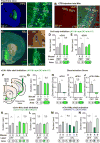
(A) Social discrimination test (SDT). (B) Representative heat map of test-mouse position during SDT. (C) Kinetics of SDT (see Methods). (D to F) Expression of AAV8-CaMKII:eArchT-EYFP (green) in vHPC or dHPC of wild-type mice. Asterisk indicates optic fiber tip. (G, H) Total duration in the sniffing area of familiar mouse-A or novel mouse-B during SDT with inhibition of vHPC (G, O, n = 17) or dHPC (H, P, n = 14). (I) Resident-intruder test. (J, K) Total sniffing duration by the resident to intruder (J, vHPC inhibition; K, dHPC inhibition) in familiar intruder group (left) and novel intruder group (right) (n = 7, each group). (L to N) Total duration in sniffing area during SDT observed in wild-type mice bilaterally injected with AAV8-CaMKII-eArchT-EYFP into vHPC and implanted with optical fibers targeting NAc (L, Q, n = 23), OB (M, R, n = 16), or BLA (N, S, n = 13). (O to S) Comparison of discrimination scores. Green bars, laser-ON; Grey bars, laser-OFF. Significance for multiple comparisons: paired t-test, *p < 0.05; **p < 0.01; ***p < 0.001, n.s., not significant. Data presented as mean ± S.E.M.
Fig. 2
Fig. 2. vCA1-NAc circuit in SDT
(A) Coronal vHPC sections of a Trpc4-Cre mouse injected with AAV9-hsyn:DIO-eArchT-EYFP into vCA1, stained with anti-GFP (green) and DAPI (blue). (B) CTB-Alexa555 (red) injection into NAc and stained with anti-GFP (green) and DAPI (blue). (C) Coronal NAc sections of a Trpc4-Cre mouse injected with AAV9-hsyn:DIO-eArchT-EYFP into vCA1, stained with anti-GFP (green), anti-TH (orange), and DAPI (blue). NAc shell (sh); NAc core (co); Septum (sep); Striatum (str). Boxed areas in (A, C) are magnified. (D, E) Cell body inhibition of vCA1 and dCA1 during SDT in Trpc4-Cre and Wfs1-Cre mice, respectively. (F) Manipulation of vCA1-NAc shell projections. (G, K to N) SDT in Trpc4-Cre mice during vCA1-NAc manipulation. vCA1 injections of AAV9-hsyn:DIO-eArchT-EYFP (G, n =14; K and L, each n = 12) and AAV9-EF1α:DIO-ChR2-EYFP (M and N, each n = 14). (H to J) Comparison of discrimination scores. Bottom, targeting area for the laser-stimulation. mouse-A, familiar mouse; mouse-B and -B′, novel mouse. Green bars, green laser-ON; blue bars, blue laser-ON; grey bars, laser-OFF. Significance for multiple comparisons: paired t-test, *p < 0.05; **p < 0.01; n.s., not significant. Data presented as mean ± S.E.M.
Fig. 3
Fig. 3. Ca 2+ events in vCA1
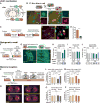
(A) Microendoscope. AAV5-hsyn:DIO-GCaMP6f injection into vCA1 of Trpc4-Cre mice or dCA1 of Wfs1-Cre mice. (B, C) Coronal sections of vCA1 and dCA1 stained with anti-GFP (green, for GCaMP6f) and DAPI (blue). (D) Stacked image acquired during a 15 min microendocope recording. (E) Top, Relative fluorescence changes (ΔF/F) for five vCA1 pyramidal neurons. Bottom, Time-lapse image sequence of GCaMP6f fluorescence in an individual neuron. (F) Top, Experimental protocol for microendoscope recording. Bottom, Relative fluorescence changes during 15 min recording. (G) Representative mouse-A neuron, mouse-B neuron, and neither neuron. Head-position at each Ca2+ event (red dots). (H) Proportion of mouse-A, mouse-B, or neither neurons in vCA1 and dCA1, before and after familiarization (3 days or 2 h) (chi-square test, *p < 0.05). (I to L, N, O) Comparison of the preference scores of mouse-A neurons (I, J, N, red dots) and all recorded neurons (K, L, O, blue dots) between After-1 (30 min) and Before sessions (I, K; linear regression, p = 0.208), between After-1 (30 min) and After-2 (30 min) (J, L, linear regression, p = 0.0002; Spearman rank correlation test, r = 0.65), and between After-1 (30 min) and After-1 (24 h) (N, O, linear regression, p = 0.0019; Spearman rank correlation test, r = 0.23). (M, P) Comparison of Ca2+ event probabilities of mouse-A neurons (ANOVA; post-hoc, Scheffe, ***p < 0.001, **p < 0.01). Data presented as mean ± S.E.M.
Fig. 4
Fig. 4. Social memory engrams
(A) Activity-dependent labeling method. (B) Injection of AAV9-TRE:FT-Slow into vCA1 of c-fos:tTA mice. Top, fluorescent color alteration of FT-Slow. Bottom, coronal vCA1 sections 12 h (left) and 72 h (right) after induction. Representative images of a blue form (pseudo green color)- or red form (red)-expressing cell. (C) Protocol for visualizing two activated neuronal populations. (D) FT-Slow blue form-, red form-, and double (yellow)-positive cells in vCA1 (left) with magnified image (right). (E) Percentage of reactivated cells when the test-mouse was exposed to the same mouse-A twice (A/A) or mouse-A then mouse-B (A/B) (n = 3, each group). (F) Protocol for optogenetic recall of social memory engram. (G) vCA1 section of c-fos:tTA mice injected with AAV9-TRE:ChR2-EYFP showing ChR2-labeling by social interaction. (H to J) SDT with or without activation of engram cells. blue bars, blue laser-ON; grey bars, laser-OFF. Bottom, targeting area for the laser-stimulation. (K) Protocol for memory inception. (L, M, O, P) Proportion of total duration in the sniffing area of mouse-A or mouse-B (yellow bars, pre-test; blue bars, shock-test; red bars, cocaine-test). AAV9-TRE:ChR2-EYFP (H, J, L, M) or AAV9-TRE:EYFP (I, O, P) injected c-fos:tTA mice. (N) Heat map representing nose position of test-mice. H, n = 10; I, n = 6; J, n = 10, L, n = 10; M, n = 14; O, n = 7; P, n = 7; Significance for multiple comparisons: paired t-test (H to I) and ANOVA, post-hoc, Scheffe (L, M, O, P), *p < 0.05; **p < 0.01; n.s., not significant. Data presented as mean ± S.E.M.

Comment in

  • Social memory goes viral.
    Saxena K, Morris RG. Saxena K, et al. Science. 2016 Sep 30;353(6307):1496-1497. doi: 10.1126/science.aai7788. Science. 2016. PMID: 27708089 No abstract available.

Similar articles

Cited by

References

    1. McGraw LA, Young LJ. Trends Neurosci. 2010;33:103–109. - PMC - PubMed
    1. Okuyama T, et al. Science. 2014;343:91–94. - PubMed
    1. Smith CN, et al. Proc Natl Acad Sci U S A. 2014;111:9935–9940. - PMC - PubMed
    1. Quiroga RQ, Reddy L, Kreiman G, Koch C, Fried I. Nature. 2005;435:1102–1107. - PubMed
    1. Viskontas IV, Quiroga RQ, Fried I. Proc Natl Acad Sci U S A. 2009;106:21329–21334. - PMC - PubMed

Publication types