. 2016 Oct 18;113(42):E6437-E6446.
doi: 10.1073/pnas.1603321113. Epub 2016 Oct 5.

Superantigens hyperinduce inflammatory cytokines by enhancing the B7-2/CD28 costimulatory receptor interaction

Affiliations

Superantigens hyperinduce inflammatory cytokines by enhancing the B7-2/CD28 costimulatory receptor interaction

Revital Levy et al. Proc Natl Acad Sci U S A. .

Abstract

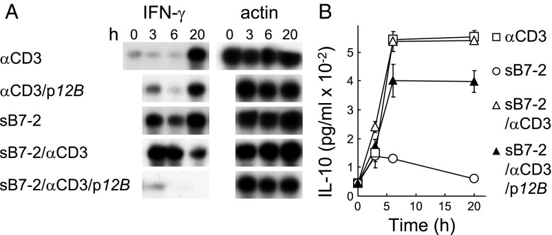
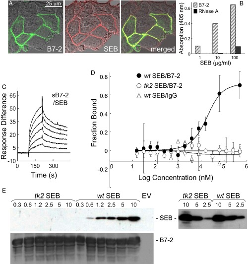
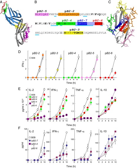
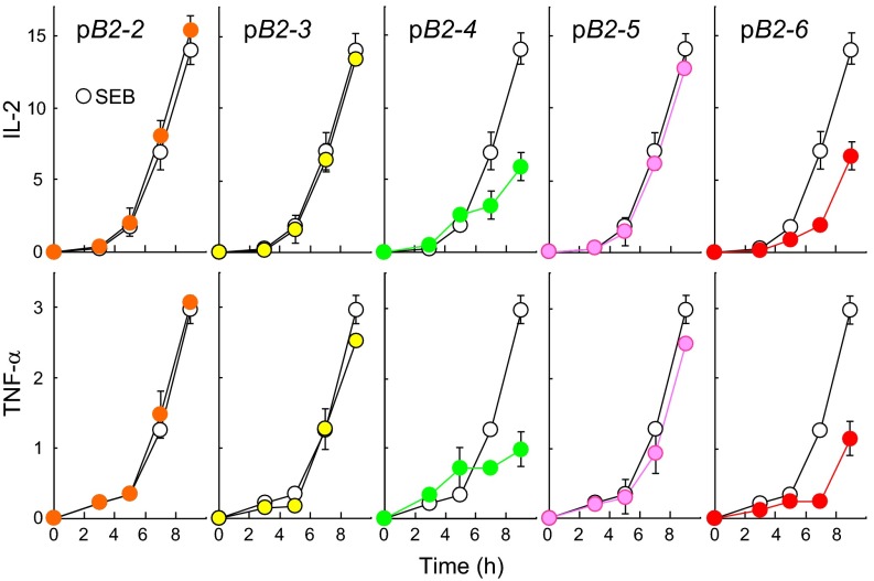
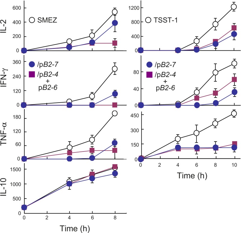
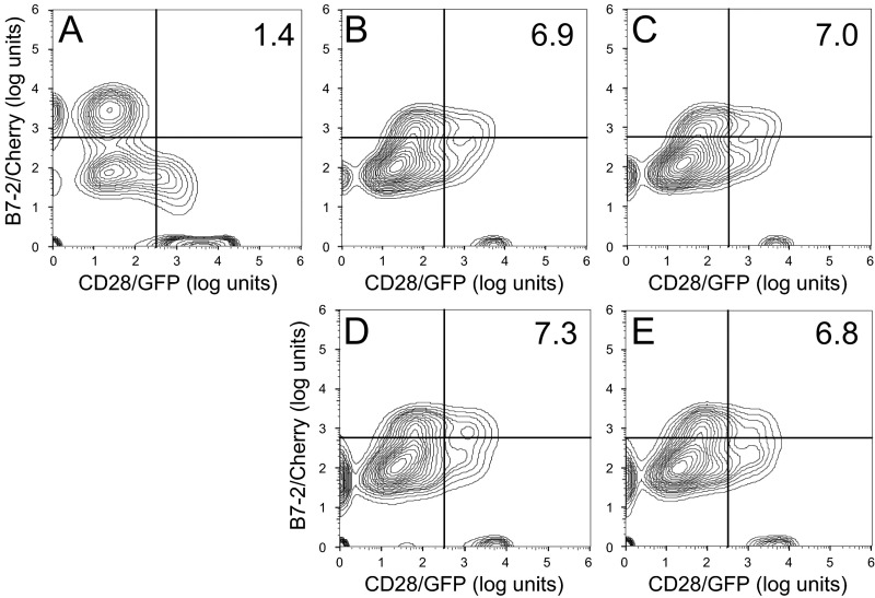
Full T-cell activation requires interaction between the costimulatory receptors B7-2 and CD28. By binding CD28, bacterial superantigens elicit harmful inflammatory cytokine overexpression through an unknown mechanism. We show that, by engaging not only CD28 but also its coligand B7-2 directly, superantigens potently enhance the avidity between B7-2 and CD28, inducing thereby T-cell hyperactivation. Using the same 12-aa β-strand-hinge-α-helix domain, superantigens engage both B7-2 and CD28 at their homodimer interfaces, areas remote from where these coreceptors interact, implying that inflammatory signaling can be controlled through the receptor homodimer interfaces. Short B7-2 dimer interface mimetic peptides bind diverse superantigens, prevent superantigen binding to cell-surface B7-2 or CD28, attenuate inflammatory cytokine overexpression, and protect mice from lethal superantigen challenge. Thus, superantigens induce a cytokine storm not only by mediating the interaction between MHC-II molecule and T-cell receptor but also, critically, by promoting B7-2/CD28 coreceptor engagement, forcing the principal costimulatory axis to signal excessively. Our results reveal a role for B7-2 as obligatory receptor for superantigens. B7-2 homodimer interface mimotopes prevent superantigen lethality by blocking the superantigen-host costimulatory receptor interaction.

Keywords: B7-2 dimer interface; costimulatory receptor; cytokine storm; superantigen.


Conflict of interest statement

The authors declare no conflict of interest.

Figures

Fig. 1.
Fig. 1.
Superantigen mimetic peptide blocks B7-2 signaling. (A and B) p12B inhibits sB7-2/αCD3-mediated induction of IFN-γ mRNA, but not of IL-10. Human PBMCs were incubated with αCD3 monoclonal antibody (0.1 μg/mL), sΒ7-2 (1 μg/mL), or both, with or without p12B (10 μg/mL). mRNA was quantitated by RNase protection analysis; β-actin mRNA indicates equal loading of RNA (A). Secreted IL-10 was determined (B).
Fig. 2.
Fig. 2.
Superantigen binds B7-2 directly. (A) Colocalization of SEB with cell-surface B7-2. Representative field of confocal microscopy is shown. HEK 293T cells were transfected to express B7-2–GFP fusion protein (green) and after 24 h incubated for 30 min with Alexa-Fluor-633–labeled SEB (red). (B) Binding of SEB to immobilized sB7-2 (5 μg/mL), but not to immobilized ribonuclease A (5 μg/mL), determined with anti–SEB-HRP antibody (0.2 μg/mL) in an ELISA. (C) Representative SPR responses for binding of B7-2-Fc in twofold increments from 0.031 μM to immobilized SEB. (D) SEB and B7-2 interact directly as free molecules. Representative MST responses are shown for binding of labeled wt or tk2 SEB (240 nM) to increasing concentrations of B7-2-Fc or for binding of wt SEB to IgG Fc (error bars, SEM; n = 3). (E) Binding of SEB to cell-surface B7-2. HEK 293T cells were transfected to express B7-2 or with empty vector (EV) and after 36 h incubated for 1 h with recombinant wt or tk2 mutant SEB (μg/mL). Cells were washed three times with cold PBS before lysis. Western blots of equal amounts of total cell protein (Bradford assay) with 0.1 μg/mL αSEB antibody followed by 0.2 μg/mL horseradish peroxidase-conjugated donkey anti-mouse IgG (Top) or with 0.1 μg/mL αB7-2 antibody followed by 0.2 μg/mL horseradish peroxidase-conjugated donkey anti-goat IgG (Bottom); autoradiograms are from representative experiments. On the Right, both tk2 and wt recombinant SEB (μg/mL) were detected by Western blot with the αSEB antibody.
Fig. S1.
Fig. S1.
Control proteins do not bind SEB. (A and B) Representative surface plasmon resonance responses for binding of IgG in four twofold increments from 0.25 μM (A) and for binding of 0.1, 0.3, and 1 μM ribonuclease A (B) to immobilized SEB, using the chip of Fig. 2C.
Fig. 3.
Fig. 3.
SEB uses its β-strand(8)/hinge/α-helix(4) domain to bind B7-2. (A and B) Representative SPR responses for binding of B7-2-Fc in twofold increments from 0.031 μM to immobilized p12C (YNKKKATVQELD) (A) or randomly scrambled p12Csc (EKAKYTQLVKDN) (B). (C) Mutation of SEB β-strand(8)/hinge/α-helix(4) domain peptides reduces binding of B7-2. SPR responses to 2 μM B7-2-Fc are compared for immobilized p12C, p12YK (ANAKKATVQELD), and p12KD (YNAKKATVQELA) (underlined residues were mutated).
Fig. 4.
Fig. 4.
Peptide mimetics of the B7-2 homodimer interface are superantigen antagonists. (A) In the CTLA-4/B7-2 complex (1I85.pdb) (1), B7-2 is shown in gray and CTLA-4 in blue, with homodimer interface domains of CTLA-4 in red (YVIDPE) and green (VVLASS) and its B7 binding site (MYPPPY) in yellow. Residues in the crystallographic B7-2 homodimer interface (1) are shown in magenta and pink; those in magenta are represented in mimetic peptides. (B) Peptide mimetics of the B7-2 dimer interface. In the extracellular domain of B7-2, residues in the dimer interface are underlined in cyan; residues that contact CTLA-4 are in boldface. Peptide sequences are marked with colors; in pB2-7, blue-gray residues overlap with pB2-4 and pB2-6, respectively. (C) Location of peptides from B in the B7-2 structure (1I85.pdb), in corresponding colors. (D) Peptides pB2-4 (EKFDSVHSKYM) and pB2-6 (DSDSWTLR) antagonize induction of IFN-γ by SEB. PBMCs were induced with SEB (10 ng/mL), in the absence (open circles) or presence (filled circles) of 0.1 μg/mL peptide as shown. Secreted IFN-γ was determined (pg/mL ×10−2). (E) Synergy between pB2-4 and pB2-6. PBMCs were induced with SEB (0.1 ng/mL), in the absence or presence of 0.01 μg/mL pB2-4, pB2-6, or both. Secreted IL-2, IFN-γ, TNF-α, and IL-10 were determined. (F) Superantigen antagonist activity of B7-2 dimer interface mimetic peptide pB2-7. PBMCs were induced with SEB (0.1 ng/mL) alone or together with 0.1 μg/mL of pB2-7 (MGRTSFDSDS) or 0.01 μg/mL of each pB2-4 and pB2-6. Secreted IL-2, IFN-γ, TNF-α, and IL-10 were determined.
Fig. S2.
Fig. S2.
Peptide mimetics of the B7-2 homodimer interface are superantigen antagonists. Peptides pB2-4 and pB2-6 antagonize induction of IL-2 and TNF-α by SEB. PBMCs were activated by SEB (10 ng/mL) in the absence (open circles) or presence (filled circles) of 0.1 μg/mL peptide as shown. Secreted IL-2 and TNF-α were determined. Colors correspond to those in Fig. 4B. Data are from the experiment shown in Fig. 4D.
Fig. S3.
Fig. S3.
Superantigen antagonist activity of B7-2 homodimer interface mimetic peptide pB2-7. PBMCs were incubated with 0.01 ng/mL SMEZ (Left) or TSST-1 (Right) alone or together with 0.1 μg/mL pB2-7 or 0.01 μg/mL each pB2-4 and pB2-6. Secreted cytokines were determined.
Fig. 5.
Fig. 5.
Peptide mimetics of the B7-2 dimer interface bind SEB and block binding of SEB to cell-surface B7-2 and CD28. (A and B) Representative SPR responses for binding of SEB in twofold increments from 0.78 μM to immobilized pB2-4 (A) and pB2-6 (B); graphical fits to the binding curves are presented in red; kinetic parameters show the specificity of these interactions (Table S1). (CE) Peptide mimetics of the B7-2 or CD28 homodimer interface inhibit binding of SEB to cell-surface B7-2. HEK 293T cells were transfected to express B7-2 or with empty vector (EV) and after 36 h incubated for 1 h without addition (B7-2) or as indicated, with 5 μg/mL αB7-2 antibody, αCD28 monoclonal antibody (12), or 10 μg/mL B7-2 mimetic peptides pB2-2, pB2-4, pB2-6, or pB2-7, or CD28 mimetic peptides p1TA or p2TA (12) before further incubation for 1 h with 15 μg/mL recombinant wt SEB. Cells were washed three times with cold PBS before lysis. Western blots of equal amounts of total cell protein with αSEB antibody (Top) or αB7-2 antibody (Bottom) were done as for Fig. 2E; autoradiograms are from representative experiments. (F) Peptide mimetics of the B7-2 or CD28 homodimer interface inhibit binding of SEB to cell-surface CD28. HEK 293T cells were transfected to express CD28 (12) or with empty vector before incubation with peptides and SEB as above. Western blots show binding of SEB to CD28 (12) (Top) and expression of CD28, assayed with 0.1 μg/mL αCD28 antibody (Bottom).
Fig. S4.
Fig. S4.
B7-2 homodimer interface peptides pB2-4 and pB2-6 bind TSST-1 and SMEZ. (AD) Representative SPR responses for binding of TSST-1 in five twofold increments from 0.0625 μM and of SMEZ in four twofold increments from 0.0625 μM to immobilized pB2-4 or pB2-6, using the chips of Fig. 5 A and B. Graphical fits to the binding curves are presented in red; kinetic parameters show the specificity of these interactions (Table S1).
Fig. 6.
Fig. 6.
Peptide mimetics of the B7-2 dimer interface protect mice from lethal SEB challenge. (A) Mice (n = 5 per group) were injected with SEB (10 μg) alone or together with 1 μg of pB2-4 or pB2-6; P for survival, 0.022. (B) Mice (n = 6 per group) were injected with SEB (10 μg) alone or together with 0.2 μg of pB2-7; P for survival, 0.005. (C) Mice (n = 10 per group) were injected with SEB (5 μg) alone or together with 1 μg of pB2-2.
Fig. 7.
Fig. 7.
The superantigen strongly enhances B7-2/CD28-dependent synapse formation. (A) Effect of SEB on binding of B7-2 to cell-surface CD28. HEK 293T cells were transfected to express cell-surface CD28 (12) or with empty vector (EV). Cells were incubated with soluble B7-2 in the absence or presence of SEB at concentrations shown. Western blots show binding of B7-2 and expression of CD28 by the cells. Bound B7-2 is quantitated in the bar graphs (error bars, SEM; n = 3). (B) Effect of SEB on binding of CD28 to cell-surface B7-2. HEK 293T cells were transfected to express cell-surface B7-2 or with empty vector. Cells were incubated with soluble CD28 in the absence or presence of SEB at concentrations shown. Western blots show binding of CD28 and expression of B7-2 by the cells. Bound CD28 is quantitated in the bar graphs (error bars, SEM; n = 3). (C and D) SEB specifically enhances the B7-2/CD28 interaction. HEK 293T cells were transfected to express cell-surface CD28 (C) or B7-2 (D) or with empty vector. Cells were incubated with soluble B7-2 (C) or CD28 (D) in the absence or presence of SEB (μg/mL). Western blots show binding of B7-2 (C) and CD28 (D) and expression of CD28 and B7-2, respectively, by the cells. Representative experiments of three are shown. (E) SEB enhances B7-2/CD28-mediated intercellular synapse formation. HEK 293T cells transfected to express CD28/GFP fusion protein (green label) were incubated with HEK 293T cells transfected to express B7-2/Cherry fusion protein (red label), in the absence or presence of SEB at concentrations shown. As negative control served mutant B7-2C/Cherry, which lost the ability to bind CD28. Intercellular B7-2/CD28-dependent synapse formation was scored using flow cytometry to quantitate percent doubly labeled cells (error bars, SEM; n = 4). (FJ) Contour plots are shown for a representative experiment in E, upon incubation of cells expressing CD28/GFP with cells expressing B7-2C/Cherry (F) or B7-2/Cherry (GJ). Incubation was done in the absence of SEB (F and G) or presence of SEB at 0.1 (H), 0.3 (I), or 1 (J) μg/mL. Percent doubly labeled cells in the upper right quadrant denote B7-2/CD28 synapse formation. (K) tk2 mutant SEB fails to enhance B7-2/CD28-mediated intercellular synapse formation. Effect of tk2 SEB on intercellular B7-2/CD28 engagement was analyzed by flow cytometry as in E (error bars, SEM; n = 3).
Fig. S5.
Fig. S5.
SEB acts specifically to enhance the interaction between CD28 and B7-2. (A) Low concentrations of SEB stimulate binding of B7-2 to CD28-transfected cells. HEK 293T cells were transfected to express cell-surface CD28 or with empty vector (EV). Cells were incubated with soluble B7-2 in the absence or presence of the indicated concentrations of SEB. Western blots show binding of B7-2 and expression of CD28 by the cells. Bound B7-2 is quantitated in the bar graphs (means ± SEM; n = 3). (B) Low concentrations of SEB stimulate binding of CD28 to B7-2–transfected cells. HEK 293T cells were transfected to express cell-surface B7-2 or with empty vector. Cells were incubated with soluble CD28 in the absence or presence of the indicated concentrations of SEB. Western blots show binding of CD28 and expression of B7-2 by the cells. Bound CD28 is quantitated in the bar graphs (means ± SEM; n = 3).
Fig. S6.
Fig. S6.
tk2 mutant SEB fails to enhance the interaction between CD28 and B7-2. (AE) Contour plots are shown for a representative experiment in Fig. 7K, upon incubation of cells expressing CD28/GFP with cells expressing B7-2C/Cherry (A) or B7-2/Cherry (BE). Incubation was done in the absence of tk2 SEB (A and B) or presence of tk2 SEB at 0.1 (C), 0.3 (D), or 1 (E) μg/mL. Percent doubly labeled cells in the upper right quadrant denote B7-2/CD28 synapse formation.
Fig. 8.
Fig. 8.
Schematic model for superantigen action, requiring direct binding of superantigen to costimulatory receptors B7-2 and CD28. Two superantigen molecules (cyan) (SEC3, a close relative of SEB) in complex with TCR Vβ (green) and MHC-II (red) extracellular domains (1jck.pdb, wherein SEB/MHC-II structure is superimposed on SEC3/TCR Vβ structure) (30) engage, through their freely accessible β-strand/hinge/α-helix domain (magenta) (12), the homodimer interface of B7-2 on the right (dimer interface residues within pB2-4, pB2-6, and pB2-7 are in brown) and the homodimer interface of CD28 on the left, respectively. Because the CD28/B7-2 complex structure remains unresolved, CD28 (1YJD.pdb) (15) was superimposed on CTLA-4 in the CTLA-4/B7-2 complex (1I85.pdb) (Fig. 4A); green and red CD28 dimer interface residues correspond to p2TA and p1TA (only HVK was resolved), respectively. Extracellular domains of CD28 and TCR are oriented such that they enter the T cell at the top, and those of the B7-2 and MHC-II molecule are oriented such that they enter the antigen-presenting cell at the bottom.

Similar articles

Cited by

References

    1. Schwartz JC, Zhang X, Fedorov AA, Nathenson SG, Almo SC. Structural basis for co-stimulation by the human CTLA-4/B7-2 complex. Nature. 2001;410(6828):604–608. - PubMed
    1. Sharpe AH, Freeman GJ. The B7-CD28 superfamily. Nat Rev Immunol. 2002;2(2):116–126. - PubMed
    1. Riley JL, June CH. The CD28 family: A T-cell rheostat for therapeutic control of T-cell activation. Blood. 2005;105(1):13–21. - PubMed
    1. Lindsten T, et al. Characterization of CTLA-4 structure and expression on human T cells. J Immunol. 1993;151(7):3489–3499. - PubMed
    1. Collins AV, et al. The interaction properties of costimulatory molecules revisited. Immunity. 2002;17(2):201–210. - PubMed

Publication types

MeSH terms