. 2016 Nov 22;7(47):76534-76550.
doi: 10.18632/oncotarget.12266.

Resistance to PARP inhibitors by SLFN11 inactivation can be overcome by ATR inhibition

Affiliations

Resistance to PARP inhibitors by SLFN11 inactivation can be overcome by ATR inhibition

Junko Murai et al. Oncotarget. .

Abstract

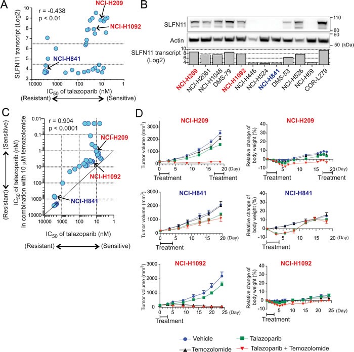
Poly(ADP-ribose) polymerase inhibitors (PARPIs) kill cancer cells by trapping PARP1 and PARP2. Talazoparib, the most potent PARPI inhibitor (PARPI), exhibits remarkable selectivity among the NCI-60 cancer cell lines beyond BRCA inactivation. Our genomic analyses reveal high correlation between response to talazoparib and Schlafen 11 (SLFN11) expression. Causality was established in four isogenic SLFN11-positive and -negative cell lines and extended to olaparib. Response to the talazoparib-temozolomide combination was also driven by SLFN11 and validated in 36 small cell lung cancer cell lines, and in xenograft models. Resistance in SLFN11-deficient cells was caused neither by impaired drug penetration nor by activation of homologous recombination. Rather, SLFN11 induced irreversible and lethal replication inhibition, which was independent of ATR-mediated S-phase checkpoint. The resistance to PARPIs by SLFN11 inactivation was overcome by ATR inhibition, mechanistically because SLFN11-deficient cells solely rely on ATR activation for their survival under PARPI treatment. Our study reveals that SLFN11 inactivation, which is common (~45%) in cancer cells, is a novel and dominant resistance determinant to PARPIs.

Keywords: ATR; BRCA; PARP inhibitor; PARP-trapping; homologous recombination.


Conflict of interest statement

CONFLICTS OF INTEREST

Y. Feng, G. K. Yu, Y. Ru, and Y. Shen are employees of and have ownership interest in BioMarin Pharmaceutical Inc.. No potential conflicts of interest were disclosed by the other authors.

Figures

Figure 1
Figure 1. SLFN11 expression is highly correlated with sensitivity to talazoparib
A. Mean-centered bar charts [20] representing SLFN11 expression (left), and sensitivity to talazoparib (middle left), olaparib (middle right) and veliparib (right) in the NCI-60. Color codes correspond to tissue of origin annotated on the sides [20]. Pearson's correlation coefficient (r) and two-sided P value (p) between SLFN11 transcripts and talazoparib or olaparib or veliparib are shown above each chart. The SLFN11-negative cell lines used for further analysis are in blue font (MDA_MB-231, HCT-116, HT29 and K-562), and the SLFN11-positive cell lines in red (SF-295, CCRF-CEM, MOLT4, and DU-145). B. Western blots of whole cell extract for the indicated cell lines and antibodies. Transcript level of SLFN11 obtained from the NCI-60 (SF-295, DU145, MDA_MB-231, HCT-116 and HT29 cell lines) and the Cancer Cell Line Encyclopedia (EW8 and A673 cell lines) database in the indicated cell lines are shown with bar graph. C. Viability curves of the indicated cell lines after continuous treatment for 72 hours with the indicated PARPIs. ATPlite assay was used to measure cell viability. The viability of untreated cells was set as 100%. Error bars represent standard deviation (SD, n ≥ 3). Drug IC90 values μM are tabulated at the right bottom. EW8 and A673 are Ewing's sarcoma cell lines
Figure 2
Figure 2. SLFN11 inactivation confers resistance to talazoparib and olaparib
A. Viability curves of the indicated parent and SLFN11-del cell lines in response to talazoparib or olaparib. Viability was determined as Figure 1C. Error bars represent SD (n ≥ 3). B. Viability curves of the indicated pairs of parental (red) and SLFN11-del (blue) cells treated with temozolomide alone (circle) or with temozolomide plus 10 nM talazoparib (+T, triangle). Viability of untreated cells was set as 100%. Error bars represent SD (n ≥ 3)
Figure 3
Figure 3. Comparable induction of DNA damage and homologous recombination regardless of SLFN11 status
A. PARP-DNA complexes were analyzed in parallel in parental (left) and SLFN11-del cells (right) in three cell line pairs (CCRF-CEM, MOLT4 and DU145) by Western blotting using chromatin-bound fractions. Cells were treated without drug (0) or with talazoparib (1 μM) plus methyl methanesulfonate MMS (0.001%) to enhance PARP trapping detection [8] for the indicated times. Blots were probed with the indicated antibodies. B. S-phase damage induction by talazoparib. The indicated cells were treated without or with talazoparib (1 μM) for 12 hours. γH2AX levels were analyzed by flow cytometry. DNA content stained with propidium iodide (PI) is on the x-axis and γH2AX levels measured by FITC signal the y-axis (logarithmic scale). The average population (%) of γH2AX-positive cells from 3 independent experiments is indicated at the top left in each panel. C. γH2AX and RAD51 foci formation in DU145 parental and SLFN11-del cells treated with or without talazoaprib (1 μM) for 3 hours. Representative confocal microscopy images are shown. Quantification was done with the ImageJ software (NIH). N = 99-115. ***p < 0.0001. D. BRCA2 functions in parallel with SLFN11. Transfection using control siRNA (siCtl) and BRCA2 siRNA (siB2/ siBRCA2) was done in the indicated cell lines. The suppression of BRCA2 mRNA was established by RT-PCR two days after transfection (top). Colony formation assay in DU145 cells (middle), and 72 hours viability assay in EW8 cells (bottom) were performed. Error bars represent SD (n ≥ 3)
Figure 4
Figure 4. Enhanced activity of PARPIs by the ATR inhibitor (VE-821) in SLFN11-del cells
A. Representative cell cycle analyses of DU145 parental and SLFN11-del cells treated as indicated for 24 or 48 hours. Experimental protocols are shown to the right. Vertical dashed lines correspond to 2N and 4N DNA contents. B. Cytotoxicity for the indicated cell lines in response to talazoparib and olaparib combined with or without 1 μM VE-821 (+V). Viability was normalized to untreated parental and SLFN11-del cells. Plots and experiments were done as in Figure 1C. Error bars represent SD (n = 3)
Figure 5
Figure 5. SLFN11 expression is correlated with sensitivity to talazoparib as single-agent or combined with temozolomide in small cell lung cancer (SCLC) cells in vitro and in vivo
A. Correlation between SLFN11 expression (mRNA) and IC50 of talazoparib across SCLC cell lines. Pearson coefficient correlation: r=0.438, p<0.01. B. Selected SCLC cell lines were examined for SLFN11 transcript and protein levels. Western blots of whole cell extract for the indicated cell lines and antibodies are compared to the SLFN11 transcript level obtained from Broad CCLE database. C. Correlation between response to talazoparib in combination with 10 μM temozolomide (y-axis) and response to talazoparib as single agent (x-axis) across the SCLC cell lines. Pearson coefficient correlation: r = 0.9041, p < 0.0001. D. Mouse xenograft experiments using NCI-H209 (high SLFN11, high MGMT), NCI-H841 (low SLFN11, high MGMT) and NCI-H1092 (high SLFN11, low MGMT). Mice bearing tumor (volume ~125 mm3) were treated with vehicle, temozolomide, talazoparib, or the combination of both drugs. Treatment schedule is annotated in the graphs (see materials and methods). Tumor volume (left) and relative change of body weight (right) are plotted. Error bars represent SEM (n = 8)
Figure 6
Figure 6. Summary scheme proposing the role of SLFN11 in parallel to ATR and homologous recombination (BRCA1/2)
Red boxes indicate disadvantage factors for cell survival, while blue boxes indicate supportive factors for cell survival. See Discussion for details.

References

    1. Rouleau M, Patel A, Hendzel MJ, Kaufmann SH, Poirier GG. PARP inhibition: PARP1 and beyond. Nat Rev Cancer. 2010;10:293–301. doi: 10.1038/nrc2812. - DOI - PMC - PubMed
    1. Shen Y, Rehman FL, Feng Y, Boshuizen J, Bajrami I, Elliott R, Wang B, Lord CJ, Post LE, Ashworth A. BMN 673, a novel and highly potent PARP1/2 inhibitor for the treatment of human cancers with DNA repair deficiency. Clin Cancer Res. 2013;19:5003–15. doi: 10.1158/1078-0432.CCR-13-1391. - DOI - PMC - PubMed
    1. Benjamin RC, Gill DM. ADP-ribosylation in mammalian cell ghosts. Dependence of poly(ADP-ribose) synthesis on strand breakage in DNA. J Biol Chem. 1980;255:10493–501. doi: - PubMed
    1. Durkacz BW, Omidiji O, Gray DA, Shall S. (ADP-ribose)n participates in DNA excision repair. Nature. 1980;283:593–6. doi: - PubMed
    1. Bryant HE, Schultz N, Thomas HD, Parker KM, Flower D, Lopez E, Kyle S, Meuth M, Curtin NJ, Helleday T. Specific killing of BRCA2-deficient tumours with inhibitors of poly(ADP-ribose) polymerase. Nature. 2005;434:913–7. doi: 10.1038/nature03443. - DOI - PubMed

MeSH terms