. 2016 Nov 15;7(46):75685-75697.
doi: 10.18632/oncotarget.12353.

Differential effects of peptidoglycan on colorectal tumors and intestinal tissue post-pelvic radiotherapy

Affiliations

Differential effects of peptidoglycan on colorectal tumors and intestinal tissue post-pelvic radiotherapy

Gen Li et al. Oncotarget. .

Abstract

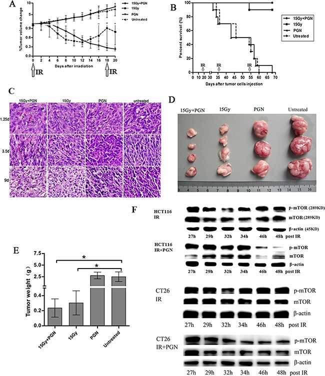
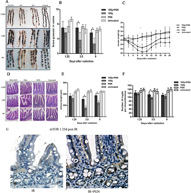
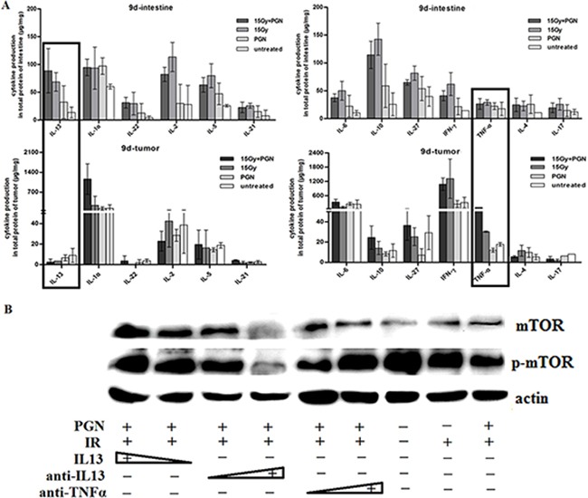
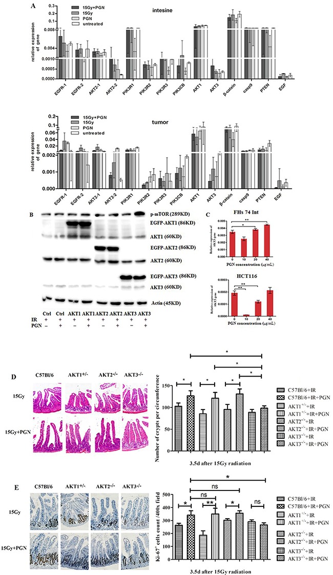
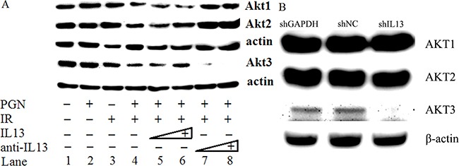
Immediate medical intervention is required after pelvic tumor radiotherapy to protect the radiosensitive intestine and also to mitigate tumor growth. Toll-like receptors (TLRs) have been shown to promote tissue repair processes. Here, we analyzed the effect observed upon combining the TLR2 agonist, peptidoglycan (PGN), with radiation therapy on tumors as well as intestinal tissue, both in vitro and in vivo. In contrast to radiotherapy alone, PGN when combined with ionizing radiation (IR) elicited enhanced antitumor effects and also reduced the IR-induced intestinal damage. Mechanistic studies showed that PGN first induced an IL13 response in the irradiated intestine, but was decreased in tumor cell models screened by Th1/Th2 FlowCytomix assay and validated by the application of IL13 and anti-IL13 neutralizing antibodies. Next, PGN stimulated Akt3, but not Akt1/2, as was verified by AKT1/2/3 plasmid transfection assay and in AKT1/2/3 knockout mice in vivo. Akt3 expression was inhibited in 20 μg/mL PGN-treated tumor cells and in 1.5 mg/kg PGN-treated mouse tumor models. However, Akt3 was raised via IL13 in the irradiated intestine and human intestinal cell line after the same treatment. Finally, PGN activated mTOR via IL13/AKT3 in the intestine and restored intestinal structure and function. As an adjuvant to radiotherapy, PGN inhibited tumorigenesis by suppression of mTOR activity. To summarize, the IL13/AKT3/mTOR pathway was lessened in PGN-treated irradiated tumors but was raised in the normal intestine tissue. This distinct effect of PGN on normal and tumor tissues during pelvic radiotherapy suggests that PGN may be a promising adjuvant therapy to radiation.

Keywords: IL13-AKT3-mTOR pathway; colorectal tumor; intestine; pelvic radiotherapy; peptidoglycan.


Conflict of interest statement

CONFLICTS OF INTEREST

The authors declare that they have no conflict of interest.

Figures

Figure 1
Figure 1. PGN synergizes with bowel irradiation to elicit enhanced antitumor responses in the CT26 model of colorectal cancer
Mice received either PBS (n=19) or 1.5 mg/kg PGN (n=19) once every 19 days i.p., 15 Gy abdominal IR (n=20) given every 18 days or a combination of PGN and 15 Gy abdominal IR (n=20, PGN was given 24 h after IR). A. Tumor volumes were plotted from the first day of local radiotherapy (day 0). Growth curves are shown as a mean±standard deviation (n=6 per group, *p<0.05 relative to 15 Gy abdominal IR). B. Survival curves for above cohorts up to 70 days (n=10 per group, arrows indicate abdominal IR on days 0, 18, 36, and 54). C. H&E staining of tumors on 1.25, 3.5, and 9 days after IR. In these representative pictures, black arrows mark apoptotic cells. D. Tumor images at termination (15 Gy+PGN, n=4; 15 Gy, n=4; PGN, n=3; untreated, n=3). E. Tumor weights of above cohorts 9 days after the initial IR (*p<0.05 relative to untreated group). F. Western blots for mTOR and phospho-mTOR expression following 15 Gy IR or 15 Gy IR + 20 μg/ml PGN in HCT116 and CT26 cells, at indicated time points.
Figure 2
Figure 2. Peptidoglycan helped to sustain the structure and function of the irradiated intestine
A. A representative image of stools in the colon. B. Feces discharged from the bowels were counted at 1.25, 3.5, and 9 days after IR (n=3 per group). C. Body weights after radiotherapy were recorded every other day in each group for a total of 20 days (n=6 per group). D. Representative images of the pathologic and morphologic changes of the small intestinal villi in each group at 1.25, 3.5, and 9 days after IR using H&E staining (200× magnification). E. Villi height of each group (n=3) were measured and compared at 1.25, 3.5, and 9 days after IR. F. Number of intestinal crypts along the jejunum circumference (10 circuits in total) in each group at 1.25, 3.5, and 9 days after IR was counted. Data are shown as the mean of 10 circuits±standard deviation. Asterisk indicates p<0.05. G. immunohistochemistry of mouse intestinal tissue 1.25 days after 15 Gy IR. Arrows indicate the mTOR positive crypts (400×).
Figure 3
Figure 3. Contrasting effect of PGN treatment on the intestine and tumor following irradiation
A. Representative images of Ki67 immunohistochemical staining of tissues of the jejunum and tumor at 3.5 days after local IR (200× magnification). B. Ki67-positive cells were counted in 10 unduplicated fields visualized at 400× magnification. Data are shown as the mean of 10 view fields±standard deviation (*, p<0.05; **, p<0.01).
Figure 4
Figure 4. IL13 played a significant role in PGN's differential effect on irradiated intestine and tumor
A. Flowcytomix assays using bead technology were utilized to detect cytokine production (μg cytokine/mg tissue weight) in intestinal and tumor tissues that were untreated, treated with PGN alone, radiation alone, or radiation + PGN at 9 days after IR. B. Western blot analysis of mTOR and phospho-mTOR expression in HCT116 cells following treatment with radiation alone or radiation + PGN in the presence or absence of IL13 (0.8 and 1.2 ng/mL), IL13 neutralizing antibody (0.12 and 0.2 μg/mL), or TNF-α neutralizing antibody (0.04 and 0.08 μg/mL).
Figure 5
Figure 5. AKT3 was implicated in PGN's distinct effects on intestinal and tumor cell proliferation after IR
A. EGFR (two transcripts), AKT2 (two transcripts), PIK3R1, PIK3R2, PIK3R3, β-catenin, AKT3, AKT1, Casp9, PTEN, PIK3CB, and EGF expression were detected by real-time PCR. Data are shown as the mean of 2(Ct, actin-Ct, target)±standard deviation. B. Western blots for AKT1/2/3 and p-mTOR in AKT1/2/3 overexpressing HCT116 cells. ‘EGFP-AKT' represents exogenously expressed AKT whereas ‘AKT’ represents endogenously expressed protein. β-actin was used as loading control. C. Real-time PCR analysis of AKT3 expression in FHS 74 Int and HCT116 cells following treatment with 0, 10, 20, and 40 μg/mL PGN (*, p<0.05; **, p<0.01). D. H&E staining of the cross section intestines of AKT1+/−, AKT2−/−, AKT3−/−, and control C57Bl/6 mice, irradiated or treated with PGN after IR. The crypts per circumference were counted. PGN had no effect on the number of crypts in irradiated AKT3−/− mice. E. Ki67 immunohistochemical staining showed that the number of Ki67+ crypt did not increase when irradiated AKT3−/− mice were treated with PGN. n=6 in each group; magnification: 400×. *, p <0.05; **, p <0.01.
Figure 6
Figure 6. IL13 preferentially stimulated AKT3
A. Western blot analysis of AKT1/2/3 expression in untreated, PGN alone-treated, radiation alone-treated, or radiation + PGN treated HCT116 cells. Lanes 5 and 6 reflect addition of 0.8 and 1.2 ng/mL IL13, respectively. The concentrations of IL13 neutralizing antibody in lanes 7 and 8 were 0.12 and 0.2 μg/mL, respectively. β-actin was used as a loading control. B. shIL13/shNC/shGAPDH plasmids were transfected into HCT116 cells, as indicated. AKT1/2/3 was detected by western blot after 48 h. β-actin was used as a loading control.

References

    1. Andreyev HJN. Gastrointestinal Problems after Pelvic Radiotherapy: the Past, the Present and the Future. Clinical Oncology. 2007;19:790–799. - PubMed
    1. Sermeus A, Leonard W, Engels B, De Ridder M. Advances in radiotherapy and targeted therapies for rectal cancer. World J Gastroenterol. 2014;20:1–5. - PMC - PubMed
    1. Andreyev HJN. The gastrointestinal complications of pelvic radiotherapy: are they of any importance? Gut. 2005;54:1051–1054. - PMC - PubMed
    1. Andreyev J. Gastrointestinal symptoms after pelvic radiotherapy: a new understanding to improve management of symptomatic patients. Lancet Oncol. 2007;8:1007–1017. - PubMed
    1. Takemura N, Kawasaki T, Kunisawa J, Sato S, Lamichhane A, Kobiyama K, Aoshi T, Ito J, Mizuguchi K, Karuppuchamy T, Matsunaga K, Miyatake S, Mori N, et al. Blockade of TLR3 protects mice from lethal radiation-induced gastrointestinal syndrome. Nat Commun. 2014;5:3492. - PMC - PubMed

MeSH terms