. 2016 Nov 1;7(44):72197-72210.
doi: 10.18632/oncotarget.12363.

A strong host response and lack of MYC expression are characteristic for diffuse large B cell lymphoma transformed from nodular lymphocyte predominant Hodgkin lymphoma

Affiliations

A strong host response and lack of MYC expression are characteristic for diffuse large B cell lymphoma transformed from nodular lymphocyte predominant Hodgkin lymphoma

Bianca Schuhmacher et al. Oncotarget. .

Abstract

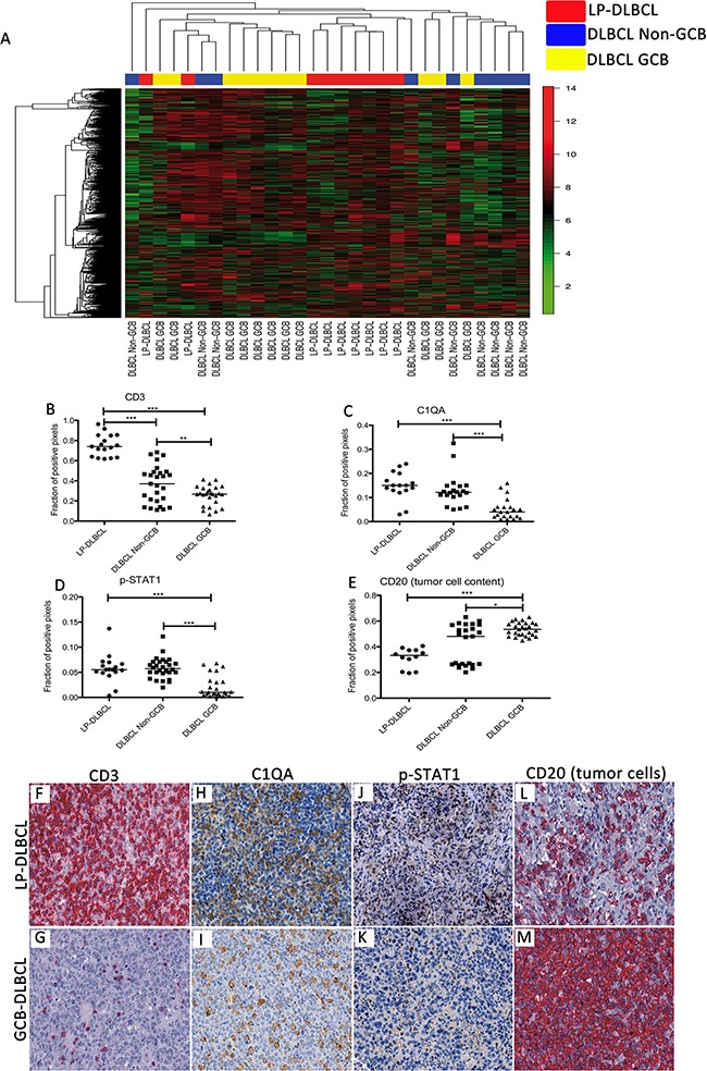
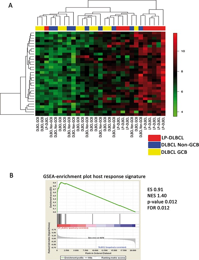
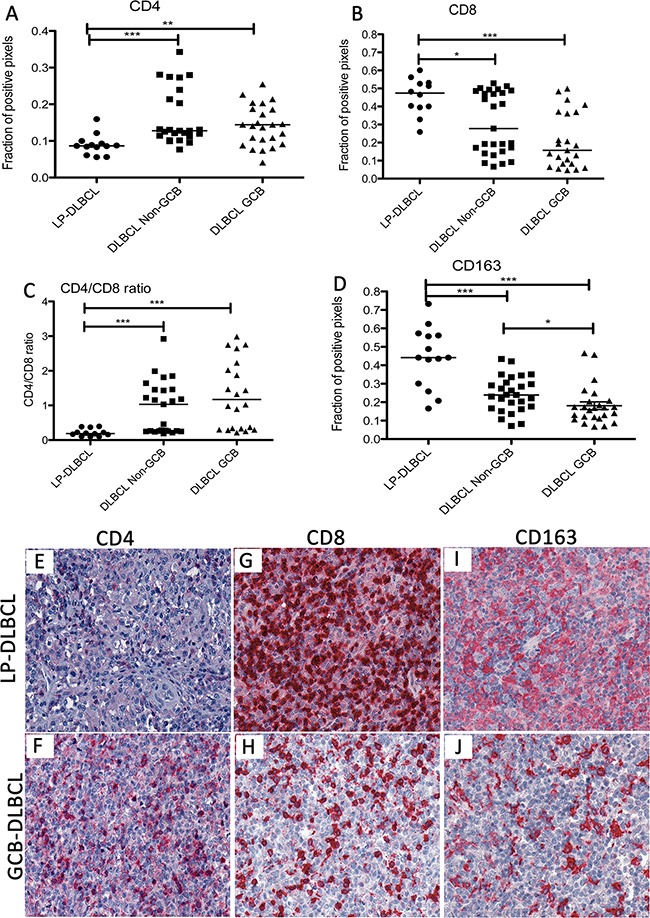
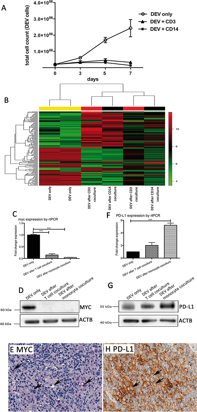
Nodular lymphocyte predominant Hodgkin lymphoma (NLPHL) is an indolent lymphoma, but can transform into diffuse large B cell lymphoma (DLBCL), showing a more aggressive clinical behavior. Little is known about these cases on the molecular level. Therefore, the aim of the present study was to characterize DLBCL transformed from NLPHL (LP-DLBCL) by gene expression profiling (GEP). GEP revealed an inflammatory signature pinpointing to a specific host response. In a coculture model resembling this host response, DEV tumor cells showed an impaired growth behavior. Mechanisms involved in the reduced tumor cell proliferation included a downregulation of MYC and its target genes. Lack of MYC expression was also confirmed in 12/16 LP-DLBCL by immunohistochemistry. Furthermore, CD274/PD-L1 was upregulated in DEV tumor cells after coculture with T cells or monocytes and its expression was validated in 12/19 cases of LP-DLBCL. Thereby, our data provide new insights into the pathogenesis of LP-DLBCL and an explanation for the relatively low tumor cell content. Moreover, the findings suggest that treatment of these patients with immune checkpoint inhibitors may enhance an already ongoing host response in these patients.

Keywords: T cell/histiocyte rich large B cell lymphoma; diffuse large B cell lymphoma; gene expression profiling; host response; nodular lymphocyte predominant Hodgkin lymphoma.


Conflict of interest statement

CONFLICTS OF INTEREST

The authors report no potential conflict of interest.

Figures

Figure 1
Figure 1. Gene expression profiling recognizes LP-DLBCL as a core group among all DLBCL
A. Unsupervised hierarchical clustering of gene expression data of LP-DLBCL (red), GCB type DLBCL (yellow) and Non-GCB type DLBCL (blue). We considered 1203 probe sets with a standard deviation > 2. B. Quantification of the fraction of CD3-positive pixels reveals significantly higher numbers in LP-DLBCL compared with Non-GCB and GCB DLBCL (***p<0.0001, **p=0.0093 paired t-test). C. Quantification of the fraction of C1QA-positive pixels reveals significantly higher numbers in LP-DLBCL compared with GCB DLBCL (***p<0.0001, Mann-Whitney-test). D. Quantification of the fraction of pSTAT1-positive pixels reveals significantly higher numbers in LP-DLBCL compared with GCB DLBCL (***p≤0.0003, Mann-Whitney-test). E. The fraction of CD20-positive pixels was significantly lower in LP-DLBCL compared with GCB type DLBCL (***p<0.0001, * p=0.0221, Mann-Whitney-test). F. Representative example of CD3 immunostaining of a LP-DLBCL (200x). G. Representative example of CD3 immunostaining of a GCB type DLBCL (200x). H. Representative example of C1QA immunostaining of a LP-DLBCL (200x). I. Representative example of C1QA immunostaining of a GCB type DLBCL (200x). J. Representative example of pSTAT1 immunostaining of a LP-DLBCL (200x). K. Representative example of pSTAT1 immunostaining of a GCB type DLBCL (200x). L. Representative example of CD20 immunostaining of a LP-DLBCL (200x). M. Representative example of CD20 immunostaining of a GCB type DLBCL (200x).
Figure 2
Figure 2. LP-DLBCL is characterized by a strong inflammatory infiltrate
A. Supervised clustering based on the host inflammatory response signature identified in THRLBCL [19]. B. Gene set enrichment analysis shows a significant enrichment of the host inflammatory response signature [19] in LP-DLBCL compared with all other DLBCL. ES enrichment score, NES normalized enrichment score, FDR false discovery rate.
Figure 3
Figure 3. The inflammatory infiltrate in LP-DLBCL shows a low CD4/CD8 ratio and a high content of macrophages
A. The fraction of CD4-positive pixels was significantly lower in LP-DLBCL compared with Non-GCB and GCB type DLBCL (***p<0.0001, **p=0.0035, Mann-Whitney-test). B. Quantification of the fraction of CD8-positive pixels reveals significantly higher numbers in LP-DLBCL compared with Non-GCB and GCB DLBCL (***p=0.0002, *p=0.0169, Mann-Whitney-test). C. The CD4/CD8 ratio is significantly decreased in the microenvironment of LP-DLBCL compared with Non-GCB and GCB DLBCL (***p≤0.0003, Mann-Whitney-test). D. Quantification of the fraction of CD163-positive pixels reveals significantly higher numbers in LP-DLBCL compared with Non-GCB and GCB DLBCL (***p≤0.0003, *p=0.0109, Mann-Whitney-test). E. Representative example of LP-DLBCL in CD4 immunostaining (200x). F. Representative example of a GCB type DLBCL in CD4 immunostaining (200x). G. Representative example of LP-DLBCL in CD8 immunostaining (200x). H. Representative example of a GCB type DLBCL in CD8 immunostaining (200x). I. Representative example of LP-DLBCL in CD163 immunostaining (200x). J. Representative example of a GCB type DLBCL in CD163 immunostaining (200x).
Figure 4
Figure 4. Growth of the NLPHL cell line DEV is impaired in the presence of T cells or monocytes
A. Growth curves of the NLPHL cell line DEV in coculture with T cells or monocytes compared to a corresponding monoculture. B. Unsupervised GEP clustering of DEV cells in monoculture and DEV cells isolated after 5 days from coculture with T cells or monocytes. Two representative replicates of several experiments were analyzed for changes in GEP. We considered 158 probe sets with a standard deviation > 2 for the cluster analysis. C. MYC mRNA expression determined by Taqman realtime RT-PCR in DEV cells after coculture with T cells or monocytes, relative to GAPDH and relative to DEV cells from monoculture (***p<0.0001, paired t-test). D. Western blot of MYC protein in representative samples of DEV cells after coculture with T cells or monocytes compared to a corresponding monoculture. ACTB was used as loading control. E. Example of an LP-DLBCL with lack of MYC expression in the majority of the tumor cells (200x). F. CD274/PD-L1 mRNA expression determined by Taqman realtime RT-PCR in DEV cells after coculture with T cells or monocytes, relative to GAPDH and relative to DEV cells from monoculture (***p<0.0001, paired t-test). G. Western blot of PD-L1 protein in representative samples of DEV cells after coculture with T cells or monocytes compared to a corresponding monoculture. ACTB was used as loading control. H. Example of an LP-DLBCL with membrane bound CD274/PD-L1 expression in the tumor cells (200x).

References

    1. Swerdlow SH, International Agency for Research on Cancer, World Health Organization . WHO classification of tumours of haematopoietic and lymphoid tissues. 4. Vol. 439 Lyon, France: International Agency for Research on Cancer; 2008.
    1. Wright G, Tan B, Rosenwald A, Hurt EH, Wiestner A, Staudt LM. A gene expression-based method to diagnose clinically distinct subgroups of diffuse large B cell lymphoma. Proc Natl Acad Sci USA. 2003;100:9991–96. doi: 10.1073/pnas.1732008100. - DOI - PMC - PubMed
    1. Rosenwald A, Wright G, Chan WC, Connors JM, Campo E, Fisher RI, Gascoyne RD, Muller-Hermelink HK, Smeland EB, Giltnane JM, Hurt EM, Zhao H, Averett L, et al. The use of molecular profiling to predict survival after chemotherapy for diffuse large-B-cell lymphoma. N Engl J Med. 2002;346:1937–47. doi: 10.1056/NEJMoa012914. - DOI - PubMed
    1. Young RM, Wu T, Schmitz R, Dawood M, Xiao W, Phelan JD, Xu W, Menard L, Meffre E, Chan WC, Jaffe ES, Gascoyne RD, Campo E, et al. Survival of human lymphoma cells requires B-cell receptor engagement by self-antigens. Proc Natl Acad Sci USA. 2015;112:13447–54. doi: 10.1073/pnas.1514944112. - DOI - PMC - PubMed
    1. Hans CP, Weisenburger DD, Greiner TC, Gascoyne RD, Delabie J, Ott G, Müller-Hermelink HK, Campo E, Braziel RM, Jaffe ES, Pan Z, Farinha P, Smith LM, et al. Confirmation of the molecular classification of diffuse large B-cell lymphoma by immunohistochemistry using a tissue microarray. Blood. 2004;103:275–82. doi: 10.1182/blood-2003-05-1545. - DOI - PubMed

MeSH terms