

Distinct biological effects of low-dose radiation on normal and cancerous human lung cells are mediated by ATM signaling
- PMID: 27708248
- PMCID: PMC5342128
- DOI: 10.18632/oncotarget.12379
Distinct biological effects of low-dose radiation on normal and cancerous human lung cells are mediated by ATM signaling
Abstract
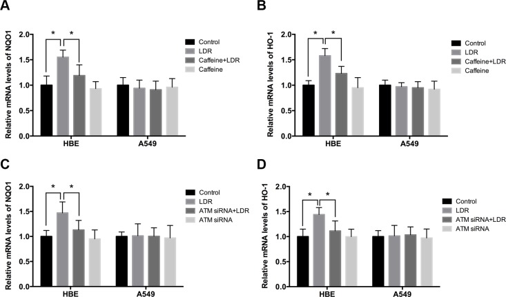
Low-dose radiation (LDR) induces hormesis and adaptive response in normal cells but not in cancer cells, suggesting its potential protection of normal tissue against damage induced by conventional radiotherapy. However, the underlying mechanisms are not well established. We addressed this in the present study by examining the role of the ataxia telangiectasia mutated (ATM) signaling pathway in response to LDR using A549 human lung adenocarcinoma cells and HBE135-E6E7 (HBE) normal lung epithelial cells. We found that LDR-activated ATM was the initiating event in hormesis and adaptive response to LDR in HBE cells. ATM activation increased the expression of CDK4/CDK6/cyclin D1 by activating the AKT/glycogen synthase kinase (GSK)-3β signaling pathway, which stimulated HBE cell proliferation. Activation of ATM/AKT/GSK-3β signaling also increased nuclear accumulation of nuclear factor erythroid 2-related factor 2, leading to increased expression of antioxidants, which mitigated cellular damage from excessive reactive oxygen species production induced by high-dose radiation. However, these effects were not observed in A549 cells. Thus, the failure to activate these pathways in A549 cells likely explains the difference between normal and cancer cells in terms of hormesis and adaptive response to LDR.
Keywords: ATM; biological effects; low-dose radiation; lung cancer cells; normal lung cells.
Conflict of interest statement
None.
Figures


Similar articles
-
Ionizing radiation can induce GSK-3beta phosphorylation and NF-kappaB transcriptional transactivation in ATM-deficient fibroblasts.Cell Signal. 2008 Apr;20(4):602-12. doi: 10.1016/j.cellsig.2007.10.022. Epub 2007 Nov 6. Cell Signal. 2008. PMID: 18243662
-
Mitochondrial reactive oxygen species perturb AKT/cyclin D1 cell cycle signaling via oxidative inactivation of PP2A in lowdose irradiated human fibroblasts.Oncotarget. 2016 Jan 19;7(3):3559-70. doi: 10.18632/oncotarget.6518. Oncotarget. 2016. PMID: 26657292 Free PMC article.
-
Low-Dose Radiation Promotes Dendritic Cell Migration and IL-12 Production via the ATM/NF-KappaB Pathway.Radiat Res. 2018 Apr;189(4):409-417. doi: 10.1667/RR14840.1. Epub 2018 Feb 8. Radiat Res. 2018. PMID: 29420126
-
Luteolin induces G1 arrest in human nasopharyngeal carcinoma cells via the Akt-GSK-3β-Cyclin D1 pathway.Cancer Lett. 2010 Dec 8;298(2):167-75. doi: 10.1016/j.canlet.2010.07.001. Epub 2010 Jul 23. Cancer Lett. 2010. PMID: 20655656
-
Influence of Individual Radiosensitivity on the Hormesis Phenomenon: Toward a Mechanistic Explanation Based on the Nucleoshuttling of ATM Protein.Dose Response. 2020 May 8;18(2):1559325820913784. doi: 10.1177/1559325820913784. eCollection 2020 Apr-Jun. Dose Response. 2020. PMID: 32425719 Free PMC article. Review.
Cited by
-
Hormetic Response to Low-Dose Radiation: Focus on the Immune System and Its Clinical Implications.Int J Mol Sci. 2017 Jan 27;18(2):280. doi: 10.3390/ijms18020280. Int J Mol Sci. 2017. PMID: 28134809 Free PMC article. Review.
-
Reduction of Delayed Homologous Recombination by Induction of Radioadaptive Response in RaDR-GFP Mice (Yonezawa Effect): An Old Player With a New Role.Dose Response. 2019 Mar 4;17(1):1559325819833840. doi: 10.1177/1559325819833840. eCollection 2019 Jan-Mar. Dose Response. 2019. PMID: 30858771 Free PMC article.
-
Biological basis of radiation protection needs rejuvenation.Int J Radiat Biol. 2017 Oct;93(10):1056-1063. doi: 10.1080/09553002.2017.1294773. Epub 2017 Mar 13. Int J Radiat Biol. 2017. PMID: 28287035 Free PMC article. Review.
-
Changes in the Proliferation Rate, Clonogenicity, and Radiosensitivity of Cultured Cells During and After Continuous Low-Dose-Rate Irradiation.Dose Response. 2019 Apr 22;17(2):1559325819842733. doi: 10.1177/1559325819842733. eCollection 2019 Apr-Jun. Dose Response. 2019. PMID: 31040760 Free PMC article.
-
Role of Mitochondria in Radiation Responses: Epigenetic, Metabolic, and Signaling Impacts.Int J Mol Sci. 2021 Oct 13;22(20):11047. doi: 10.3390/ijms222011047. Int J Mol Sci. 2021. PMID: 34681703 Free PMC article. Review.
References
-
- Mettler FA, Sinclair WK, Anspaugh L, Edington C, Harley JH, Ricks RC, Selby PB, Webster EW, Wyckoff HO. The 1986 and 1988 UNSCEAR (United Nations Scientific Committee on the Effects of Atomic Radiation) reports: findings and implications. Health Phys. 1990;58:241–250. - PubMed
-
- Luckey TD. Physiological benefits from low levels of ionizing radiation. Health Phys. 1982;43:771–789. - PubMed
-
- Feinendegen LE. Evidence for beneficial low level radiation effects and radiation hormesis. The British journal of radiology. 2005;78:3–7. - PubMed
-
- Smith H. Radiation hormesis in relation to radiation protection. Chin Med J (Engl) 1994;107:615–623. - PubMed
MeSH terms
Substances
LinkOut - more resources
Full Text Sources
Other Literature Sources
Medical
Research Materials
Miscellaneous
