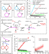
A modular platform for one-step assembly of multi-component membrane systems by fusion of charged proteoliposomes
- PMID: 27708275
- PMCID: PMC5059690
- DOI: 10.1038/ncomms13025
A modular platform for one-step assembly of multi-component membrane systems by fusion of charged proteoliposomes
Abstract
An important goal in synthetic biology is the assembly of biomimetic cell-like structures, which combine multiple biological components in synthetic lipid vesicles. A key limiting assembly step is the incorporation of membrane proteins into the lipid bilayer of the vesicles. Here we present a simple method for delivery of membrane proteins into a lipid bilayer within 5 min. Fusogenic proteoliposomes, containing charged lipids and membrane proteins, fuse with oppositely charged bilayers, with no requirement for detergent or fusion-promoting proteins, and deliver large, fragile membrane protein complexes into the target bilayers. We demonstrate the feasibility of our method by assembling a minimal electron transport chain capable of adenosine triphosphate (ATP) synthesis, combining Escherichia coli F1Fo ATP-synthase and the primary proton pump bo3-oxidase, into synthetic lipid vesicles with sizes ranging from 100 nm to ∼10 μm. This provides a platform for the combination of multiple sets of membrane protein complexes into cell-like artificial structures.
Figures


Similar articles
-
Detergent-free Ultrafast Reconstitution of Membrane Proteins into Lipid Bilayers Using Fusogenic Complementary-charged Proteoliposomes.J Vis Exp. 2018 Apr 5;(134):56909. doi: 10.3791/56909. J Vis Exp. 2018. PMID: 29683454 Free PMC article.
-
Delivery of membrane proteins into small and giant unilamellar vesicles by charge-mediated fusion.FEBS Lett. 2016 Jul;590(14):2051-62. doi: 10.1002/1873-3468.12233. Epub 2016 Jun 21. FEBS Lett. 2016. PMID: 27264202
-
Modulating Liposome Surface Charge for Maximized ATP Regeneration in Synthetic Nanovesicles.ACS Synth Biol. 2024 Dec 20;13(12):4061-4073. doi: 10.1021/acssynbio.4c00487. Epub 2024 Nov 26. ACS Synth Biol. 2024. PMID: 39592139 Free PMC article.
-
Single-Vesicle Assays Using Liposomes and Cell-Derived Vesicles: From Modeling Complex Membrane Processes to Synthetic Biology and Biomedical Applications.Chem Rev. 2018 Sep 26;118(18):8598-8654. doi: 10.1021/acs.chemrev.7b00777. Epub 2018 Aug 28. Chem Rev. 2018. PMID: 30153012 Review.
-
Strategies in the reassembly of membrane proteins into lipid bilayer systems and their functional assay.J Bioenerg Biomembr. 1983 Dec;15(6):321-34. doi: 10.1007/BF00751053. J Bioenerg Biomembr. 1983. PMID: 18251429 Review.
Cited by
-
Building protein networks in synthetic systems from the bottom-up.Biotechnol Adv. 2021 Jul-Aug;49:107753. doi: 10.1016/j.biotechadv.2021.107753. Epub 2021 Apr 12. Biotechnol Adv. 2021. PMID: 33857631 Free PMC article. Review.
-
A Microfluidic Platform for Sequential Assembly and Separation of Synthetic Cell Models.ACS Synth Biol. 2021 Nov 19;10(11):3105-3116. doi: 10.1021/acssynbio.1c00371. Epub 2021 Nov 11. ACS Synth Biol. 2021. PMID: 34761904 Free PMC article.
-
Single Cell-like Systems Reveal Active Unidirectional and Light-Controlled Transport by Nanomachineries.ACS Nano. 2021 Apr 27;15(4):6747-6755. doi: 10.1021/acsnano.0c10139. Epub 2021 Mar 16. ACS Nano. 2021. PMID: 33724767 Free PMC article.
-
Detergent-free Ultrafast Reconstitution of Membrane Proteins into Lipid Bilayers Using Fusogenic Complementary-charged Proteoliposomes.J Vis Exp. 2018 Apr 5;(134):56909. doi: 10.3791/56909. J Vis Exp. 2018. PMID: 29683454 Free PMC article.
-
Experimental platform for the functional investigation of membrane proteins in giant unilamellar vesicles.Soft Matter. 2022 Aug 10;18(31):5877-5893. doi: 10.1039/d2sm00551d. Soft Matter. 2022. PMID: 35916307 Free PMC article.
References
-
- Rigaud J. L. & Levy D. Reconstitution of membrane proteins into liposomes. Method Enzymol. 372, 65–86 (2003). - PubMed
-
- Hansen J. S. et al.. Formation of giant protein vesicles by a lipid cosolvent method. ChemBioChem 12, 2856–2862 (2011). - PubMed
-
- Varnier A. et al.. A simple method for the reconstitution of membrane proteins into giant unilamellar vesicles. J. Membrane Biol. 233, 85–92 (2010). - PubMed
Publication types
MeSH terms
Substances
Grants and funding
LinkOut - more resources
Full Text Sources
Other Literature Sources

