Review
. 2017 Jan;14(1):30-38.
doi: 10.1038/nrcardio.2016.163. Epub 2016 Oct 6.

Neurohormonal activation in heart failure with reduced ejection fraction

Affiliations
Review

Neurohormonal activation in heart failure with reduced ejection fraction

Justin Hartupee et al. Nat Rev Cardiol. 2017 Jan.

Abstract

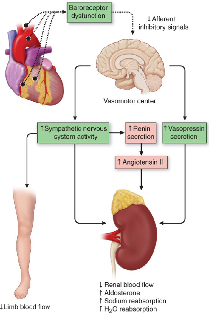
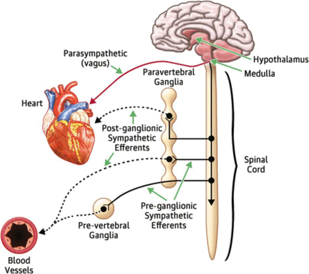
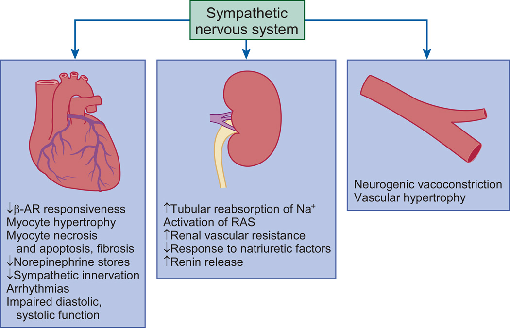
Heart failure with reduced ejection fraction (HFrEF) develops when cardiac output falls as a result of cardiac injury. The most well-recognized of the compensatory homeostatic responses to a fall in cardiac output are activation of the sympathetic nervous system and the renin-angiotensin-aldosterone system (RAAS). In the short term, these 'neurohormonal' systems induce a number of changes in the heart, kidneys, and vasculature that are designed to maintain cardiovascular homeostasis. However, with chronic activation, these responses result in haemodynamic stress and exert deleterious effects on the heart and the circulation. Neurohormonal activation is now known to be one of the most important mechanisms underlying the progression of heart failure, and therapeutic antagonism of neurohormonal systems has become the cornerstone of contemporary pharmacotherapy for heart failure. In this Review, we discuss the effects of neurohormonal activation in HFrEF and highlight the mechanisms by which these systems contribute to disease progression.


Figures

Figure 1
Figure 1. Activation of neurohormonal systems in heart failure
Decreased cardiac output in patients with heart failure with reduced EF results in the unloading of high-pressure baroceptors (black circles) in the left ventricle, carotid sinus, and aortic arch. This unloading leads to generation of afferent signals to the central nervous system (CNS) that, in turn, lead to activation of efferent sympathetic nervous system pathways that innervate the heart, kidney, peripheral vasculature, and skeletal muscles. This unloading also leads to afferent signals to the CNS that stimulate cardioregulatory centers in the brain that stimulate the release of arginine vasopression from the posterior pituitary. (Modified from Mann, D.L. & Chakinala, M. Heart failure and cor pulmonale in Harrison's Principles of Internal Medicine (Kasper, D.L., Fauci, A.S., Hauser, S.L., Longo, D.L., Jameson, J.L. & Loscalzo, J,) 1500–1506 (McGraw Hill Medical, 2015).
Figure 2
Figure 2. Effects of sympathetic nervous system activation
Increased sympathetic nervous system (SNS) activity contributes to the pathophysiology of heart failure through multiple mechanisms involving cardiac, renal, and vascular function. In the heart, increased SNS outflow leads to desensitization of the βAR, myocyte hypertrophy, necrosis, apoptosis, and fibrosis. In the kidneys, increased sympathetic activation induces arterial and venous vasoconstriction, activation of the RAAS, increase in salt and water retention, and an attenuated response to natriuretic peptides. In the peripheral vessels, increased SNS activity induces neurogenic vasoconstriction and vascular hypertrophy. (From Hassefuss, G. & Mann, D.L. Pathophysiology of heart failure in Braunwald's Heart Disease: A Textbook of Cardiovascular Medicine (Mann, D.L., Zipes, D., Libby P.L. & Bonow, R.L.) 454–472 (Elsevier/Saunders, 2014). βAR, β-adrenergic receptors; RAAS, renin–angiotensin–aldosterone system.
Figure 3
Figure 3. Cardiac and cellular remodelling in response to haemodynamic overloading
The pattern of remodelling depends on the nature of the inciting stimulus. a | When overload is predominantly caused by an increase in pressure (e.g. with systemic hypertension or aortic stenosis), the increase in systolic wall stress leads to the parallel addition of sarcomeres and widening of the cardiac myocytes, resulting in ‘concentric hypertrophy’. When the overload is predominantly caused by an increase in ventricular volume, the increase in diastolic wall stress leads to the series addition of sarcomeres, lengthening of cardiac myocytes, and left ventricular dilatation, which is referred to as ‘eccentric hypertrophy’. b | Phenotypically distinct changes occur in the morphology of the myocyte in response to the type of haemodynamic overload. When the overload is predominantly caused by an increase in pressure the increase in systolic wall stress leads to the parallel addition of sarcomeres and widening of the cardiac myocytes. When the overload is predominantly caused by an increase in ventricular volume, the increase in diastolic wall stress leads to the series addition of sarcomeres, and thus lengthening of cardiac myocytes. The expression of maladaptive embryonic genes is increased in both eccentric and concentric hypertrophy, but not in physiologic myocyte hypertrophy that occurs with exercise. (From Hassefuss, G. & Mann, D.L. Pathophysiology of heart failure in Braunwald's Heart Disease: A Textbook of Cardiovascular Medicine (Mann, D.L., Zipes, D., Libby P.L. & Bonow, R.L.) 454–472 (Elsevier/Saunders, 2014).

References

    1. Mann DL. Innate Immunity and the Failing Heart: The Cytokine Hypothesis Revisited. Circ. Res. 2015;116:1254–1268. doi:CIRCRESAHA.116.302317 [pii];10.1161/CIRCRESAHA.116.302317 [doi] - PMC - PubMed
    1. Rouleau JL, et al. Activation of neurohumoral systems following acute myocardial infarction. Am. J. Cardiol. 1991;68:80D–86D. - PubMed
    1. Packer M. The Neurohormonal Hypothesis: a theory to explain the mechanism of disease progression in heart failure. J. Am. Coll. Cardiol. 1992;20:248–254. - PubMed
    1. Piepoli M, et al. Contribution of muscle afferents to the hemodynamic, autonomic, and ventilatory responses to exercise in patients with chronic heart failure: effects of physical training. Circulation. 1996;93:940–952. - PubMed
    1. Giannoni A, et al. Combined increased chemosensitivity to hypoxia and hypercapnia as a prognosticator in heart failure. J Am Coll Cardiol. 2009;53:1975–1980. - PubMed

Publication types

MeSH terms