. 2016 Oct 6:6:34477.
doi: 10.1038/srep34477.

Exposure to the Functional Bacterial Amyloid Protein Curli Enhances Alpha-Synuclein Aggregation in Aged Fischer 344 Rats and Caenorhabditis elegans

Affiliations

Exposure to the Functional Bacterial Amyloid Protein Curli Enhances Alpha-Synuclein Aggregation in Aged Fischer 344 Rats and Caenorhabditis elegans

Shu G Chen et al. Sci Rep. .

Abstract

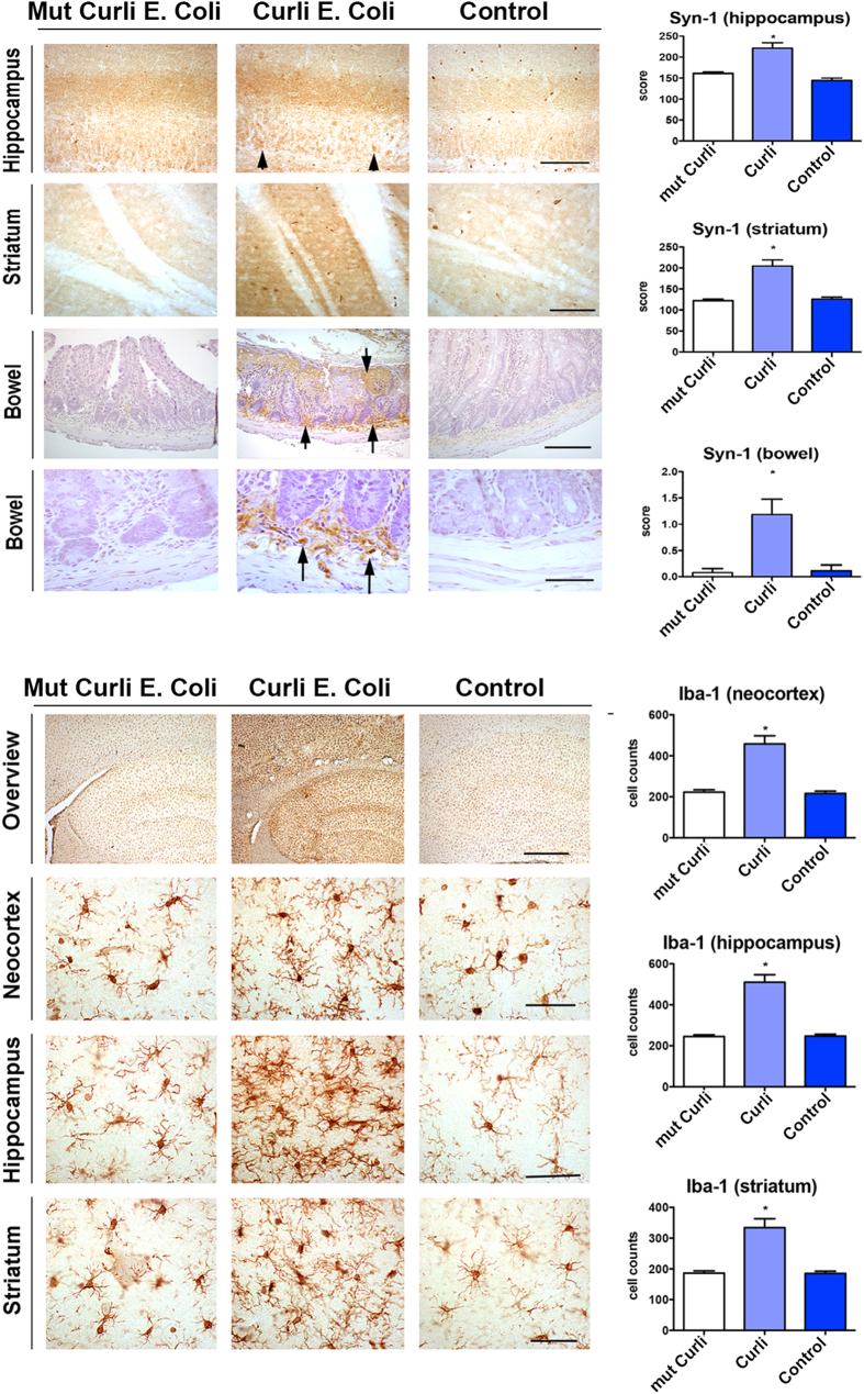
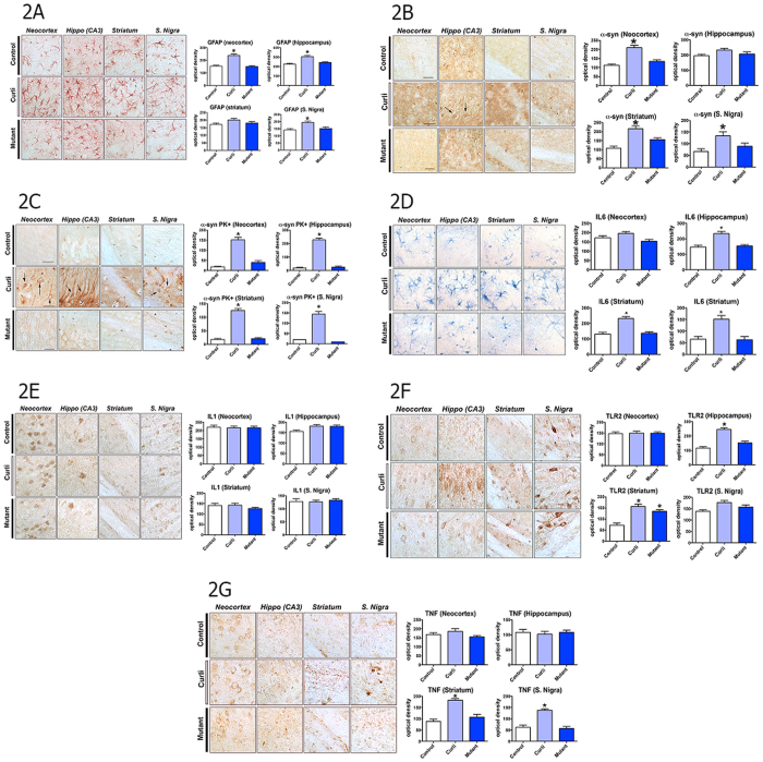
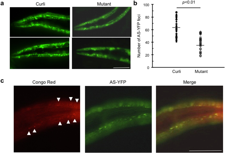
Misfolded alpha-synuclein (AS) and other neurodegenerative disorder proteins display prion-like transmission of protein aggregation. Factors responsible for the initiation of AS aggregation are unknown. To evaluate the role of amyloid proteins made by the microbiota we exposed aged rats and transgenic C. elegans to E. coli producing the extracellular bacterial amyloid protein curli. Rats exposed to curli-producing bacteria displayed increased neuronal AS deposition in both gut and brain and enhanced microgliosis and astrogliosis compared to rats exposed to either mutant bacteria unable to synthesize curli, or to vehicle alone. Animals exposed to curli producing bacteria also had more expression of TLR2, IL-6 and TNF in the brain than the other two groups. There were no differences among the rat groups in survival, body weight, inflammation in the mouth, retina, kidneys or gut epithelia, and circulating cytokine levels. AS-expressing C. elegans fed on curli-producing bacteria also had enhanced AS aggregation. These results suggest that bacterial amyloid functions as a trigger to initiate AS aggregation through cross-seeding and also primes responses of the innate immune system.


Figures

Figure 1
Figure 1. Rats exposed to bacteria expressing curli have enhanced AS deposition in hippocampus, striatum and gut neurons and increased growth of microglia.
(a) Alpha-synuclein (Syn-1) immunohistochemical staining in gut, striatum and hippocampus. Gut neurons containing AS deposits are indicated with arrows, and hippocampal neurons containing AS are indicated with arrowheads. Data are for animals exposed to mutant bacteria unable to produce curli (Mut Curli E. coli), curli-producing wild type bacteria (Curli E. coli) and Control (exposed only to vehicle). For the striatum and hippocampus images the bars are 20 μm. For the bowel images the upper bar for the upper row of three bowel images is 250 μm and the lower bar for the lower row of three bowel images is 150 μm (higher magnification). Right panels represent quantification of the staining. *p < 0.05 when compared to control. (b) Iba1 (allograft inflammatory factor) staining of neocortex, hippocampus and striatum. The upper bar is 250 μm and the lower bars are 20 μm. The top images are lower magnification and the bottom three panels are higher magnification. *p < 0.05 when compared to control.
Figure 2
Figure 2. Rats exposed to bacteria expressing curli have increased astroglial growth, enhanced deposition of aggregated AS, and increased expression in brain of IL6, TLR2 and TNF.
Immunohistochemical staining in neocortex, hippocampus, striatum and substantia nigra. Right panels represent quantification of the staining. Data are for animals exposed to bacteria unable to produce curli (Mut Curli E. coli), curli producing wild type bacteria (Curli E coli) and Control (exposed only to vehicle). Right panels represent quantification of the staining (A) Glial fibrillary acidic protein (GFAP) staining of neocortex, hippocampus (CA3) striatum and substantia nigra. *p < 0.05. (B) Alpha synuclein (α-syn) staining, without proteinase K treatment. *p < 0.05. (C) Alpha synuclein (α-syn) staining following proteinase K treatment (PK+). Arrows represent deposits of proteinase K resistant AS in neurons. *p < 0.05. (D) Interleukin 6 (IL6) staining in neocortex, hippocampus (CA3) striatum and Substantia nigra. *p < 0.05. (E) Interleukin1 (IL1) staining in neocortex, hippocampus (CA3) striatum and Substantia nigra. (F) Toll like receptor 2 (TLR2) staining in neocortex, hippocampus (CA3) striatum and Substantia nigra. *p < 0.05. (G) Tissue necrosis factor (TNF) staining in neocortex, hippocampus (CA3) striatum and Substantia nigra. *p < 0.05. For images (AG) the bars are 20 um.
Figure 3
Figure 3. C. elegans exposed to E. coli expressing curli has enhanced AS aggregation.
(a) Fluorescence microscopy of the anterior region of C. elegans expressing AS-YFP in the body wall muscle. Transgenic nematodes (Punc-54::AS::YFP) were age-synchronized and were fed for three days on lawns of either curli-producing E. coli or its mutant strain unable to express curli (non-curli mutant). Fluorescence microscopy was performed on immobilized live nematodes and imaging was acquired under identical conditions for the nematode groups fed with different bacterial strains. C. elegans exposed to curli expressing bacteria (Curli) contained increased numbers of larger and brighter foci of AS-YFP as compared to those exposed to non-curli expressing mutant bacteria (Mutant). Scale bar = 100 μm. (b) Quantitative analysis of AS-YFP foci. The number of YFP foci in the head region (from the base of the second pharyngeal bulb to the nose) of C. elegans was plotted for a group of 15 animals each exposed to either curli-producing E. coli (Curli) or its corresponding mutant (Mutant). The horizontal lines represent the average number of AS-YFP foci. p < 0.01, unpaired t test. (c) Congo red staining of C. elegans expressing AS-YFP exposed to curli-producing E. coli. Congo red stained deposits (arrowheads) in the head region of C. elegans expressing AS-YFP in the body wall muscles colocalized with AS-YFP aggregates. Scale bar = 100 μm.

References

    1. Walker L. C. & Jucker M. Amyloid by default. Nature neuroscience 14, 669–670 (2011). - PMC - PubMed
    1. Desplats P. et al. Inclusion formation and neuronal cell death through neuron-to-neuron transmission of alpha-synuclein. Proceedings of the National Academy of Sciences of the United States of America 106, 13010–13015 (2009). - PMC - PubMed
    1. Prusiner S. B. Cell biology. A unifying role for prions in neurodegenerative diseases. Science 336, 1511–1513 (2012). - PMC - PubMed
    1. Friedland R. P. Mechanisms of molecular mimicry involving the microbiota in neurodegeneration. Journal of Alzheimer’s disease: JAD 45, 349–362 (2015). - PubMed
    1. Soto C. Transmissible proteins: expanding the prion heresy. Cell 149, 968–977 (2012). - PMC - PubMed

Publication types

MeSH terms