. 2016 Oct 6:6:34314.
doi: 10.1038/srep34314.

Role of areca nut induced JNK/ATF2/Jun axis in the activation of TGF-β pathway in precancerous Oral Submucous Fibrosis

Affiliations

Role of areca nut induced JNK/ATF2/Jun axis in the activation of TGF-β pathway in precancerous Oral Submucous Fibrosis

Ila Pant et al. Sci Rep. .

Abstract

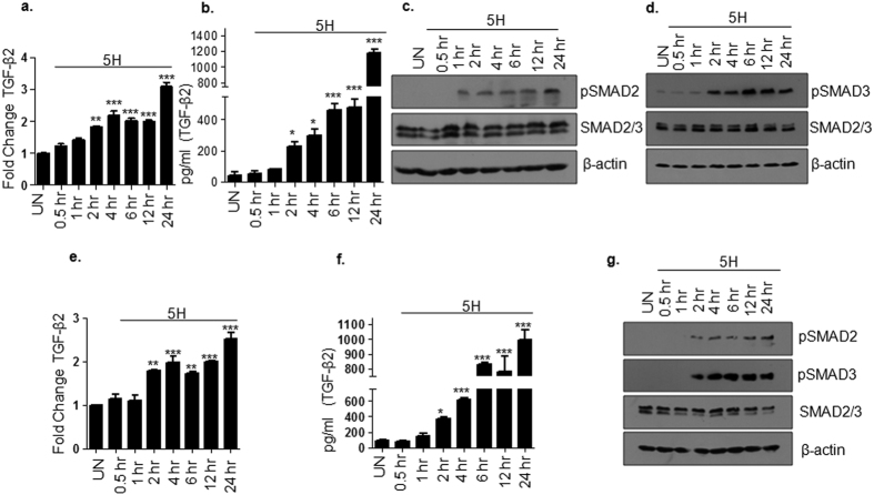
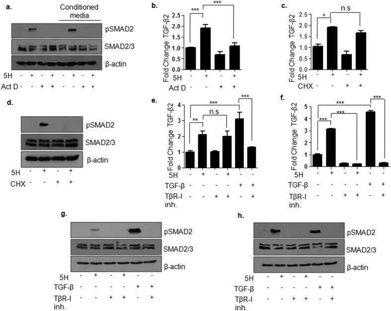
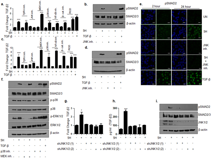
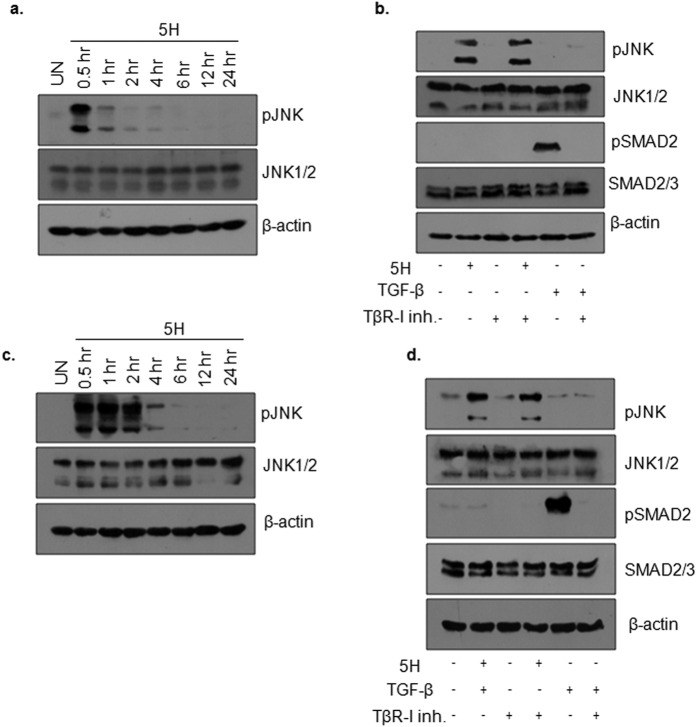
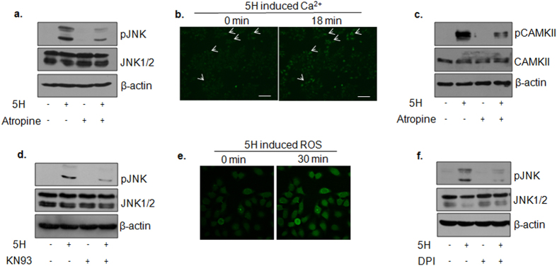
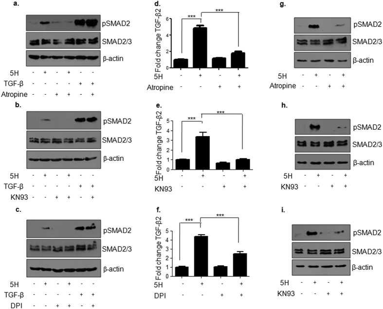
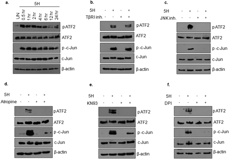
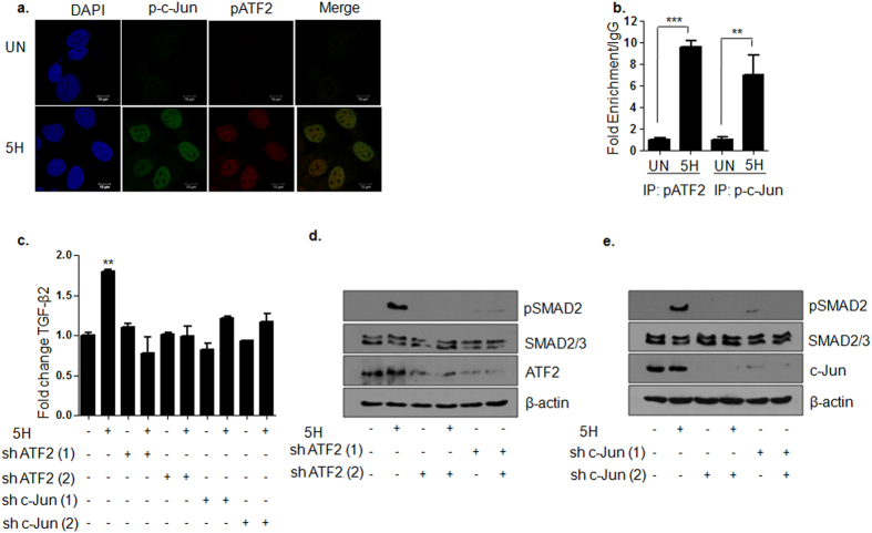
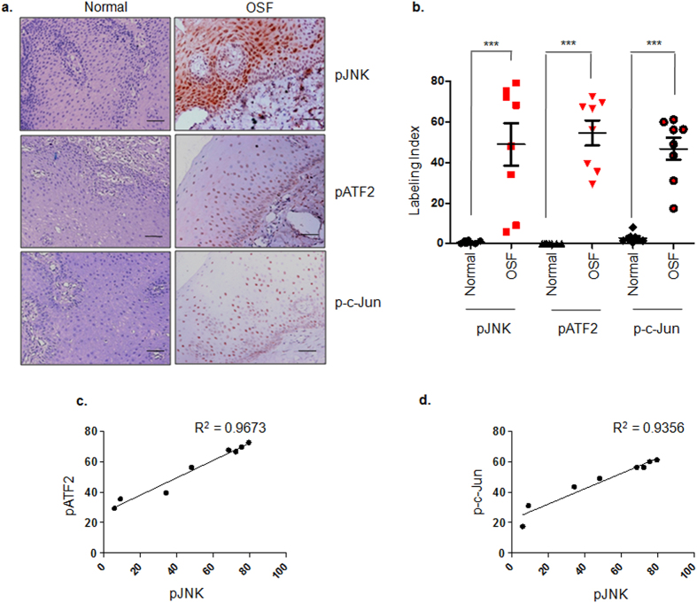
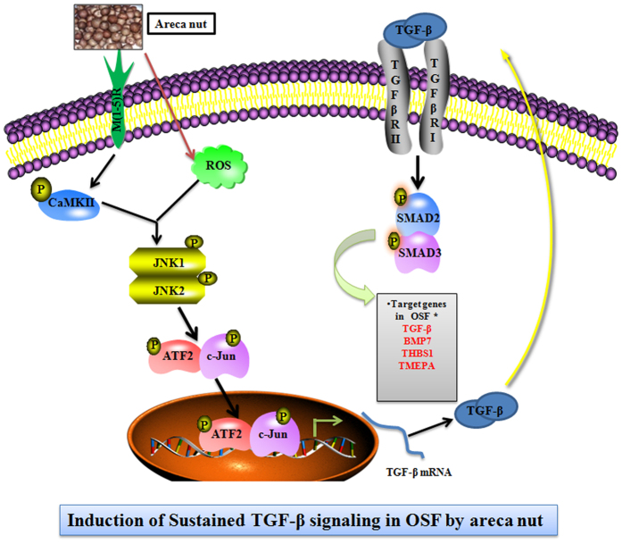
Oral submucous fibrosis (OSF) is potentially premalignant with progressive and irreversible extracellular matrix deposition accompanied by epithelial atrophy and like other fibrotic disorders, is primarily a TGF-β driven disease. OSF is caused by prolonged chewing of areca nut. Our previous studies reported a pivotal role for TGF-β activation and its effects contributing to OSF. However, the mechanism for activation of TGF-β signaling in OSF is still unknown. In this study we demonstrate activation of TGF-β signaling with sub-cytotoxic dose of areca nut in epithelial cells and discovered a key role for pJNK in this process. In good correlation; pJNK was detected in OSF tissues but not in normal tissues. Moreover, activation of JNK was found to be dependent on muscarinic acid receptor induced Ca2+/CAMKII as well as ROS. JNK dependent phosphorylation of ATF2/c-Jun transcription factors resulted in TGF-β transcription and its signaling. pATF2/p-c-Jun were enriched on TGF-β promoter and co-localized in nuclei of epithelial cells upon areca nut treatment. In corroboration, OSF tissue sections also had nuclear pATF2 and p-c-Jun. Our results provide comprehensive mechanistic details of TGF-β signaling induced by etiological agent areca nut in the manifestation of fibrosis which can lead to new therapeutic modalities for OSF.


Figures

Figure 1
Figure 1. Activation of TGF-β pathway by areca nut in epithelial cells.
(a) qRT-PCR data representing fold change in TGF-β2 transcript in HaCaT cells upon areca nut treatment (5H; 5 μg/ml) at various time points compared to untreated cells. (b) Bar graph representing TGF-β2 protein in HaCaT cells induced by areca nut treatment (5H; 5 μg/ml) at various time points compared to untreated cells. (c,d) Immunoblots depicting areca nut (5H; 5 μg/ml) induced phosphorylation of SMAD2 (c) and SMAD3 (d) in HaCaT cells at various time points compared to untreated cells. (e) qRT-PCR data representing fold change in TGF-β2 transcript in HPL1D cells upon areca nut treatment (5H; 5 μg/ml) at various time points compared to untreated cells. (f) Bar graph representing TGF-β2 protein in HPL1D cells induced by areca nut treatment (5H; 5 μg/ml) at various time points compared to untreated cells. (g) Immunoblots representing areca nut (5H; 5 μg/ml) induced phosphorylation of SMAD2 and SMAD3 in HPL1D cells at various time points compared to untreated cells. β-actin is used as loading control in all the immunoblots. ***,**,* represent p values ≤ 0.0001, 0.001, 0.01 respectively.
Figure 2
Figure 2. Areca nut mediated activation of TGF-β pathway is dependent on transcription in HaCaT cells.
(a) Immunoblot of HaCaT extracts probed with pSMAD2 antibodies. First four lanes depict pSMAD2 levels upon 2 hour treatment with areca nut with or without actinomycin D. Last four lanes of the immunoblot depict pSMAD2 levels upon further treatment of untreated serum starved HaCaT cells for 2 hours with conditioned media of areca nut treated cells with or without actinomycin D (Act D). (b) TGF-β2 transcript levels at 2 hours post areca nut and/or actinomycin D (Act D) treatment. (c,d) TGF-β2 transcript and pSMAD2 levels at 2 hours post areca nut and/or cycloheximide (CHX) treatment. TGF-β2 transcript at 2 hours (e) and 24 hours (f) post areca nut or TGF-β with or without TβR-I inhibitor treatment. pSMAD2 levels at 2 hours (g) and 24 hours (h) post areca nut or TGF-β with or without TβR-I inhibitor treatment. β-actin is used as loading control in all the immunoblots. (5H; 5 μg/ml areca nut extract). ***,**,* represent p values ≤ 0.0001; 0.001; 0.01 respectively.
Figure 3
Figure 3. Activation of TGF-β pathway by areca nut is dependent on JNK in HaCaT cells.
(a,b) Regulation of TGF-β2 transcript (bar graph) and pSMAD2 (immunoblot) respectively by 5H treatment with or without the indicated inhibitors at 2 hour time point. (c,d) Regulation of TGF-β2 transcript (bar graph) and pSMAD2 (immunoblot) respectively by 5H treatment with or without the indicated inhibitors at 24 hour time point. (e) Immunofluorescence data depicting induction of nuclear localized pSMAD2 by 5H at 2 and 24 hours which is compromised in the presence of JNK inhibitor. (f) Immunoblot representing pSMAD2 levels upon 24 hour treatment of 5H with or without MEK or p38 inhibitor. Levels of p-p38, pERK1/2 served as positive controls for the inhibitors. TGF-β treatment is used as positive control in (a–f). Graphs respresenting TGF-β2 transcript (g) and protein (h) at 2 hours upon transient knockdown of JNK1/2 by using two different combinations of shRNAs (1 & 2) with or without areca nut treatment. (i) Immunoblot representing pSMAD2 at 2 hours upon transient knockdown of JNK1/2 using two different combinations of shRNAs (1 & 2) with or without areca nut treatment. β-actin is used as loading control in all the immunoblots. ***,**,* represent p values ≤ 0.0001; 0.001; 0.01 respectively. (5H; 5 μg/ml areca nut extract).
Figure 4
Figure 4. Areca nut activates JNK in epithelial cells.
(a) Immnunoblot representing kinetics of pJNK induction by 5H in HaCaT cells at the indicated time points. (b) Immunoblot representing pJNK and pSMAD2 levels upon 0.5 hour treatment of 5H or TGF-β with or without TβR-I inhibitor on HaCaT cells. (c) Immnunoblot representing kinetics pJNK induction by areca nut in HPL1D cells at the indicated time points. (b) Immunoblot representing pJNK and pSMAD2 levels upon 0.5 hour treatment of 5H or TGF-β with or without TβR-I inhibitor on HPL1D cells. β-actin is used as loading control in all the immunoblots. (5H; 5 μg/ml areca nut extract, TβR-I inhibitor; 10 μM TGF-β receptor I inhibitor).
Figure 5
Figure 5. Areca nut induced JNK activation is dependent on Ca2+/CAMKII and ROS in HaCaT cells.
(a) Immunoblot representing compromise in areca nut induced pJNK levels in the presence of muscarinic acid receptor inhibitor (atropine) at 0.5 hours. (b) Representative images from Supplementary video 1 depicting areca nut induced calcium release. White arrows depict the cells with higher calcium levels post areca nut treatment. Magnification factor; 10X, Scale bar = 100 μm. (c) Immunoblot representing compromise in areca nut induced pCAMKII levels in the presence of muscarinic acid receptor inhibitor (atropine) at 0.5 hours. (d) Immunoblot representing compromise in areca nut induced pJNK levels in the presence of CAMKII inhibitor (KN93) at 0.5 hours. (e) Representative images for live confocal imaging of areca nut induced ROS at 0 and 30 minutes. Magnification factor; 63X. (f) Immunoblot representing compromise in areca nut induced pJNK levels in the presence of ROS inhibitor (DPI) at 0.5 hours. β-actin is used as loading control in all the immunoblots.
Figure 6
Figure 6. Activation of TGF-β signaling by areca nut is dependent on Ca2+/CAMKII and ROS in HaCaT cells.
(a–c) Immunoblots representing pSMAD2 levels upon 2 hour treatment of areca nut or TGF-β with or without the indicated inhibitors (atropine, KN93 and DPI). (d–f) Bar graphs showing induction of TGF-β2 transcript upon 24 hour treatment of areca nut which is compromised in the presence of the indicated inhibitors (atropine, KN93 and DPI). (g–i) Immunoblots representing pSMAD2 levels upon 24 hour treatment of areca nut with or without the indicated inhibitors (atropine, KN93 and DPI). β-actin is used as loading control in all the immunoblots. Error bars represent mean ± SEM from n = 3. p ≤ 0.01, 0.001, 0.0001 is represented as *,**,*** respectively. (5H; 5 μg/ml areca nut extract).
Figure 7
Figure 7. Areca nut activates ATF2 and Jun in HaCaT cells.
(a) Protein expression data for phospho and total proteins of ATF2 and c-Jun upon areca nut treatment for the indicated time points. (b,c) Immunoblots depicting induction of phospho ATF2 and phospho c-Jun by areca nut is independent of TβR-I (b) but dependent on JNK (c) activity. (d–f) Immunoblots representing compromise in areca nut induced pATF2/p-c-Jun upon treatment with (d) atropine; (e) KN93 and (f) DPI. β-actin is used as loading control in all the immunoblots.
Figure 8
Figure 8. ATF2/Jun mediate areca nut induced TGF-β pathway activation in HaCaT cells.
(a) Representative immunofluorescence images of areca nut induced nuclear localized pATF2 and p-c-Jun at 2 hours. Magnification: 63X; Zoom: 1.8; Scale bar = 10 μm. (b) Bar graph of qPCR results of chIP assay showing fold enrichment of pATF2 and p-c-Jun on TGF-β2 promoter at 2 hours upon areca nut treatment compared to untreated cells. (c) Bar graph showing fold change at 2 hour of areca nut induced TGF-β2 and its compromise upon transient knock down of ATF2 or c-Jun. (d,e) Immunoblots representing pSMAD2 levels at 2 hour areca nut treatment and its compromise upon transient knock down of ATF2 or c-Jun. β-actin is used as loading control in all the immunoblots. ***,** represent p values ≤ 0.0001; 0.001 respectively. (5H; 5 μg/ml areca nut extract).
Figure 9
Figure 9. pJNK, pATF2 and p-c-Jun are up regulated in OSF tissues.
(a) Representative images of immunohistochemistry performed on normal (n = 8) and OSF (n = 8) tissues for evaluating activation of JNK (pJNK), ATF2 (pATF2) and c-Jun (p-c-Jun). pJNK, pATF2 and p-c-Jun levels were observed to be high in OSF tissues as compared to normal tissues. Magnification factor = 20X; scale bar = 50 μm. (b) Scatter graph depicting labeling index for each of the tissue samples scored for pJNK, pATF2 and p-c-Jun staining. Unpaired t-test was performed for statistical significance between the median labeling index of two groups (Normal and OSF tissues). (c) Pearson’s correlation graph for pATF2 (Y axis) and pJNK (X axis) staining in OSF tissues. (d) Pearson’s correlation graph for p-c-Jun (Y axis) and pJNK (X axis) staining in OSF tissues. *** represents p value ≤ 0.0001.
Figure 10
Figure 10. Schematic representation of key findings from the study.
Areca nut acts on muscarinic acid receptors to release calcium and activate CAMKII. It also induces intracellular reactive oxygen species (ROS). CAMKII and ROS together activate JNK which subsequently phosphorylates ATF2 and c-Jun transcription factors. The two transcription factors induce TGF-β2 promoter. The translated TGF-β protein can now activate the canonical SMAD signaling pathway and auto-induce TGF-β and other targets *22 in epithelial cells.

Similar articles

Cited by

References

    1. IARC. Tobacco habits other than smoking; betel quid and areca-nut chewing; and some related nitrosamines. IARC Working Group. Lyon, 23–30 October 1984. IARC Monogr Eval Carcinog Risk Chem to Hum 37, 1–268 (1985). - PubMed
    1. Lee C. H. et al.. Intercountry prevalences and practices of betel-quid use in south, southeast and eastern Asia regions and associated oral preneoplastic disorders: an international collaborative study by Asian betel-quid consortium of south and east Asia. Int J Cancer 129, 1741–1751 (2011). - PubMed
    1. IARC. Betel-quid and areca-nut chewing and some areca-nut derived nitrosamines. IARC Monogr Eval Carcinog Risk Hum 85, 1–334 (2004). - PMC - PubMed
    1. Chu N. S. Effects of Betel chewing on the central and autonomic nervous systems. J Biomed Sci 8, 229–236 (2001). - PubMed
    1. Bouchner B. J. & Mannan N. Metabolic effects of the consumption of Areca catechu. Addict Biol 7, 103–110 (2002). - PubMed

Publication types

MeSH terms