

Human chorionic gonadotropin (hCG) concentrations during the late first trimester are associated with fetal growth in a fetal sex-specific manner
- PMID: 27709449
- PMCID: PMC5374189
- DOI: 10.1007/s10654-016-0201-3
Human chorionic gonadotropin (hCG) concentrations during the late first trimester are associated with fetal growth in a fetal sex-specific manner
Abstract
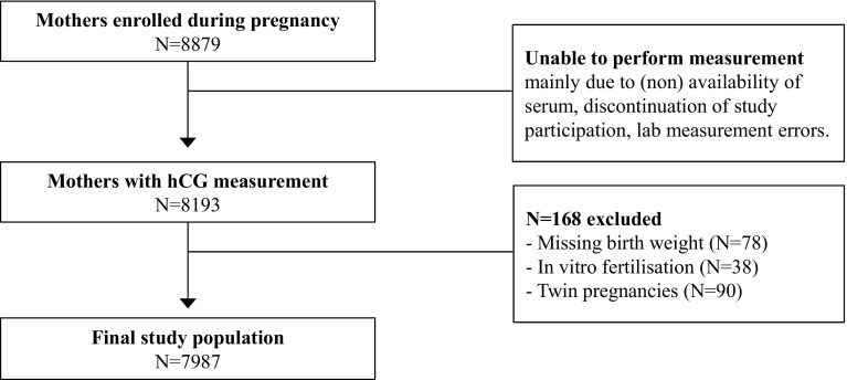
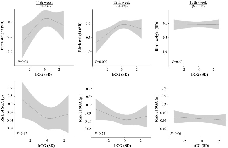
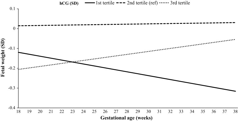
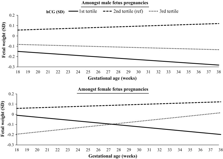
Human chorionic gonadotropin (hCG) is a pregnancy-specific hormone that regulates placental development. hCG concentrations vary widely throughout gestation and differ based on fetal sex. Abnormal hCG concentrations are associated with adverse pregnancy outcomes including fetal growth restriction. We studied the association of hCG concentrations with fetal growth and birth weight. In addition, we investigated effect modification by gestational age of hCG measurement and fetal sex. Total serum hCG (median 14.4 weeks, 95 % range 10.1-26.2), estimated fetal weight (measured by ultrasound during 18-25th weeks and >25th weeks) and birth weight were measured in 7987 mother-child pairs from the Generation R cohort and used to establish fetal growth. Small for gestational age (SGA) was defined as a standardized birth weight lower than the 10th percentile of the study population. There was a non-linear association of hCG with birth weight (P = 0.009). However, only low hCG concentrations measured during the late first trimester (11th and 12th week) were associated with birth weight and SGA. Low hCG concentrations measured in the late first trimester were also associated with decreased fetal growth (P = 0.0002). This was the case for both male and female fetuses. In contrast, high hCG concentrations during the late first trimester were associated with increased fetal growth amongst female, but not male fetuses. Low hCG in the late first trimester is associated with lower birth weight due to a decrease in fetal growth. Fetal sex differences exist in the association of hCG concentrations with fetal growth.
Keywords: Birth weight; Fetal growth; Fetal sex; Trophoblast; hCG.
Conflict of interest statement
Conflict of interest
The authors have nothing to disclose.
Ethics approval
The general design, all research aims and the specific measurements in the Generation R study have been approved by the Medical Ethical Committee of the Erasmus Medical Center, Rotterdam. Written informed consent was obtained from all participants.
Figures
References
Publication types
MeSH terms
Substances
LinkOut - more resources
Full Text Sources
Other Literature Sources
Medical
