. 2017 Mar;35(3):824-837.
doi: 10.1002/stem.2512. Epub 2016 Nov 17.

EGF-Amphiregulin Interplay in Airway Stem/Progenitor Cells Links the Pathogenesis of Smoking-Induced Lesions in the Human Airway Epithelium

Affiliations

EGF-Amphiregulin Interplay in Airway Stem/Progenitor Cells Links the Pathogenesis of Smoking-Induced Lesions in the Human Airway Epithelium

Wu-Lin Zuo et al. Stem Cells. 2017 Mar.

Abstract

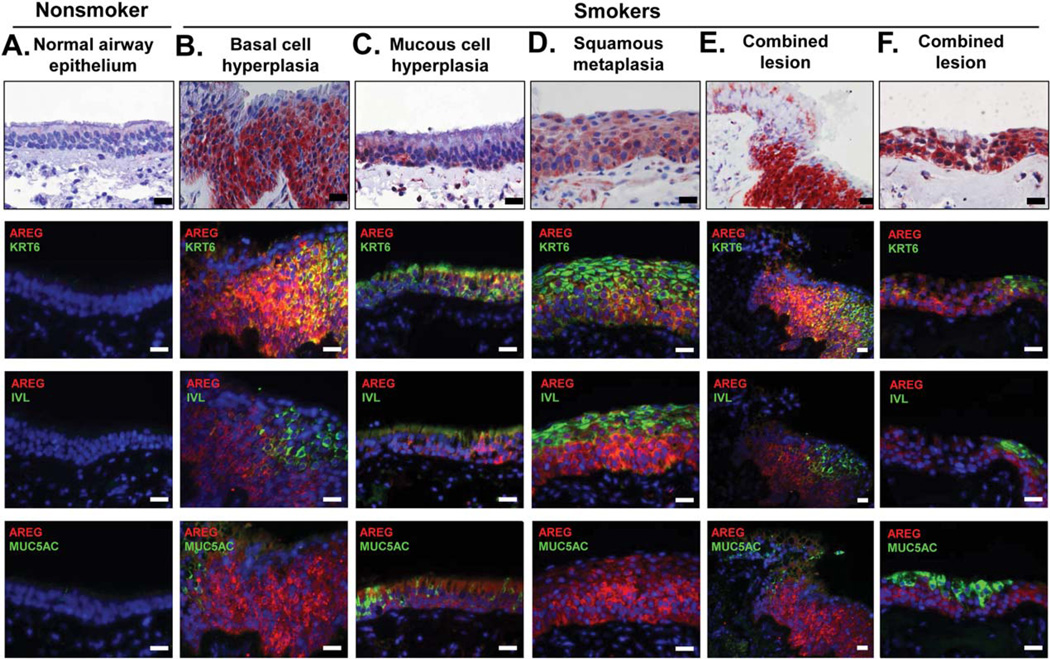
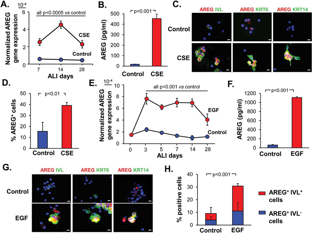
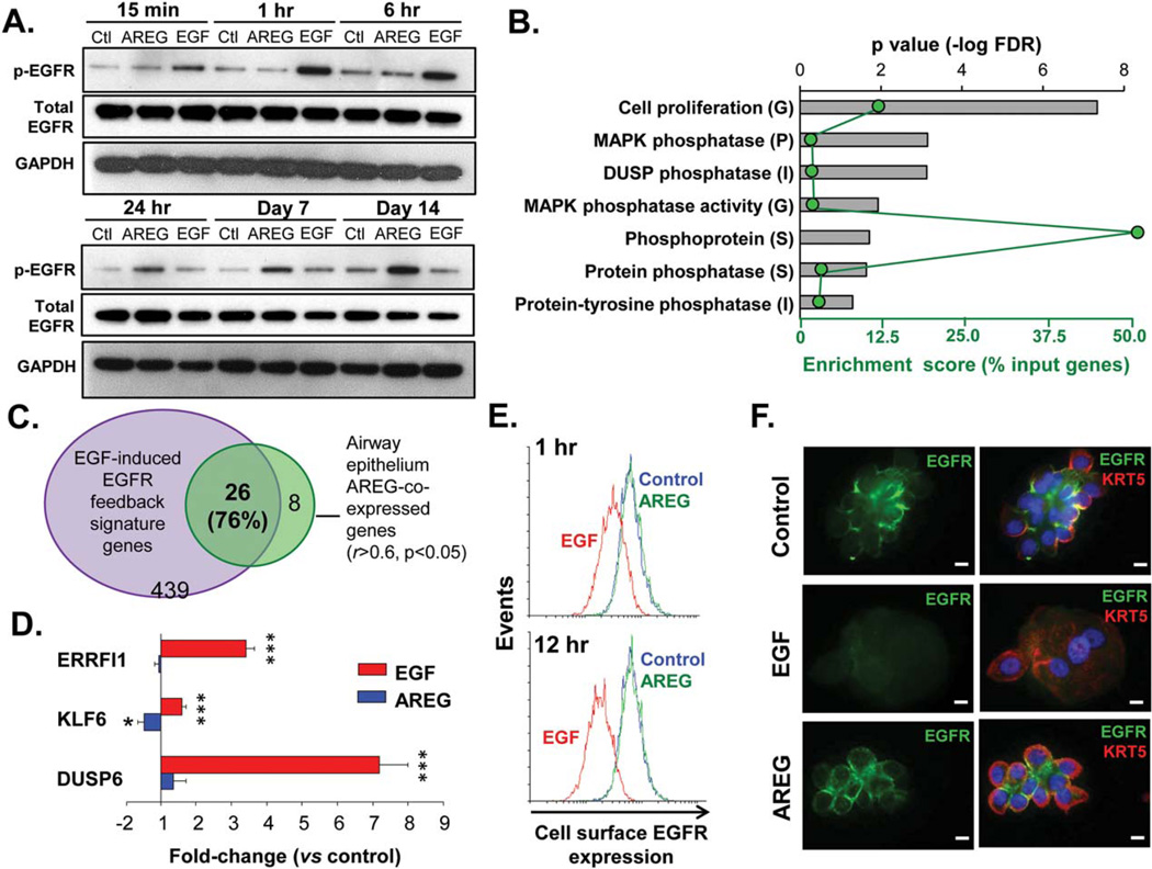
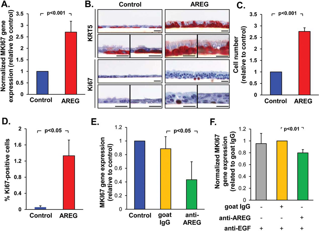
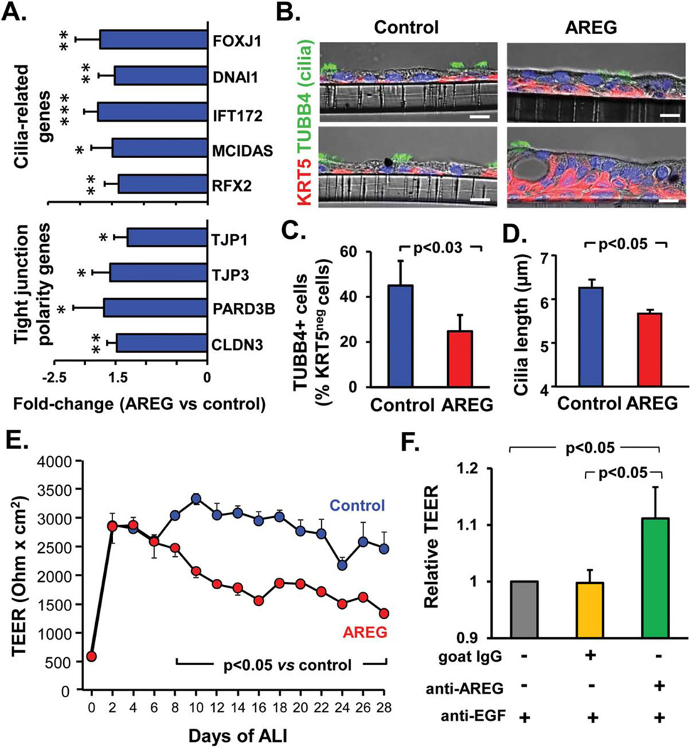
The airway epithelium of cigarette smokers undergoes dramatic remodeling with hyperplasia of basal cells (BC) and mucus-producing cells, squamous metaplasia, altered ciliated cell differentiation and decreased junctional barrier integrity, relevant to chronic obstructive pulmonary disease and lung cancer. In this study, we show that epidermal growth factor receptor (EGFR) ligand amphiregulin (AREG) is induced by smoking in human airway epithelium as a result of epidermal growth factor (EGF)-driven squamous differentiation of airway BC stem/progenitor cells. In turn, AREG induced a unique EGFR activation pattern in human airway BC, distinct from that evoked by EGF, leading to BC- and mucous hyperplasia, altered ciliated cell differentiation and impaired barrier integrity. Further, AREG promoted its own expression and suppressed expression of EGF, establishing an autonomous self-amplifying signaling loop in airway BC relevant for promotion of EGF-independent hyperplastic phenotypes. Thus, EGF-AREG interplay in airway BC stem/progenitor cells is one of the mechanisms that mediates the interconnected pathogenesis of all major smoking-induced lesions in the human airway epithelium. Stem Cells 2017;35:824-837.

Keywords: Airway basal cells; Chronic obstructive pulmonary disease; Epidermal growth factor receptor; Hyperplasia; Metaplasia.


Conflict of interest statement

Disclosure of Potential Conflict of Interest

The authors indicate no potential conflicts of interest.

Figures

Figure 1
Figure 1
Upregulation of AREG in the airway epithelium of smokers. Airway samples with indicated histologic phenotypes analyzed using immunohistochemistry (upper row) or immunofluorescence (IF) for AREG and markers of squamous (IVL), BC/IC (KRT6), and mucous-producing (MUC5AC) cells. Each vertical panel (A–F) shows representative images of the adjacent or nearby sections from the same specimen. In IF, nuclei are stained with DAPI (blue). Scale bars-20 µm. See Supporting Information Figure S1 for more examples and quantification data. Abbreviations: AREG, amphiregulin; IVL, involucrin; KRT6, keratin 6.
Figure 2
Figure 2
Cigarette smoke and EGF induce AREG in the airway epithelium. (A): Normalized AREG gene expression in the epithelium derived from airway basal cells (BC) treated every other day from day 0 of ALI with 3% CSE or untreated (control) BC at indicated time-points of ALI (mean ± SD); representative of three experiments. (B): AREG protein levels determined by ELISA in the basolateral supernatants of the epithelium derived from BC after 14 days of ALI as described in A (mean ± SD). (C): IF analysis of cytospins of epithelial cells generated from airway BC as described in B for expression of AREG (red) and indicated squamous-related markers (green); scale bars-20 µm. (D): AREG+ cells (% of total) in the samples derived in ALI and analyzed by IF as shown in C (mean ±SD). (E): Normalized AREG gene expression in the epithelium derived from EGF-treated (10 ng/ml) and control BC at different time points of ALI (mean ± SD). See Supporting Information Figure S2E for summary of three independent experiments. (F): AREG ELISA of the basolateral supernatants of the epithelium derived from BC after 14 days of ALI as described in E (mean ±SD). (G): IF analysis of cytospins of epithelial cells generated from airway BC as described in F for expression of AREG (red) and indicated squamous-related markers (green); scale bars-20 µm. (H): Quantification of AREG+ and IVL+ cells (% of total cells) in samples described in G; p value shows significance of difference in the total AREG+ and AREG+/IVL+ cells between the groups (mean±SD). All panels represent data derived from ≥3 independent experiments. Abbreviations: ALI, air-liquid interface; AREG, amphiregulin; CSE, cigarette smoke extract; EGF, epidermal growth factor; IVL, involucrin; KRT6, keratin 6.
Figure 3
Figure 3
Distinct patterns of EGFR activation by EGF and AREG. (A): Western analysis of phospho-EGFR (Tyr1173), total EGFR and GAPDH protein levels in airway epithelial cells derived from basal cells (BC) treated with EGF or AREG (both 10 ng/ml) versus unstimulated BC at indicated time-points; representative of >3 independent experiments (see Supporting Information Fig. S3A for quantification data). (B): Top seven categories enriched in the AREG-coexpressed airway epithelial gene set (r ≥ 0.5, p < .05) annotated using Gene Ontology (G) and protein family classifications (P, Protein Information Resource; I, InterPro; S, The Human Spliceosome Protein-Protein Interaction Resource) using GATHER (for G, additional criteria: Beyes factor >6) and DAVID (for G, P, I, S); ranked by false discovery rate-corrected p value (top axis; bars); green dots represent enrichment score (% input genes; bottom axis). (C): Venn diagram: overlap of the top AREG-coexpressed airway epithelial genes (r ≥ 0.6, p < .05) with the EGFR feedback signature. (D): Fold-change in expression of the EGFR feedback genes (see text for full gene names) in the epithelium derived from BC during 28 days of air-liquid interface (ALI) ± basolaterally added EGF or AREG versus control (mean ± SD; n= 5 independent experiments). (E): FACS analysis: histogram plots of the cell surface EGFR expression in control basal cells (BC) (blue) and BC treated with AREG or EGF; at 1 hours and 12 hours after stimulation; representative of three independent experiments. (F): IF images of cytospins of epithelial cells derived from BC after 14 days of ALI culture ± AREG or EGF for expression of EGFR and BC marker KRT5. Nuclei are stained with DAPI (blue). Scale bars-10 µm. See Supporting Information Figure S3C for more examples. Abbreviations: AREG, amphiregulin; DUSP, dual specificity protein phosphatase; EGFR, epidermal growth factor receptor.
Figure 4
Figure 4
AREG promotes airway basal cells (BC) proliferation and hyperplasia. (A): Expression of the MKI67 gene in the epithelium derived from airway BC after 28 days of air-liquid interface (ALI) in the presence of AREG relative to control group (mean±SD). (B): Morphology and immunohistochemistry of samples derived as described in A for BC marker KRT5 and proliferation marker Ki-67. Shown are three separate areas of the ALI cultures for each of marker; scale bars-20 µm. (C): Total cell number (normalized to the control group) and (D) % Ki-67+ cells in the samples derived from BC as described A in three randomly selected fields (mean ± SD). (E). MKI67 gene expression in the epithelium derived from BC during 14 days in ALI ± neutralizing anti-AREG antibody or goat IgG isotype control (both 1 µg/ml; normalized to control group; mean ± SD). (F): MKI67 gene expression in the airway epithelium derived from BC treated with EGF-conditioned media (EGF-CM; see text) ± neutralizing anti-AREG antibodies (1 µg/ml), and neutralizing anti-EGF antibody (0.5 µg/ml); normalized to the control IgG group (mean ± SD); for more data see Supporting Information Figure S4. All panels represent data derived from ≥3 independent experiments. Abbreviations: AREG, amphiregulin; EGF, epidermal growth factor.
Figure 5
Figure 5
AREG promotes mucous cell differentiation and hyperplasia. (A): Fold-change in expression of mucous differentiation-related genes (see text for full gene names) in the epithelium derived from basal cells (BC) after 28 days of ALI culture in the presence of AREG versus untreated control (mean ± SD). (B): Cytopreps (top panels) and sections (remaining panels) of ALI samples derived from BC as described in A analyzed using immunohistochemistry for MUC5AC (see Supporting Information Fig. S5A for more examples) or Alcian blue staining (blue–mucus); scale bars-20 µm (representative of three experiments). (C): Quantification of MUC5AC-expressing cells in samples described in B (relative to the AREG group; mean±SD). (D): % Alcian blue-positive cells in AREG versus control groups described in B (mean ± SD; n = 3 experiments); arrows show Alcian blue-positive cells. (E): Fold-change in expression of indicated mucous differentiation-related genes in the epithelium derived from BC during 14 days of ALI in the presence of neutralizing anti-AREG antibody versus goat IgG control (mean±SD). (F): Expression of indicated mucous-related genes in the epithelium derived from BC treated as described in Figure 4F; normalized to control IgG group (mean ± SD); see more data in Supporting Information Figure S5C. All panels represent data derived from ≥3 independent experiments. Abbreviations: AGR2, anterior gradient 2; AREG, amphiregulin; SPDEF, SAM pointed domain containing ETS transcription factor.
Figure 6
Figure 6
AREG alters ciliated cell differentiation and barrier integrity. (A): Fold-change in expression of the ciliated cell differentiation-related genes (FOXJ1, forkhead box protein J1; DNAI1, dynein intermediate chain 1, axonemal; IFT172, intraflagellar transport 172; MCI-DAS, multicilin, and RFX2, regulatory factor X, 2) and tight junction (TJ)- and epithelial polarity-related genes (TJ proteins (TJP) 1 and − 3, partitioning defective PARD3, and claudin CLDN3) in the epithelium derived from basal cells (BC) after 28 days of ALI ± AREG (mean 6SD). (B): Representative phase contrast IF images of sections of the airway epithelium derived in ALI as described in A stained for BC marker keratin 5 (KRT5) and cilia marker tubulin, beta 4 (TUBB4); blue–DAPI (nuclei); scale bars-10 µm. (C): % of ciliated (TUBB4-positive) cells (relative to all KRT5-negative, that is, non-BC, cells) in AREG versus control groups described in B (mean ± SD; n= 3 experiments). (D): Cilia length in epithelia derived from BC as described in A (mean ± SD). (E): TER of BC-derived epithelium at different time-points of ALI in AREG-treated versus control groups (mean ± SD; n= 4 replicates/group); see Supporting Information Figure S6B for summary of three independent experiments). (F): TER of airway BC-derived epithelium treated as described in Figure 4F measured at day 16 of ALI (mean ± SD). Panels A-D, and F represent data derived from ≥3 independent experiments. Abbreviations: ALI, air-liquid interface; AREG, amphiregulin; CSE, cigarette smoke extract; EGF, epidermal growth factor; TER, transepithelial resistance.
Figure 7
Figure 7
Effect of AREG on the AREG and EGF gene expression. (A): AREG gene expression in the epithelium derived from airway basal cells (BC) during 28 days of ALI in the presence or absence of AREG relative to the control group (mean ± SD; n = 4 experiments). (B): Sections of the airway epithelium derived from BC during 28 days in ALI in the presence (lower) or absence (upper) of AREG analyzed by IF for expression of AREG and BC marker KRT5; arrows indicate AREG+ BC; representative of three experiments; scale bars-20 µm. (C): AREG+/KRT5+ and AREG+/KRT5 cells (% of AREG+ cells in the AREG-treated group) in samples derived from three independent experiments described in B; p value shows significance of difference in % of all AREG+ cells and AREG/CK5-double+ cells between the groups. (D): Normalized AREG gene expression in the airway epithelium derived from BC during 14 days of ALI culture in the presence or absence of 3% CSE, neutralizing anti-AREG antibodies or isotype control goat IgG (mean ± SD; n = 3 experiments); quantification of AREG+ cells for these experiments is shown in Supporting Information Figure S7B. (E): Normalized EGF gene expression in AREG-stimulated and control samples from 3 independent experiments described in A and B. Abbreviations: AREG, amphiregulin; CSE, cigarette smoke extract; KRT5, keratin 5.

References

    1. Crystal RG, Randell SH, Engelhardt JF, et al. Airway epithelial cells: Current concepts and challenges. Proc Am Thorac Soc. 2008;5:772–777. - PMC - PubMed
    1. Rock JR, Randell SH, Hogan BL. Airway basal stem cells: A perspective on their roles in epithelial homeostasis and remodeling. Dis Model Mech. 2010;3:545–556. - PMC - PubMed
    1. Fahy JV, Dickey BF. Airway mucus function and dysfunction. N Engl J Med. 2010;363:2233–2247. - PMC - PubMed
    1. Hong KU, Reynolds SD, Watkins S, et al. Basal cells are a multipotent progenitor capable of renewing the bronchial epithelium. Am J Pathol. 2004;164:577–588. - PMC - PubMed
    1. Rock JR, Onaitis MW, Rawlins EL, et al. Basal cells as stem cells of the mouse trachea and human airway epithelium. Proc Natl Acad Sci USA. 2009;106:12771–12775. - PMC - PubMed