. 2016 Dec:110:45-59.
doi: 10.1016/j.biomaterials.2016.09.003. Epub 2016 Sep 5.

Bioprinting 3D microfibrous scaffolds for engineering endothelialized myocardium and heart-on-a-chip

Affiliations

Bioprinting 3D microfibrous scaffolds for engineering endothelialized myocardium and heart-on-a-chip

Yu Shrike Zhang et al. Biomaterials. 2016 Dec.

Erratum in

Abstract

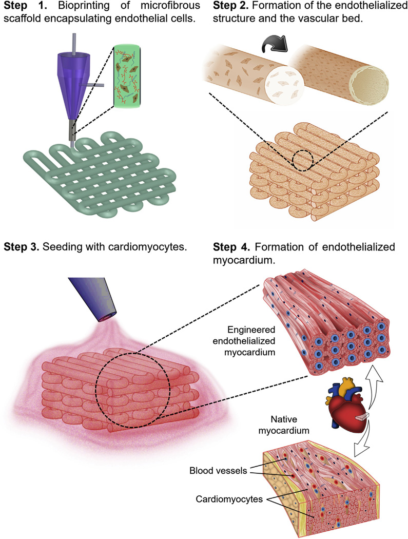
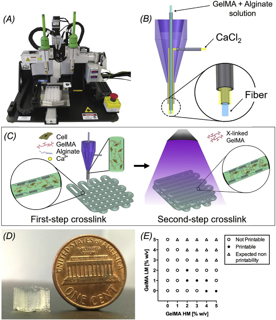
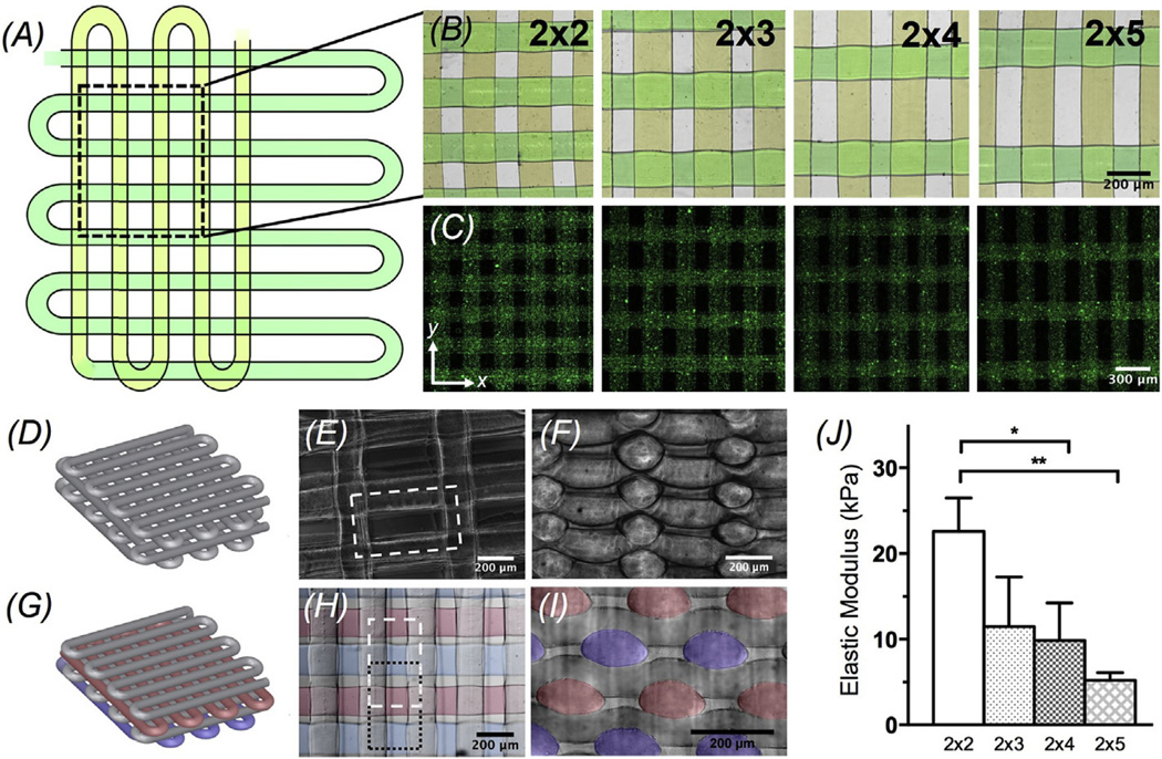
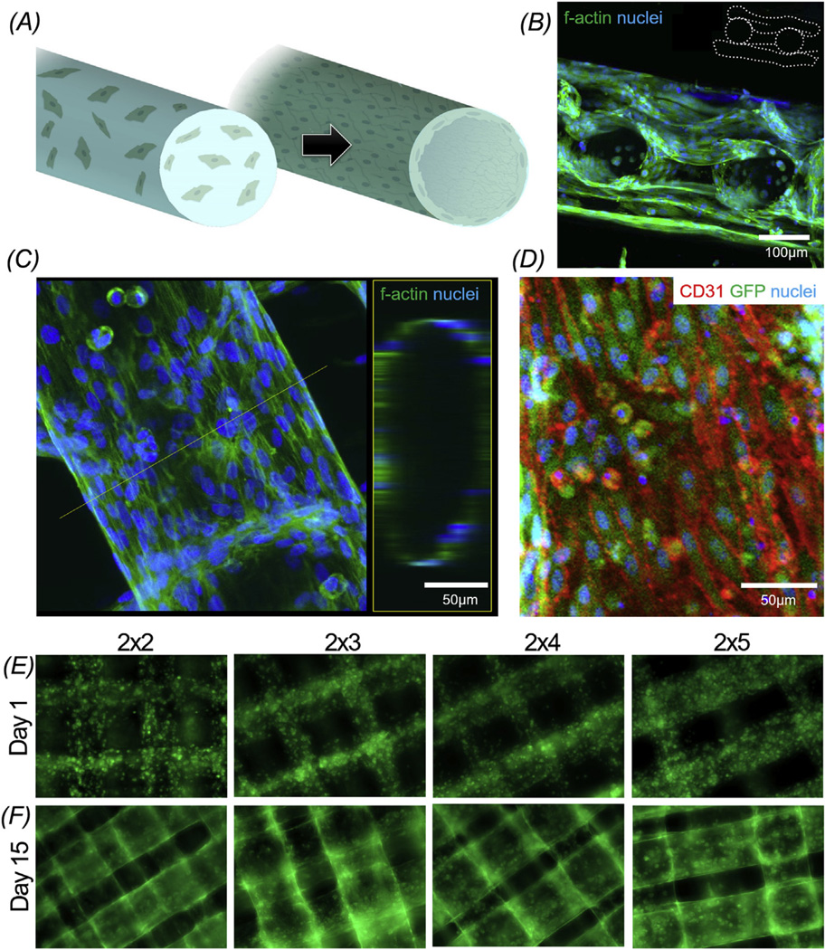
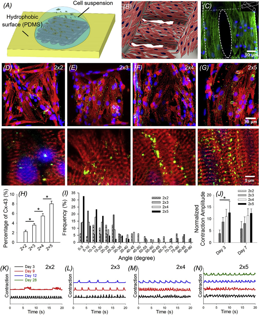
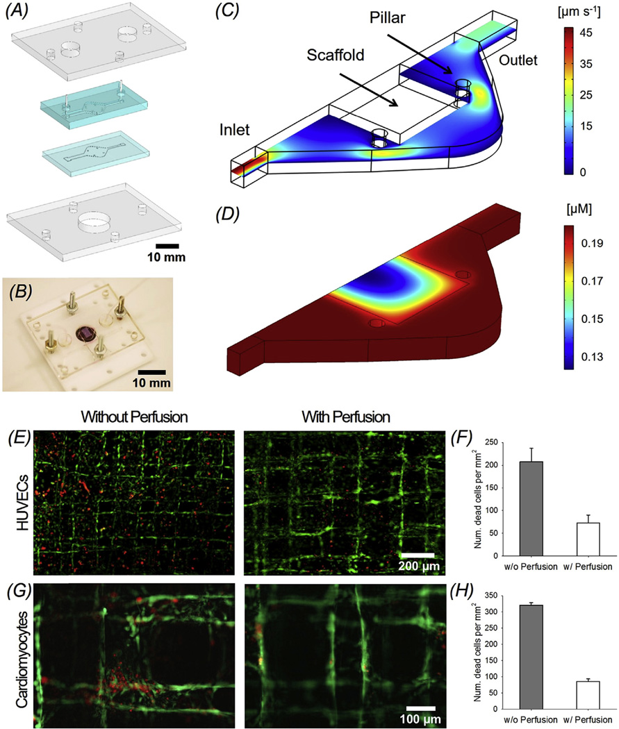
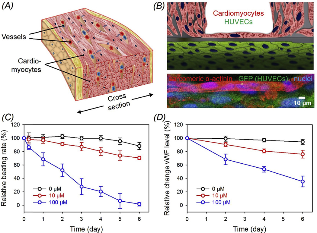
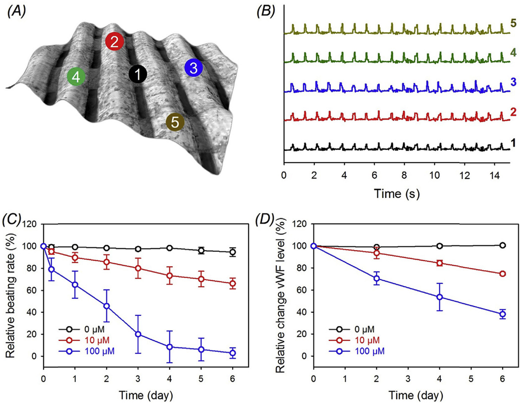
Engineering cardiac tissues and organ models remains a great challenge due to the hierarchical structure of the native myocardium. The need of integrating blood vessels brings additional complexity, limiting the available approaches that are suitable to produce integrated cardiovascular organoids. In this work we propose a novel hybrid strategy based on 3D bioprinting, to fabricate endothelialized myocardium. Enabled by the use of our composite bioink, endothelial cells directly bioprinted within microfibrous hydrogel scaffolds gradually migrated towards the peripheries of the microfibers to form a layer of confluent endothelium. Together with controlled anisotropy, this 3D endothelial bed was then seeded with cardiomyocytes to generate aligned myocardium capable of spontaneous and synchronous contraction. We further embedded the organoids into a specially designed microfluidic perfusion bioreactor to complete the endothelialized-myocardium-on-a-chip platform for cardiovascular toxicity evaluation. Finally, we demonstrated that such a technique could be translated to human cardiomyocytes derived from induced pluripotent stem cells to construct endothelialized human myocardium. We believe that our method for generation of endothelialized organoids fabricated through an innovative 3D bioprinting technology may find widespread applications in regenerative medicine, drug screening, and potentially disease modeling.

Keywords: Bioprinting; Cardiac tissue engineering; Cardiovascular toxicity; Heart-on-a-chip; Vascularization.


Figures

Fig. 1
Fig. 1
Schematics showing the procedure of fabricating endothelialized myocardium using the 3D bioprinting strategy. Step 1: bioprinting of a microfibrous scaffold using a composite bioink encapsulating endothelial cells. Step 2: formation of the vascular bed through migration of HUVECs to the peripheries of the microfibers. Step 3: seeding of cardiomyocytes into the interstitial space of the endothelialized scaffold. Step 4: formation of engineered endothelialized myocardium structurally resembling the native myocardium.
Fig. 2
Fig. 2
(A) Photograph of an Organovo Novogen MMX bioprinter. (B) Schematic of the coaxial needle where the bioink is delivered from the core and the ionic crosslinking CaCl2 solution is sheathed on the side. (C) Schematic diagrams showing the two-step crosslinking process, where the alginate component is first physically crosslinked by the CaCl2 followed by chemical crosslinking of the GelMA component using UV illumination. (D) Photograph of a bioprinted cubic microfibrous scaffold (6-mm edge length). (E) Bioink optimization where conditions of printability and non-printability for different concentrations of GelMA-HM and GelMA-LM (with a constant alginate concentration of 4 w/v%) were analyzed.
Fig. 3
Fig. 3
(A) Top view single-layer schematic of the design of the bioprinted microfibrous scaffold and corresponding (B) brightfield (pseudocolored to match the schematic) and (C) fluorescence micrographs showing the bioprinted scaffolds with different aspect ratios of unit grids. (D) 3D schematic of a scaffold without offset and corresponding (E) top-view and (F) cross-sectional micrographs. (G) 3D schematic of a scaffold with offset and corresponding (H) top-view and (I) cross-sectional micrographs showing the offset between the layers. (J) Elastic moduli of the bioprinted scaffolds with different aspect ratios of unit grids.
Fig. 4
Fig. 4
(A) Schematic representation showing the assembly of the encapsulated HUVECs inside the bioprinted microfibers into a confluent layer of endothelium. (B) Confocal fluorescence micrograph showing the cross-sectional view of a three-layer scaffold at Day 14, indicating the formation of the endothelium by the HUVECs. (C) High-resolution confocal fluorescence micrograph showing the distribution of the HUVECs in a single microfiber at Day 14. Left: projection view; Right: 3D rendering of the tubular structure at the position of the dotted line. (D) Confocal fluorescence micrograph showing the GFP-HUVECs in a single fiber for CD31, GFP, and nuclei. (E, F) Fluorescence micrographs showing the distribution and spreading of GFP-HUVECs in the bioprinted microfibrous scaffolds with different aspect ratios of unit grids at Day 1 and Day 15, respectively.
Fig. 5
Fig. 5
(A) Schematic showing the seeding procedure of cardiomyocytes onto the bioprinted microfibrous scaffolds. (B) Schematic showing a scaffold seeded with neonatal rat cardiomyocytes. (C) F-actin (green) staining showing the distribution of the cardiomyocytes on the surface of the scaffold at the location where two microfibers of adjacent layers crossed. (D–G) Immunofluorescence staining of sarcomeric α-actinin (red) and connexin-43 (Cx-43, green) of cardiomyocytes seeded on bioprinted microfibrous scaffolds with different aspect ratios of unit grids, showing varying degrees of alignment of the cardiomyocytes. Lower panels: magnified images showing the sarcomeric banding. (H) Quantification of Cx-43 expression by the cardiomyocytes on the four types of scaffolds, plotted as percentages of area coverage calculated from the fluorescence images; *p < 0.005. (I) Quantification of the angle distribution of cardiomyocytes on the four types of bioprinted microfibrous scaffolds. (J) Quantification of the contraction amplitude of the four types of bioprinted myocardial constructs. *p < 0.05. (K–N) Beating analysis of the cardiac organoid on bioprinted scaffolds with different aspect ratios of unit grids. Note that the contraction amplitudes in (K–N) were all normalized to the same height for easy comparison across the samples. (For interpretation of the references to colour in this figure legend, the reader is referred to the web version of this article.)
Fig. 6
Fig. 6
(A) Schematics showing exploded view of the design of the two-layer microfluidic bioreactor sandwiched by a pair of PMMA clamps. (B) Photograph of the bioreactor with an embedded bioprinted scaffold. (C, D) Simulation results of flow velocity and oxygen distribution, respectively, in the bioreactor chamber at a flow rate of 50 µL min−1 (E, F) Live/dead micrographs and quantified cell morbidity of bioprinted endothelialized scaffolds without and with perfusion in the bioreactors. (G, H) Live/dead micrographs and quantified cell morbidity of bioprinted cardiac organoids without and with perfusion in the bioreactors.
Fig. 7
Fig. 7
(A) Schematic showing a native myocardium containing blood vessels embedded in a matrix of cardiomyocytes. (B) Schematic and high-resolution confocal fluorescence micrograph showing an endothelialized myocardial tissue formed by seeding neonatal rat cardiomyocytes onto the bioprinted endothelialized microfibrous scaffold after 15 days of pre-endothelialization. (C, D) Relative beating of the endothelialized myocardial tissues and the levels of vWF expression by the endothelial cells, respectively, upon treatment with different dosages of doxorubicin.
Fig. 8
Fig. 8
(A) Pseudo-3D brightfield micrograph showing an all-human endothelialized myocardial tissue formed by seeding hiPSC-cardiomyocytes onto the bioprinted endothelialized scaffold after 15 days of pre-endothelialization. (B) Beating plots of the different local regions indicated in (A). Note that the contraction amplitudes were all normalized to the same height for easy comparison across the samples. (C, D) Relative beating of the endothelialized human myocardial tissues and the levels of vWF expression by the endothelial cells, respectively, upon treatment with different dosages of doxorubicin.

References

    1. Chiu L, Iyer RK, Reis LA, Nunes SS, Radisic M. Cardiac tissue engineering: current state and perspectives. Front. Biosci. 2011;17:1533–1550. - PubMed
    1. Chiu LL, Radisic M. Cardiac tissue engineering. Curr. Opin. Chem. Eng. 2013;2:41–52.
    1. Zhang YS, Aleman J, Arneri A, Bersini S, Piraino F, Shin SR, et al. From cardiac tissue engineering to heart-on-a-chip: beating challenges. Biomed. Mater. 2015;10:034006. - PMC - PubMed
    1. Bhise NS, Ribas J, Manoharan V, Zhang YS, Polini A, Massa S, et al. Organ-on-a-chip platforms for studying drug delivery systems. J. Control. Release. 2014;190:82–93. - PMC - PubMed
    1. Zhang YS, Khademhosseini A. Seeking the Right context for evaluating nanomedicine: from tissue models in petri dishes to microfluidic organs-on-a-chip. Nanomedicine. 2015;10:685–688. - PubMed

Publication types