. 2016 Oct 6;10(10):e0005047.
doi: 10.1371/journal.pntd.0005047. eCollection 2016 Oct.

Mechanistic Insight into the Host Transcription Inhibition Function of Rift Valley Fever Virus NSs and Its Importance in Virulence

Affiliations

Mechanistic Insight into the Host Transcription Inhibition Function of Rift Valley Fever Virus NSs and Its Importance in Virulence

Kaori Terasaki et al. PLoS Negl Trop Dis. .

Abstract

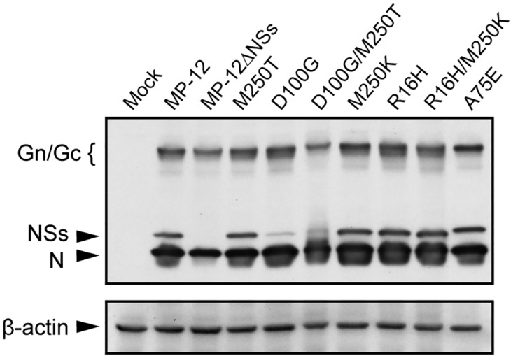
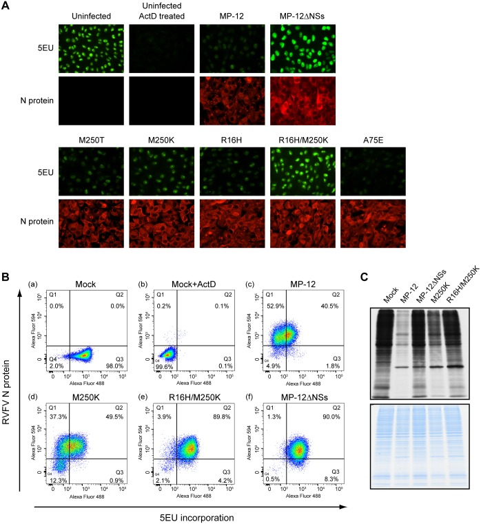
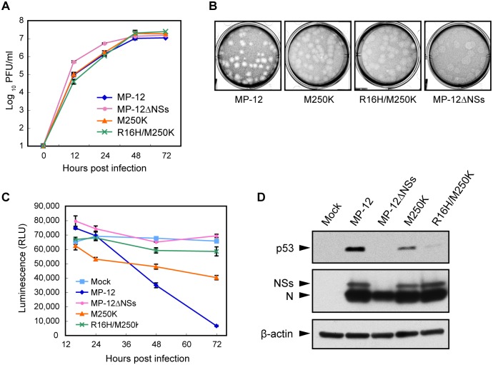
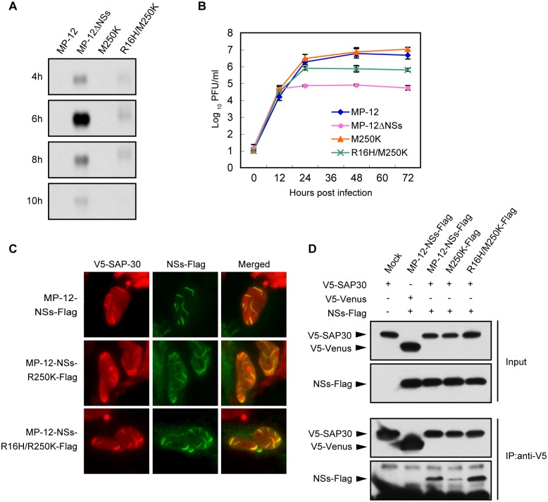
Rift Valley fever virus (RVFV), a member of the genus Phlebovirus within the family Bunyaviridae, causes periodic outbreaks in livestocks and humans in countries of the African continent and Middle East. RVFV NSs protein, a nonstructural protein, is a major virulence factor that exhibits several important biological properties. These include suppression of general transcription, inhibition of IFN-β promoter induction and degradation of double-stranded RNA-dependent protein kinase R. Although each of these biological functions of NSs are considered important for countering the antiviral response in the host, the individual contributions of these functions towards RVFV virulence remains unclear. To examine this, we generated two RVFV MP-12 strain-derived mutant viruses. Each carried mutations in NSs that specifically targeted its general transcription inhibition function without affecting its ability to degrade PKR and inhibit IFN-β promoter induction, through its interaction with Sin3-associated protein 30, a part of the repressor complex at the IFN-β promoter. Using these mutant viruses, we have dissected the transcription inhibition function of NSs and examined its importance in RVFV virulence. Both NSs mutant viruses exhibited a differentially impaired ability to inhibit host transcription when compared with MP-12. It has been reported that NSs suppresses general transcription by interfering with the formation of the transcription factor IIH complex, through the degradation of the p62 subunit and sequestration of the p44 subunit. Our study results lead us to suggest that the ability of NSs to induce p62 degradation is the major contributor to its general transcription inhibition property, whereas its interaction with p44 may not play a significant role in this function. Importantly, RVFV MP-12-NSs mutant viruses with an impaired general transcription inhibition function showed a reduced cytotoxicity in cell culture and attenuated virulence in young mice, compared with its parental virus MP-12, highlighting the contribution of NSs-mediated general transcription inhibition towards RVFV virulence.


Conflict of interest statement

The authors have declared that no competing interests exist.

Figures

Fig 1
Fig 1. Accumulation of wt and mutant NSs proteins in infected cells.
Vero E6 cells were mock infected (Mock) or infected with MP-12 or MP-12 mutants, each carrying a different NSs mutant, at an m.o.i. of 3. The infected cells were harvested at 16 h p.i. and analyzed in Western blots by using anti-MP-12 antibody or anti-β actin antibody followed by secondary antibody.
Fig 2
Fig 2. Effects of NSs mutants on host general transcription.
(A) Fluorescence microscopic analysis of newly synthesized RNAs. Vero E6 cells were mock infected (uninfected), mock infected and ActD treated (Uninfected ActD treated) or infected with MP-12 or its mutants at an m.o.i. of 3. At 16 h p.i., cells were incubated in the presence of 5EU for 1 h. The incorporated 5EU were visualized by using Alexa fluor 488 conjugated azide (5EU), and viral N protein was stained by anti-N antibody followed by Alexa fluor 594 conjugated secondary antibody (N protein). Fluorescence microscopic analysis was performed to detect newly synthesized RNAs. (B) Flow cytometric analysis of newly synthesized RNAs in infected cells. Vero E6 cells were mock infected, mock infected and ActD treated, or infected with MP-12, MP-12-M250K, or MP-12-R16H/M250K, at an m.o.i. of 3. 5EU and anti-N antibody were used to label newly synthesized RNAs and N proteins, respectively, as described in (A). Then the cells were subjected to Flow cytometric analysis. The density dot plot was divided into four quadrants (Q1, Q2, Q3, and Q4). Quadrant Q1 (Upper left quadrant): RVFV N protein positive and low activity of RNA transcription. Quadrant Q2 (Upper right quadrant): RVFV N protein positive and high activity of RNA transcription. Quadrant Q3 (Lower right quadrant): RVFV N protein negative and high activity of RNA transcription. Quadrant Q4 (Lower left quadrant): RVFV N protein negative and low activity of RNA transcription. (C) Autoradiography of radiolabeled proteins in mock infected cells and infected cells. Vero E6 cells were mock infected (mock) or infected with MP-12 or its mutants at an m.o.i. of 3. At 16 h p.i., cells were incubated with 35S-methionine/cysteine for 1 h. Whole cell lysates were resolved by SDS-PAGE and visualized by autoradiography (top panel) or Coomassie Blue staining (bottom panel).
Fig 3
Fig 3. Attenuated cytotoxicity of MP-12 NSs mutants.
(A) Growth kinetics of MP-12 and its mutants. Vero E6 cells were infected with each virus at an m.o.i. of 0.01, and the culture supernatants were collected at various times p.i. Virus titers were determined by plaque assays. (B) Plaque morphology of the MP-12 and mutant viruses in VeroE6 cells at 3 day p.i. (C) Viability of cells infected with MP-12 or its mutants infected cells. Vero E6 cells were mock infected or infected with each virus at an m.o.i. of 3. Cell viability was determined by measuring cellular ATP. (D) Abundance of p53 in infected cells. Vero E6 cells were infected with each virus at an m.o.i. of 3 and harvested at 24 h p.i. Whole cell lysate was subjected to Western blot analysis. Anti-p53 antibody, anti-MP-12 antibody and anti-β-actin antibody were used as the primary antibody for the top, middle and bottom panels, respectively.
Fig 4
Fig 4. Effects of M250K and R16H/M250K mutations on PKR stability in infected cells.
HeLa cells were infected with MP-12 or mutant viruses at an m.o.i. of 3. Whole-cell lysate was harvested at 8 h p.i. and subjected to Western blot analysis by using anti-PKR antibody, anti-MP-12 antibody and anti-β-actin antibody as the primary antibody for the top, middle and bottom panels, respectively.
Fig 5
Fig 5. Inhibition of IFN-β mRNA expression by MP-12 NSs mutants.
(A) MRC-5 cells were infected with MP-12 or its mutants at an m.o.i of 3. Total RNA was collected at various time p.i. as indicated. IFN-β mRNA was detected by Northern blotting analysis using the IFN-β mRNA-specific RNA probe. (B) Growth kinetics of MP-12 and its mutants in MRC-5 cells. MRC-5 cells were infected with each virus at an m.o.i. of 0.01, and the culture supernatants were collected at various times p.i. Virus titers were determined by using a plaque assay in Vero E6 cells. (C) Co-immunostaining for NSs and SAP30. HeLa cells were transfected with plasmid expressing human SAP30 carrying an N-terminal V5 tag and infected with MP-12 carrying a Flag-tagged NSs or mutant MP-12 carrying Flag-tagged NSs mutant at 16 h post transfection. At 6 h p.i., the cells were fixed, immunostained with an anti-V5 tag and anti-Flag tag antibodies, and subjected to fluorescent microscopic examination. (D) Co-immunoprecipitation of NSs and SAP30. HeLa cells were transfected with the plasmid encoded V5-tagged SAP30 or V5-tagged Venus protein. At 16 h post transfection, cells were mock infected or infected with the MP-12 carrying Flag-tagged NSs or mutant MP-12 carrying Flag-tagged NSs mutant. At 8 h p.i., the cells were lysed and subjected to immunoprecipitation by using anti-V5 antibody. Precipitates were examined by using Western blotting with anti-V5 tag antibody (top and third panels) or anti-Flag tag antibody (second and bottom panels). The top two panels show Western blot analysis of the input lysates.
Fig 6
Fig 6. Effect of mutations in NSs on its interaction with TFIIH subunits.
(A) HeLa cells were co-transfected with NSs expression plasmid or empty vector (EV) along with reporter plasmid encoding Renilla luciferase. Luciferase activities (relative light units) were measured at 24h post transfection (top panel). Whole cell lysates were prepared from the transfected cells described above and subjected to Western blot analysis by using anti-MP-12 and anti-β-actin antibodies (bottom panel). (B) HeLa cells were mock infected (Mock) or infected with MP-12 or its mutants at an m.o.i. of 3 and harvested at 16 h p.i. Whole-cell lysates were subjected to Western blot analysis by using anti-p62 antibody and p44 antibody, anti-XPD antibody, anti-MP-12 antibody or anti-β-actin antibody as primary antibodies. (C) HeLa cells were mock infected (Mock) or infected with the MP-12 carrying Flag-tagged NSs or NSs or mutant MP-12 carrying Flag-tagged NSs mutant at an m.o.i. of 3. At 8 h p.i., the cells were lysed and subjected to immunoprecipitation by using anti-p44 antibody. Precipitates were subjected to Western blotting and analyzed by using anti-p44 antibody (third panel) and anti-Flag tag antibody (bottom). The top two panels show Western blot analysis of the input lysates by using anti-p44 antibody (top panel) and anti-Flag tag antibody (second panel). (D) HeLa cells were mock infected (Mock) or infected with the MP-12 carrying Flag-tagged NSs or NSs or mutant MP-12 carrying Flag-tagged NSs mutant at an m.o.i. of 3. At 5 h p.i., the cells were lysed and subjected to immunoprecipitation by using anti-p62 antibody. Precipitates were subjected to Western blotting and analyzed by anti-p62 antibody (third panel) and anti-Flag tag antibody (bottom). The top two panels show Western blot analysis of the input lysates using anti-p62 antibody (top panel) and anti-Flag tag antibody (second panel). (E) HeLa cells were transfected with the plasmid encoding V5-tagged full-length FBXO3 or V5-tagged Venus protein and infected with the MP-12 carrying Flag-tagged NSs or mutant MP-12 carrying Flag-tagged NSs mutant at 16 h post transfection. At 8 h p.i., the cells were lysed and subjected to immunoprecipitation using anti-V5 antibody. Precipitates were subjected to Western blotting analysis by using anti-V5 antibody (third panel) and anti-Flag tag antibody (bottom panel). The top two panels show Western blot analysis of the input lysates using anti-V5 antibody (top panel) and anti-Flag tag antibody (second panel).
Fig 7
Fig 7. Attenuated virulence of MP-12 NSs mutants.
Eighteen-day-old CD1 mice were intraperitoneally inoculated with 104 PFU of MP-12 (n = 11), MP-12-M250K (n = 10) or MP-12-R16H/M250K (n = 10) or inoculated with HBSS (n = 11). The mice were observed for 22 days after inoculation.

References

    1. Bird BH, Nichol ST. Breaking the chain: Rift Valley fever virus control via livestock vaccination. Curr Opin Virol. 2012;2(3):315–23. 10.1016/j.coviro.2012.02.017 - DOI - PubMed
    1. Ikegami T, Makino S. The Pathogenesis of Rift Valley Fever. Viruses. 2011;3(5):493–519. 10.3390/v3050493 - DOI - PMC - PubMed
    1. Won S, Ikegami T, Peters CJ, Makino S. NSm and 78-kilodalton proteins of Rift Valley fever virus are nonessential for viral replication in cell culture. J Virol. 2006;80(16):8274–8. 10.1128/JVI.00476-06 - DOI - PMC - PubMed
    1. Kading RC, Crabtree MB, Bird BH, Nichol ST, Erickson BR, Horiuchi K, et al. Deletion of the NSm virulence gene of Rift Valley fever virus inhibits virus replication in and dissemination from the midgut of Aedes aegypti mosquitoes. PLoS Negl Trop Dis. 2014;8(2):e2670 10.1371/journal.pntd.0002670 - DOI - PMC - PubMed
    1. Kreher F. The Rift Valley fever accessory proteins NSm and P78/NSm-GN are distinct determinants of virus propagation in vertebrate and invertebrate hosts. Emerg Microbes Infect. 2014;3(e71). 10.1038/emi.2014.71 - DOI - PMC - PubMed

MeSH terms