. 2016 Oct 6;12(10):e1005926.
doi: 10.1371/journal.ppat.1005926. eCollection 2016 Oct.

Sphingosine 1-Phosphate Activation of EGFR As a Novel Target for Meningitic Escherichia coli Penetration of the Blood-Brain Barrier

Affiliations

Sphingosine 1-Phosphate Activation of EGFR As a Novel Target for Meningitic Escherichia coli Penetration of the Blood-Brain Barrier

Xiangru Wang et al. PLoS Pathog. .

Abstract

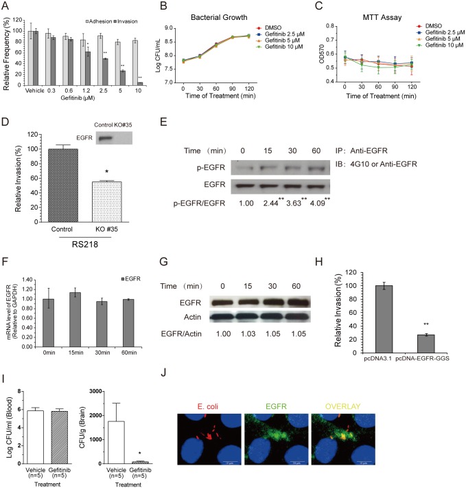
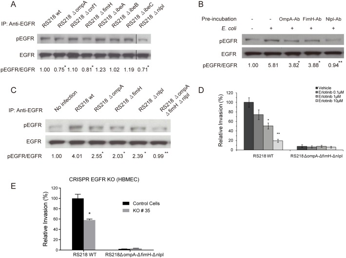
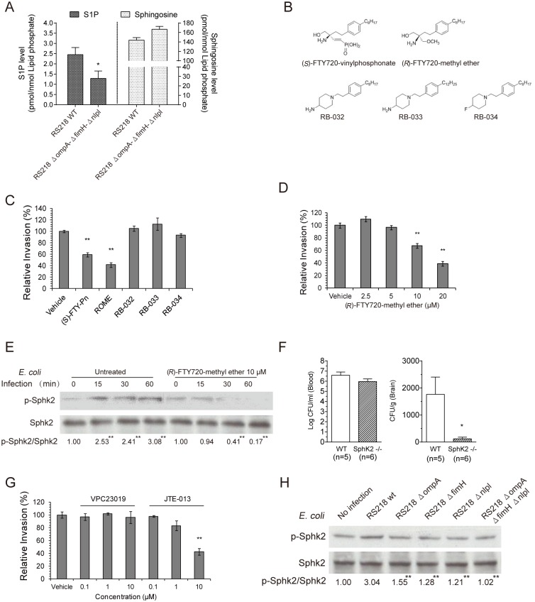
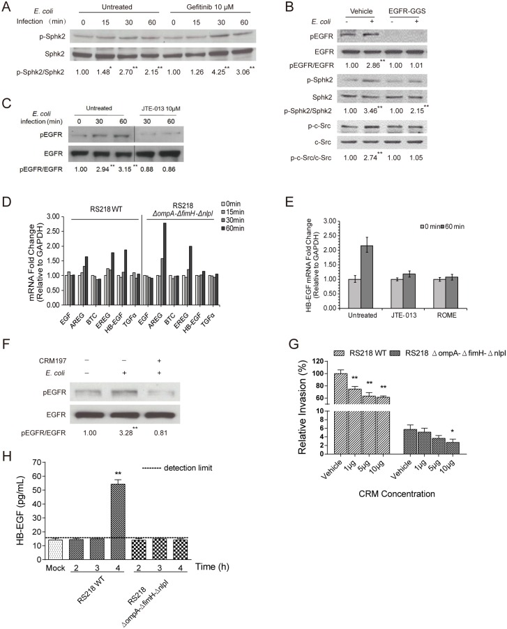
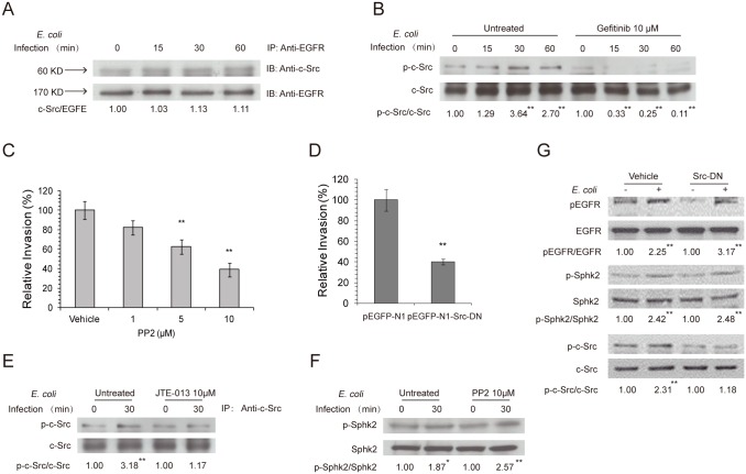
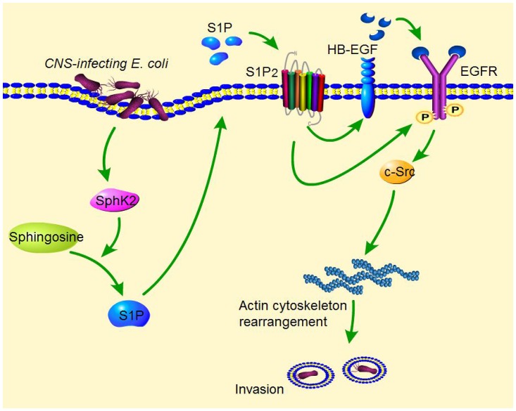
Central nervous system (CNS) infection continues to be an important cause of mortality and morbidity, necessitating new approaches for investigating its pathogenesis, prevention and therapy. Escherichia coli is the most common Gram-negative bacillary organism causing meningitis, which develops following penetration of the blood-brain barrier (BBB). By chemical library screening, we identified epidermal growth factor receptor (EGFR) as a contributor to E. coli invasion of the BBB in vitro. Here, we obtained the direct evidence that CNS-infecting E. coli exploited sphingosine 1-phosphate (S1P) for EGFR activation in penetration of the BBB in vitro and in vivo. We found that S1P was upstream of EGFR and participated in EGFR activation through S1P receptor as well as through S1P-mediated up-regulation of EGFR-related ligand HB-EGF, and blockade of S1P function through targeting sphingosine kinase and S1P receptor inhibited EGFR activation, and also E. coli invasion of the BBB. We further found that both S1P and EGFR activations occurred in response to the same E. coli proteins (OmpA, FimH, NlpI), and that S1P and EGFR promoted E. coli invasion of the BBB by activating the downstream c-Src. These findings indicate that S1P and EGFR represent the novel host targets for meningitic E. coli penetration of the BBB, and counteracting such targets provide a novel approach for controlling E. coli meningitis in the era of increasing resistance to conventional antibiotics.


Conflict of interest statement

The authors have declared that no competing interests exist.

Figures

Fig 1
Fig 1. Meningitic E. coli exploits EGFR for its penetration of the BBB in vitro and in vivo.
(A) The EGFR-selective inhibitor gefitinib inhibits meningitic E. coli RS218 invasion of HBMEC in a dose-dependent manner, but does not affect its adhesion. * p<0.05, ** p<0.01. (B) Bacterial growth was not affected by the treatment with gefitinib. Overnight bacterial culture was 1:100 transferred into fresh medium with or without gefitinib at indicated concentrations, and further incubated for 2 h. Viable bacterial counts were determined by series dilution and plating at 30 min interval. (C) Gefitinib did not lead to an inhibition of cell proliferation when used at the indicated concentrations. (D) Meningitic E. coli invasion of the EGFR knock-out HBMEC (KO#35) was significantly decreased compared to the invasion of the control cells. * p<0.05. (E) A time-dependent activation of EGFR occurs in response to E. coli RS218 in HBMEC. The ratio of p-EGFR and EGFR was calculated based on densitometry analysis. ** indicates the difference was significant compared to time 0 (p<0.01). (F) EGFR mRNA transcription levels did not change in response to E. coli RS218 in HBMEC, as assessed by real-time PCR analysis. GAPDH was used as the endogenous reference. Representative results from three individual experiments are shown. (G) EGFR protein expression levels were not affected in response to E. coli RS218 in HBMEC. Actin was probed in the same lysate and used as a loading control. (H) RS218 invasion was significantly reduced in HBMEC transfected with the dominant-negative EGFR construct pcDNA-EGFR-GGS compared with pcDNA3.1 control vector-transfected HBMEC. ** p<0.01. (I) E. coli RS218 penetration into the brain was significantly inhibited by administration of gefitinib (10 mg/kg) compared with vehicle treatment. In contrast, the magnitudes of bacteremia did not differ between the recipients of gefitinib and vehicle control. * p <0.05. (J) Co-localization of E. coli strain RS218 and EGFR is demonstrated in HBMEC. Scale bar = 10 μm.
Fig 2
Fig 2. OmpA, FimH, and NlpI proteins are involved in meningitic E. coli-induced activation of EGFR.
(A) E. coli mutants with deletion of ompA, fimH or nlpI exhibited lower EGFR activation compared with wild-type RS218 in HBMEC monolayer. * p<0.05 compared to wild type bacteria. (B) Antibodies directed against OmpA, FimH, and NlpI decreased EGFR activation in response to E. coli in HBMEC. The bacteria were preincubated with the antibodies (with 1:10 dilution) individually for 1 h, and then added to HBMEC and incubated for 30 min for assessment of EGFR activation. * p<0.05, ** p<0.01 compared to E. coli infection without antibody incubation. (C) EGFR activation in response to E. coli was not discernible with the triple deletion mutant (RS218ΔompAΔfimHΔnlpI), similar to that of the uninfected control HBMEC. * p<0.05, ** p<0.01 compared with the E. coli RS218 wild-type infection. (D) Erlotinib inhibited the wild-type strain RS218 invasion of HBMEC in a dose-dependent manner, while it did not affect the HBMEC invasion by the triple mutant strain with deletion of ompA, fimH, and nlpI. * p<0.05, ** p<0.01. (E) E. coli wild-type strain RS218 invasion was significantly decreased in EGFR knock-out HBMEC (KO#35 cells) (* p<0.05), while the triple deletion mutant’s invasion did not differ between knock-out and control cells.
Fig 3
Fig 3. SphK2-S1P-S1P2 mediates meningitic E. coli penetration of the BBB in vitro and in vivo.
(A) S1P generation was significantly higher in HBMEC incubated with wild-type RS218 compared with the triple mutant deleted of ompA, fimH and nlpI. Correspondingly, the sphingosine level was lower in HBMEC incubated with wild-type RS218 than those incubated with the triple deletion mutant. * p<0.05. (B) Structures of (S)-FTY720-vinylphosphonae (SphK1 and SphK2 inhibitor), (R)-FTY720-methyl ether (selective SphK2 inhibitor), RB-032 and RB-033 (selective SphK1 inhibitors), and RB-034 (inactive analogue). (C) Both (S)-FTY720-vinylphosphonae (SphK1 and SphK2 inhibitor, shown as (S)-FTY-Pn) and (R)-FTY720-methyl ether (SphK2 inhibitor, shown as ROME) significantly inhibited RS218 invasion of HBMEC, while the SphK1 inhibitors (RB-032 and RB-033) and inactive analogue (RB-034) did not exhibit any inhibition. ** p<0.01. The inhibitors were all used at 10 μM. (D) (R)-FTY720-methyl ether inhibited E. coli RS218 invasion of HBMEC in a dose-dependent manner. ** p<0.01. (E) E. coli RS218 activated SphK2 in a time-dependent manner in HBMEC, while such activation was abolished by pretreatment with 10 μM (R)-FTY720-methyl ether. ** p<0.01. (F) E. coli penetration into the brain was significantly less in SphK2 −/− mice compared with wild-type mice. In contrast, the levels of bacteremia did not differ between the two groups of mice. (G) JTE-013 (S1P2 antagonist) significantly inhibited E. coli invasion of HBMEC, while VPC23019 (S1P1 and S1P3 antagonist) did not exhibit any inhibition. ** p<0.01. (H) The mutants with deletion of ompA, fimH, or nlpI as well as the triple mutant (ΔompAΔfimHΔnlpI) induced significantly lower levels of SphK2 activation in HBMEC, compared with wild-type RS218. ** p<0.01.
Fig 4
Fig 4. SphK2-S1P-S1P2 is upstream of EGFR activation in meningitic E. coli invasion of HBMEC and contributes to HB-EGF mediated transactivation of EGFR.
(A) Activation of SphK2 in response to RS218 did not differ between HBMEC with and without gefitinib pretreatment. * p<0.05, ** p<0.01. (B) SphK2 activation was not affected in HBMEC expressing dominant-negative EGFR, while EGFR activation was, as expected, abolished in HBMEC expressing dominant-negative EGFR. Activation of c-Src occurred in response to E. coli in vector-transfected HBMEC, but did not occur in HBMEC expressing dominant-negative EGFR. ** p<0.01. (C) JTE-013 (S1P2 antagonist) inhibited EGFR activation in response to E. coli in HBMEC. ** p<0.01. (D) Real-time PCR analysis of the expression of EGFR ligands in response to wild-type E. coli RS218 or the triple deletion mutant in HBMEC. Representative results from three independent assays are shown. GAPDH was used as an endogenous reference. (E) Pretreatment of HBMEC with JTE-013 or (R)-FTY720-methyl ether (shown as ROME) prevented HB-EGF up-regulation (analyzed by real-time PCR) in response to RS218. (F) Pretreatment of HBMEC with CRM197 prevented EGFR activation in response to RS218. ** p<0.01. (G) CRM197 dose-dependently inhibited RS218 invasion of HBMEC, while only the highest dosage of CRM197 significantly affected HBMEC invasion by the triple mutant. ** p<0.01. (H) The release of HB-EGF from HBMEC infected with the triple deletion mutant for up to 4 h was below the detection limit, while HB-EGF release was significantly increased by approximately 3-fold from the cells infected with wild-type RS218 at 4 h, ** p<0.01.
Fig 5
Fig 5. S1P and EGFR promote meningitic E. coli invasion of HBMEC monolayer via exploiting c-Src.
(A) Association of c-Src with EGFR in response to E. coli in HBMEC, as shown by co-immunoprecipitation of HBMEC lysates with an anti-EGFR antibody. (B) c-Src activation occurred in response to E. coli in a time-dependent manner in HBMEC, but was abolished by pretreatment with gefitinib. (C) Pretreatment of HBMEC with PP2 (Src inhibitor) exhibited a dose-dependent inhibition of E. coli RS218 invasion. ** p<0.01. (D) E. coli RS218 invasion was significantly reduced in HBMEC expressing the dominant-negative Src construct, pEGFP-N1-Src-DN, compared with the vector (pEGFP-N1)-transfected HBMEC. ** p<0.01. (E, F) Pretreatment of HBMEC with JTE-013 (S1P2 antagonist) inhibited c-Src activation in response to E. coli (E), while pretreatment with PP2 (Src inhibitor) did not affect SphK2 activation (F). * p<0.05, ** p<0.01. (G) E. coli activation of EGFR and SphK2 was not affected in HBMEC expressing dominant-negative c-Src, while c-Src activation was, as expected, abolished in HBMEC transfected with dominant-negative c-Src compared with vector control-transfected HBMEC. ** p<0.01.
Fig 6
Fig 6. Schematic representation of the S1P-EGFR signaling pathway in meningitic E. coli invasion of HBMEC.
Meningitic E. coli penetration of the BBB follows the microbial-host interactions contributing to HBMEC invasion, via exploiting specific host cell signaling molecules. During the HBMEC invasion, meningitic E. coli strains activate SphK2, which catalyzes the synthesis of S1P from sphingosine. S1P is then secreted outside and acts on S1P receptor S1P2. S1P interaction with S1P2 is involved in the activation of EGFR, as well as the up-regulation and release of EGFR-related ligand HB-EGF, which is proteolytically processed by metalloproteinases. The released HB-EGF binds to the extracellular ligand-binding domain of EGFR and leads to tyrosine phosphorylation of the EGFR cytoplasmic kinase domain. This SphK2-S1P-S1P2-EGFR cascade induces the activation of c-Src tyrosine kinase, an intracellular mediator that has been shown to regulate host cell actin cytoskeleton rearrangements, leading to E. coli invasion of HBMEC.

Similar articles

Cited by

References

    1. Kim KS (2003) Pathogenesis of bacterial meningitis: from bacteraemia to neuronal injury. Nat Rev Neurosci 4: 376–385. 10.1038/nrn1103 - DOI - PubMed
    1. Kim KS (2008) Mechanisms of microbial traversal of the blood-brain barrier. Nat Rev Microbiol 6: 625–634. 10.1038/nrmicro1952 - DOI - PMC - PubMed
    1. Kim KS (2010) Acute bacterial meningitis in infants and children. Lancet Infect Dis 10: 32–42. 10.1016/S1473-3099(09)70306-8 - DOI - PubMed
    1. Kim KS (2012) Current concepts on the pathogenesis of Escherichia coli meningitis: implications for therapy and prevention. Curr Opin Infect Dis 25: 273–278. 10.1097/QCO.0b013e3283521eb0 - DOI - PubMed
    1. Kim KS, Itabashi H, Gemski P, Sadoff J, Warren RL, et al. (1992) The K1 capsule is the critical determinant in the development of Escherichia coli meningitis in the rat. J Clin Invest 90: 897–905. 10.1172/JCI115965 - DOI - PMC - PubMed

MeSH terms