. 2016 Oct 6;11(10):e0162532.
doi: 10.1371/journal.pone.0162532. eCollection 2016.

Signaling of Prostaglandin E Receptors, EP3 and EP4 Facilitates Wound Healing and Lymphangiogenesis with Enhanced Recruitment of M2 Macrophages in Mice

Affiliations

Signaling of Prostaglandin E Receptors, EP3 and EP4 Facilitates Wound Healing and Lymphangiogenesis with Enhanced Recruitment of M2 Macrophages in Mice

Kanako Hosono et al. PLoS One. .

Abstract

Lymphangiogenesis plays an important role in homeostasis, metabolism, and immunity, and also occurs during wound-healing. Here, we examined the roles of prostaglandin E2 (PGE2) receptor (EP) signaling in enhancement of lymphangiogenesis in wound healing processes. The hole-punch was made in the ears of male C57BL/6 mice using a metal ear punch. Healing process and lymphangiogenesis together with macrophage recruitment were analyzed in EP knockout mice. Lymphangiogenesis was up-regulated in the granulation tissues at the margins of punched-hole wounds in mouse ears, and this increase was accompanied by increased expression levels of COX-2 and microsomal prostaglandin E synthase-1. Administration of celecoxib, a COX-2 inhibitor, suppressed lymphangiogenesis in the granulation tissues and reduced the induction of the pro-lymphangiogenic factors, vascular endothelial growth factor (VEGF) -C and VEGF-D. Topical applications of selective EP receptor agonists enhanced the expressions of lymphatic vessel endothelial hyaluronan receptor-1 and VEGF receptor-3. The wound-healing processes and recruitment of CD11b-positive macrophages, which produced VEGF-C and VEGF-D, were suppressed under COX-2 inhibition. Mice lacking either EP3 or EP4 exhibited reduced wound-healing, lymphangiogenesis and recruitment of M2 macrophages, compared with wild type mice. Proliferation of cultured human lymphatic endothelial cells was not detected under PGE2 stimulation. Lymphangiogenesis and recruitment of M2 macrophages that produced VEGF-C/D were suppressed in mice treated with a COX-2 inhibitor or lacking either EP3 or EP4 during wound healing. COX-2 and EP3/EP4 signaling may be novel targets to control lymphangiogenesis in vivo.


Conflict of interest statement

The authors have declared that no competing interests exist.

Figures

Fig 1
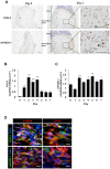
Fig 1. Lymphangiogenesis at the margins of healing wounds in ear skin.
(A) Representative images of the wound granulation tissues at the edges of the hole-punch injuries and time course of closure of wounds. Scale bars: 200 μm. Data are expressed as the mean ± SEM (n = 6). ***P < 0.001 versus day 0. (B) Immunofluorescent staining of LYVE-1 (green) and CD31 (red) in whole-mounted ear skin samples on day 7 post-injury. Scale bars: 200 μm. (C, D) Temporal changes in LYVE-1 (C) and VEGFR-3 (D) mRNA expression levels in the granulation tissues of the wound margins. Data are expressed as the mean ± SEM (n = 12). *P < 0. 05 and ***P < 0.001 versus day 0. (E) Representative images of LYVE-1 (green) and CD11b (red) immunostaining of the day 2 wound granulation tissues. The nuclei were stained with DAPI (blue). Scale bars: 50 μm.
Fig 2
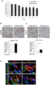
Fig 2. Expression levels of COX-2 and mPGES-1 in the wound granulation tissues.
(A) Representative images of COX-2 and mPGES-1 immunostaining in the wound granulation tissues on day 0 (left) and 3 (right). Scale bars: 50 μm. (B, C) Temporal changes in COX-2 (B) and mPGES-1 (C) mRNA levels in the wound granulation tissues. Data are expressed as the mean ± SEM (n = 12). *P < 0. 05 and ***P < 0.001 versus day 0. (D) Double labeling analysis of COX-2 (green) or mPGES-1 (green) and S100A4 (red) or CD11b (red) in the granulation tissues on day 3. Scale bars: 10 μm.
Fig 3
Fig 3. Effects of celecoxib on lymphangiogenesis.
(A–D) Real-time PCR analyses of the expression levels of the LYVE-1 (A), VEGFR-3 (B), VEGF-C (C), and VEGF-D (D) mRNAs in day 5 granulation tissues from mice treated with or without celecoxib (100 mg/kg). Data are expressed as the mean ± SEM (n = 12). (E) Representative images of immunostained LYVE-1 (green) and CD31 (red) in whole-mounted wound granulation tissues of control and celecoxib-treated mice on day 7. Scale bars: 200 μm. (F) Quantification of the number of sprouts in control and celecoxib-treated mice on day 7. Data are expressed as the mean ± SEM (n = 10). **P < 0.01. (G) HMVECs were cultured with PGE2, EP1–4 receptor agonist (1 or 10 nM), or rhVEGF-C (10 μg/mL) for 48 h. HMVEC proliferation was detected using a Cell Counting Kit-8 assay. Data are expressed as the mean ± SEM (n = 6). **P < 0.01 versus control (0 μg/mL).
Fig 4
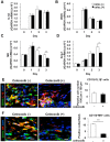
Fig 4. Reduction in the recruitment of CD11b-positive macrophages producing VEGF-C/D in wound granulation tissues by a COX-2 inhibitor.
A) Temporal changes in the closure of wounds in control and celecoxib-treated mice. Data are expressed as the mean ± SEM (n = 8). *P < 0.05. (B, C) The upper panels show representative images of S100A4-positive (B) and CD11b-positive (C) cells in immunostained day 3 wound granulation tissues from mice treated with or without celecoxib (100 mg/kg). Scale bars: 50 μm. The data are quantified in the lower panels. Data are expressed as the mean ± SEM (n = 8). ***P < 0.001. (D) Double labeling analysis of VEGF-C (green) or VEGF-D (green) and S100A4 (red) or CD11b (red) in the day 3 granulation tissues. Scale bars: 10 μm.
Fig 5
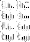
Fig 5. Reduction in the recruitment of M2 macrophages in wound granulation tissues by a COX-2 inhibitor.
(A–D) Real-time PCR analyses of the expression levels of the IL-1β (A), iNOS (B), MR (C), and Ang-1(D) mRNAs in day 3 granulation tissues from mice treated with or without celecoxib (100 mg/kg). Data are expressed as the mean ± SEM (n = 6). *P < 0.05. (E, F) The left panels show representative images of CD11b (green) (E, F) and IL-1β (red) (E) or MR (red) (F) in immunostained day 3 wound granulation tissues from mice treated with or without celecoxib (100 mg/kg). Scale bars: 10 μm. The data are quantified in the right panel. Data are expressed as the mean ± SEM (n = 6). ***P < 0.001.
Fig 6
Fig 6. Temporal changes in the expression levels of cytokine and growth factor mRNAs in wound granulation tissues.
Real-time PCR analyses of the expression levels of the MCP-1 (A), MCP-2 (B), MCP-3 (C), M-CSF (D), FGF-2 (E), EGF (F), TGF-β (G), and SDF-1 (H) mRNAs in granulation tissues from control and celecoxib-treated (100 mg/kg) mice. Data are expressed as the mean ± SEM (n = 6). *P < 0.05 and **P < 0.01 versus control mice.
Fig 7
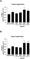
Fig 7. Enhancement of LYVE-1 and VEGFR-3 mRNA expression by EP agonists.
Real-time PCR analyses of the expression levels of the LYVE-1 (A) and VEGFR-3 (B) mRNAs in the ear skin of mice that received topical application of EP1-4 receptor agonists (50 nmol/site, once a day, from day 0–3). EP1 receptor agonist (ONO-DI-004), EP2 receptor agonist (ONO-AE1-259-01), EP3 receptor agonist (ONO-AE-248), EP4 receptor agonist (ONO-AE1-329). Data are expressed as the mean ± SEM (n = 6). *P < 0.05, **P < 0.01, and ***P < 0.001 versus vehicle.
Fig 8
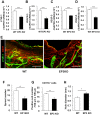
Fig 8. Suppression of lymphangiogenesis in EP3 receptor knockout mice.
(A–D) Real-time PCR analyses of the levels of the LYVE-1 (A), VEGFR-3 (B), VEGF-C (C), and VEGF-D (D) mRNAs in granulation tissues of WT and EP3–/– mice on day 5. Data are expressed as the mean ± SEM (n = 12). ***P < 0.001. (E) Representative images of immunostained LYVE-1 (green) and CD31 (red) in whole-mounted wound granulation tissues of WT and EP3–/– mice on day 7. Scale bars: 200 μm. (F) Quantification of the number of sprouts in the wound granulation tissues of WT and EP3–/– mice on day 7. Data are expressed as the mean ± SEM (WT, n = 10; EP3–/–, n = 8). **P < 0.01. (G) The numbers of CD11b-positive cells in the granulation tissues of WT and EP3–/– mice on day 3. Data are expressed as the mean ± SEM (n = 6). **P < 0.01. (H) The closure of wounds in WT and EP3–/– mice on day 3. Data are expressed as the mean ± SEM (n = 8). *P < 0.05.
Fig 9
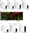
Fig 9. Suppression of lymphangiogenesis in EP4 receptor knockout mice.
(A–D) Real-time PCR analyses of the levels of the LYVE-1 (A), VEGFR-3 (B), VEGF-C (C), and VEGF-D (D) mRNAs in granulation tissues of WT and EP4–/– mice on day 5. Data are expressed as the mean ± SEM (n = 6). *P < 0.05 and ***P < 0.001. (E) Representative images of immunostained LYVE-1 (green) and CD31 (red) in whole-mounted wound granulation tissues of WT and EP4–/– mice on day 7. Scale bars: 200 μm. (F) Quantification of the number of sprouts in the wound granulation tissues of WT and EP4–/– mice on day 7. Data are expressed as the mean ± SEM (WT, n = 8; EP4–/–, n = 7).*P < 0.05. (G) The numbers of CD11b-positive cells in the granulation tissues of WT and EP4–/– mice on day 3. Data are expressed as the mean ± SEM (n = 6). *P < 0.001. (H) The closure of wounds in WT and EP4–/– mice on day 3. Data are expressed as the mean ± SEM (n = 8). *P < 0.05.
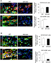
Fig 10
Fig 10. Reduction of the recruitment of M2 macrophages in wound granulation tissues in EP3 and EP4 receptor knockout mice.
(A, B) The left panels show representative images of CD11b (green) and IL-1β (red) or MR (red) in the granulation tissues of WT and EP3–/–(A) or EP4–/–(B) mice on day 3. Scale bars: 10 μm. The data are quantified in the right panel. Data are expressed as the mean ± SEM (n = 6). ***P < 0.001.

Similar articles

Cited by

References

    1. Kim H, Kataru RP, Koh GY. Regulation and implications of inflammatory lymphangiogenesis. Trends in immunology. 2012;33(7):350–6. Epub 2012/05/15. 10.1016/j.it.2012.03.006 . - DOI - PubMed
    1. Paavonen K, Puolakkainen P, Jussila L, Jahkola T, Alitalo K. Vascular endothelial growth factor receptor-3 in lymphangiogenesis in wound healing. The American journal of pathology. 2000;156(5):1499–504. Epub 2000/05/04. 10.1016/s0002-9440(10)65021-3 - DOI - PMC - PubMed
    1. Kurashige C, Hosono K, Matsuda H, Tsujikawa K, Okamoto H, Majima M. Roles of receptor activity-modifying protein 1 in angiogenesis and lymphangiogenesis during skin wound healing in mice. FASEB journal: official publication of the Federation of American Societies for Experimental Biology. 2014;28(3):1237–47. Epub 2013/12/07. 10.1096/fj.13-238998 . - DOI - PubMed
    1. Baluk P, Tammela T, Ator E, Lyubynska N, Achen MG, Hicklin DJ, et al. Pathogenesis of persistent lymphatic vessel hyperplasia in chronic airway inflammation. The Journal of clinical investigation. 2005;115(2):247–57. Epub 2005/01/26. 10.1172/jci22037 - DOI - PMC - PubMed
    1. Yao LC, McDonald DM. Plasticity of airway lymphatics in development and disease. Advances in anatomy, embryology, and cell biology. 2014;214:41–54. Epub 2013/11/28. 10.1007/978-3-7091-1646-3_4 - DOI - PMC - PubMed

MeSH terms

Substances