. 2016 Oct 6;11(10):e0163058.
doi: 10.1371/journal.pone.0163058. eCollection 2016.

Cell-Free Phospholipid Biosynthesis by Gene-Encoded Enzymes Reconstituted in Liposomes

Affiliations

Cell-Free Phospholipid Biosynthesis by Gene-Encoded Enzymes Reconstituted in Liposomes

Andrew Scott et al. PLoS One. .

Abstract

The goal of bottom-up synthetic biology culminates in the assembly of an entire cell from separate biological building blocks. One major challenge resides in the in vitro production and implementation of complex genetic and metabolic pathways that can support essential cellular functions. Here, we show that phospholipid biosynthesis, a multiple-step process involved in cell membrane homeostasis, can be reconstituted starting from the genes encoding for all necessary proteins. A total of eight E. coli enzymes for acyl transfer and headgroup modifications were produced in a cell-free gene expression system and were co-translationally reconstituted in liposomes. Acyl-coenzyme A and glycerol-3-phosphate were used as canonical precursors to generate a variety of important bacterial lipids. Moreover, this study demonstrates that two-step acyl transfer can occur from enzymes synthesized inside vesicles. Besides clear implications for growth and potentially division of a synthetic cell, we postulate that gene-based lipid biosynthesis can become instrumental for ex vivo and protein purification-free production of natural and non-natural lipids.


Conflict of interest statement

The authors have declared that no competing interests exist.

Figures

Fig 1
Fig 1. Overview of methods for cell-free transcription-translation of acyltransferase enzymes.
(a) The genes plsB and plsC coding for the GPAT and LPAAT enzymes, respectively, were expressed by in vitro transcription translation (IVTT) in the presence of SUVs. Spontaneously assembled proteoliposomes containing synthesized GPAT and LPAAT proteins were isolated by ultracentrifugation (floatation method) and the protein content was analysed by SDS-PAGE. Activity assays were performed by adding the phospholipid precursors G3P and acyl-CoA (shown in the reaction scheme is palmitoyl-CoA, p-CoA) either before or subsequent to IVTT reaction. Biosynthesis of 1,2-diacylglycerol-3-phosphate (here DPPA) occurs in a two-step acyl transfer reaction catalysed by the GPAT and LPAAT enzymes. The intermediate product 1-acylglycerol-3-phosphate (here 16:0 LPA) and two free CoA molecules are also formed. After reaction the lipid fraction was extracted and assayed by LC-MS. To quantify the enrichment of vesicles with synthesized lipids, liposomes were purified by immobilization on beads before the lipid extraction step. (b) Cell-free expression of either the plsB or plsC gene (no gene as negative control) occurred for 3 h at 37°C in the presence of 100-nm SUVs and of GreenLys reagent (tRNA-loaded fluorescent amino acid) for fluorescence labelling of translation products. Reconstituted proteoliposomes were purified and membrane-integrated proteins separated by SDS-PAGE were visualized with coomassie brilliant blue (CBB) staining and fluorescence scanning. As shown with CBB staining the PURE system background proteins (lane 7) can efficiently be eliminated by purification, while the GPAT and LPAAT protein bands were co-purified with the SUVs (lanes 9,10). Isolation of acyltransferase enzymes is also visible on the fluorescence scan (lanes 4,5). The lower bands on lanes 2,3 correspond to background signal from the GreenLys reagent. (c) Normalized chromatogram of lipids as measured by LC-MS operating in multiple reaction monitoring (MRM) mode with negative polarity. In this example, 16:0 LPA, DOPG, DPPA, DOPE and cardiolipin were clearly resolved. Since the quaternary amine has a permanent positive charge, DOPC is not well detected in the negative mode. It was also possible to detect 18:1 LPA, DOPA, DPPS, DPPG and DPPE (Figure A in S1 File).
Fig 2
Fig 2. Two-step acyl transfer reaction mediated by cell-free synthesized GPAT and LPAAT enzymes.
(a) LC-MS analysis of the GPAT and LPAT reaction products. The lipid precursors G3P and palmitoyl-CoA (p-CoA), or p-CoA and 16:0 LPA (66.6 μM each, except in two-enzyme cascade experiments, where p-CoA concentration was 133.3 μM) were added after the IVTT reactions performed in the presence of SUVs. The two enzymes were assayed separately in their respective activity buffer or together in the reducing buffer known to support GPAT activity. Negative controls in GPAT and LPAAT activity buffers were performed using the DHFR and LacI genes. For combined GPAT and LPAAT reactions, controls were conducted without G3P. Error bars in single-enzyme experiments are s.e.m. from multiple measurements of one sample. In the GPAT and LPAAT co-expression experiments data are mean and s.e.m. across four independent samples; For each repeat the sample was injected multiple times, the average value of the different injections was calculated and data are reported as the mean and standard error of independent trials. Student t-test analysis: *P<0.015, **P<0.025. (b) Acyltransferase activity as measured using a fluorescence-based assay in which released CoA reacts with a fluorogenic substrate. Negative controls for GPAT and LPAAT activity were performed using the DFHR and LacI genes, respectively. DTT was dialysed out after the IVTT reaction to create the non-reducing conditions compatible with the assay. Blank was measured from the buffer included in the fluorescence-based CoA assay kit. Data are mean values and s.e.m. of two independent experiments. Student t-test analysis: *Difference statistically not significant, **P<0.23.
Fig 3
Fig 3. One-pot IVTT and acyl transfer reactions.
The GPAT and LPAAT enzymes were either produced separately or concurrently in the presence of G3P and p-CoA substrates. The generated lipid products 16:0 LPA and DPPA were detected by LC-MS. (a) End-point measurements of 16:0 LPA and DPPA synthesized under various experimental conditions. Substrate concentrations were 500 μM G3P, 100 μM p-CoA and 100 μM 16:0 LPA. Individual and combined enzymatic reactions were carried out with (inside-out configuration) or without 400-nm liposomes during overnight incubation at 37°C. Samples with liposomes and without p-CoA served as a negative control. Both acyltransferase enzymes showed reduced activity in the absence of SUVs. Higher yield of DPPA is obtained by two-step acyl transfer catalysed by the GPAT and LPAAT enzymes co-reconstituted in proteoliposomes. Data represent mean and s.e.m. of three independent experiments. For each repeat the mean of multiple sample injections was calculated and data are reported as the mean and standard error of three independent trials. Student t-test analysis: *P<0.1, **P<0.12, ***P<0.012. (b-e) Kinetic of acyltransferase activity in single-enzyme and two-enzyme modes. For each reaction scenario the percentage of acyl-CoA conversion to final product is also indicated. Substrate concentrations were 500 μM G3P and 100 μM p-CoA (b), 500 μM G3P, 50 μM p-CoA and 50 μM 16:0 LPA (c), 500 μM G3P and 100 μM p-CoA (d,e). Produced 16:0 LPA does not accumulate beyond 3 μM in the two-step acyl transfer scheme (d) since it is consumed in the second enzymatic reaction. When GPAT and LPAAT are co-expressed, production of DPPA is initially limited by GPAT activity but then it reaches higher concentration (e) than with LPAAT only starting from purified LPA and p-CoA precursors (c). Each data point is mean and s.e.m. of two independent sample preparations. For each replicate the mean of two sample injections was calculated and data are reported as the mean and standard error of independent preparations.
Fig 4
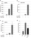
Fig 4. Inside-out acyltransferase proteoliposomes are enriched with synthesized DPPA lipid.
(a–c) LC-MS analysis of synthesized 16:0 LPA and DPPA lipids with or without liposome purification. Lipid DOPG present in the initial composition of the 400-nm vesicles was used as an internal standard to correct for the loss of lipids during purification. Lipid biosynthesis occurred in a one-pot IVTT and acyl transfer reaction starting from 500 μM G3P and 100 μM p-CoA substrates. In some samples SUV membranes were doped with a biotinylated lipid for immobilization of liposomes on streptavidin-coated magnetic beads. Inspection of the amounts of lipids detected for the different experimental conditions allowed us to discriminate between liposome-integrated and free DPPA. Data are mean and s.e.m. of three independent experiments. For each replicate the same sample was injected two times in the MS, their averaged value was calculated and data are reported as the mean and standard error across the three trials. (d) Calculation of the percentage of synthesized DPPA co-localizing with liposome membrane. The use of DOPG as an internal standard enabled the quantification of the fraction of non-immobilized or disrupted vesicles that were washed away during the purification step. Percentage values of recovered DPPA and DOPG were calculated as [counts(purif+|biotin+)–counts(purif+|biotin–)] / counts(purif–|biotin+) × 100. Then, the obtained value for DPPA was divided by that for DOPG to correct for the loss of lipids during purification (Figure C in S1 File), resulting in a value of 28% ± 14% as an estimation of synthesized DPPA that effectively localized in liposomes.
Fig 5
Fig 5. Synthesis of 18:1 LPA and DOPA from GPAT and LPAAT enzymes produced inside liposomes.
(a) Schematic of vesicle-confined experiments. PUREfrex supplemented with the plsB and plsC genes and with 500 μM G3P was encapsulated inside liposomes using gentle rehydration of a lipid film covering sub-millimetre glass beads. Lipid composition consisted of DOPC, DOPE, DOPG, cardiolipin, TexasRed-DHPE and DSPE-PEG-biotin (Table B in S1 File). Swelling occurred at 4°C to avoid reaction initiation. Gene expression outside liposomes was inhibited by protein digestion. Lipid biosynthesis was triggered by external supply of 100 μM oleoyl-CoA (o-CoA). (b) Confocal microscopy images of liposomes after swelling. Vesicles were labelled with a membrane dye (Texas-Red). Scale bar is 5 μm. (c,d) Concentration of 18:1 LPA (c) and DOPA (d) synthesized in a one-pot reaction by GPAT and LPAAT enzymes produced outside liposomes composed of DOPG, DOPE and cardiolipin (Table B in S1 File). Lipid precursors were 500 μM G3P and 100 μM o-CoA (except in negative control). Error bars indicate s.e.m. of two injections of the same sample. (e,f) Concentration of 18:1 LPA (e) and DOPA (f) produced by GPAT and LPAAT enzymes generated inside liposomes. Three experimental configurations corresponding to different localizations of protein digestion were tested. As expected, addition of Proteinase K both inside and outside liposomes totally inhibited lipid synthesis. In the absence of Proteinase K 18:1 LPA and DOPA accumulated as a result of both internal and external acyltransferase production. Liposome-confined IVTT and lipid synthesis was demonstrated by supplementing Proteinase K outside vesicles according to the reaction scheme illustrated in (a). Data are mean and s.e.m. of three independent experiments. For each replicate the same sample was injected two times in the MS, their averaged value was calculated and data are reported as the mean and standard error across the three trials.
Fig 6
Fig 6. Functional reconstitution of complete biosynthesis pathways for PE and PG lipids.
(a) Metabolic pathways that lead to the production of DPPG and DPPE starting from palmitoyl-CoA, glycerol-3-phosphate (G3P), cytidine triphosphate (CTP) and L-serine as main substrates. The two-step acyl transfer reaction and the first headgroup conversion step are common to the PE and PG pathways that then branch out into different headgroup modification reactions (see Supplementary text in S1 File for a description of the individual enzymatic steps). For the final step of PG synthesis there exist three alternative enzymes: PgpA, PgpB and PgpC, of which two (A/C) were used in this study. (b) Fluorescence scans of SDS-PAGE gels for the headgroup modifying enzymes produced in the PURE system. Fluorescently labeled lysine residues were incorporated during translation. The left gel is 15% polyacrylamide. In addition to the pssA gene product that was used as a control, the gene products of pgpA, pgpC and pgsA were synthesized. The right gel is 12% polyacrylamide and, besides the plsB gene product used as a control, the genes cdsA, pssA and psd were expressed. Size markers are in kDa. The arrowheads point to the observed protein molecular mass. The symbol “*” indicates the position of the band as expected from the nucleotide sequence of the genes (Supplementary text in S1 File). (c) Schematic of the inside-out proteoliposome reconstitution experiments and enzymatic cascade reactions, where all genes of a given pathway were expressed in PUREfrex and all specific substrates were supplied. (d) LC-MS data reporting lipid production in the PE and PG pathways under various experimental conditions. Combined gene expression and lipid biogenesis was carried out as illustrated in (c) using 25 ng of each linear DNA templates, 500 μM G3P, 100 μM palmitoyl-CoA, 1 mM CTP and 500 μM L-serine. Details of MS signatures for the different lipids are reported in Table A in S1 File. Lipids DPPE and DPPG were unambiguously detected in a pathway-specific manner. No PG is produced in the reconstituted PE pathway. Likewise, no PE was detected in the PG pathway. When the plsB gene is omitted the complete pathways are shut down. In the absence of the Psd enzyme, PE was not detected and its substrate lipid DPPS accumulated. Note that the MRM data for PS come from the MS optimizer results, not from separate experiments as used for the other compounds. Data are mean and s.e.m. of three independent experiments, except for the negative controls without plsB gene where two independent experiments were conducted. For each replicate the same sample was injected between one and four times in the MS, their averaged value was calculated and data are reported as the mean and standard error across the different trials.

Similar articles

Cited by

References

    1. Luisi PL. Toward the engineering of minimal living cells. The Anatomical Record 2002;268: 208–214. 10.1002/ar.10155 - DOI - PubMed
    1. Luisi PL, Ferri F, Stano P. Approaches to semi-synthetic minimal cells: a review. Naturwissenschaften 2006;93: 1–13. 10.1007/s00114-005-0056-z - DOI - PubMed
    1. Forster AC, Church GM. Towards synthesis of a minimal cell. Molecular Systems Biology 2006;2: 45 10.1038/msb4100090 - DOI - PMC - PubMed
    1. Noireaux V, Maeda YT, Libchaber A. Development of an artificial cell, from self-organization to computation and self-reproduction. Proceedings of the National Academy of Sciences 2011;108: 3473–3480. 10.1073/pnas.1017075108 - DOI - PMC - PubMed
    1. Schwille P. Bottom-up synthetic biology: engineering in a tinkerers world. Science 2011;333: 1252–1254. 10.1126/science.1211701 - DOI - PubMed

MeSH terms

LinkOut - more resources