. 2016 Oct 6;11(10):e0164269.
doi: 10.1371/journal.pone.0164269. eCollection 2016.

The Effect of Gender on Mesenchymal Stem Cell (MSC) Efficacy in Neonatal Hyperoxia-Induced Lung Injury

Affiliations

The Effect of Gender on Mesenchymal Stem Cell (MSC) Efficacy in Neonatal Hyperoxia-Induced Lung Injury

Ibrahim Sammour et al. PLoS One. .

Abstract

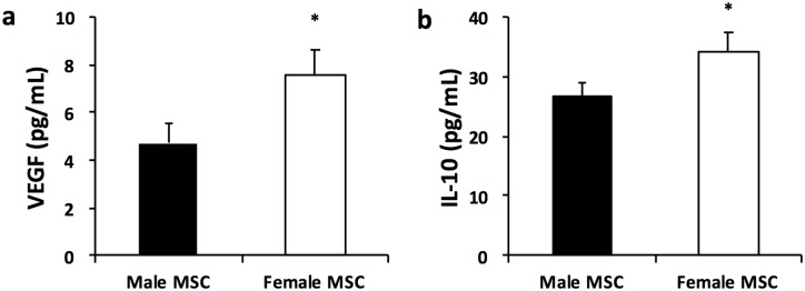
Background: Mesenchymal stem cells (MSC) improve alveolar and vascular structures in experimental models of bronchopulmonary dysplasia (BPD). Female MSC secrete more anti-inflammatory and pro-angiogenic factors as compared to male MSC. Whether the therapeutic efficacy of MSC in attenuating lung injury in an experimental model of BPD is influenced by the sex of the donor MSC or recipient is unknown. Here we tested the hypothesis that female MSC would have greater lung regenerative properties than male MSC in experimental BPD and this benefit would be more evident in males.

Objective: To determine whether intra-tracheal (IT) administration of female MSC to neonatal rats with experimental BPD has more beneficial reparative effects as compared to IT male MSC.

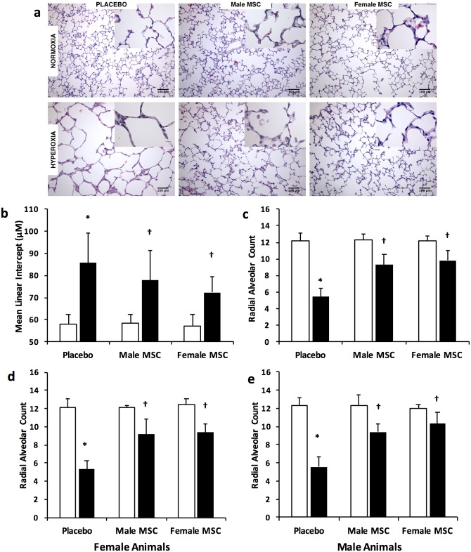
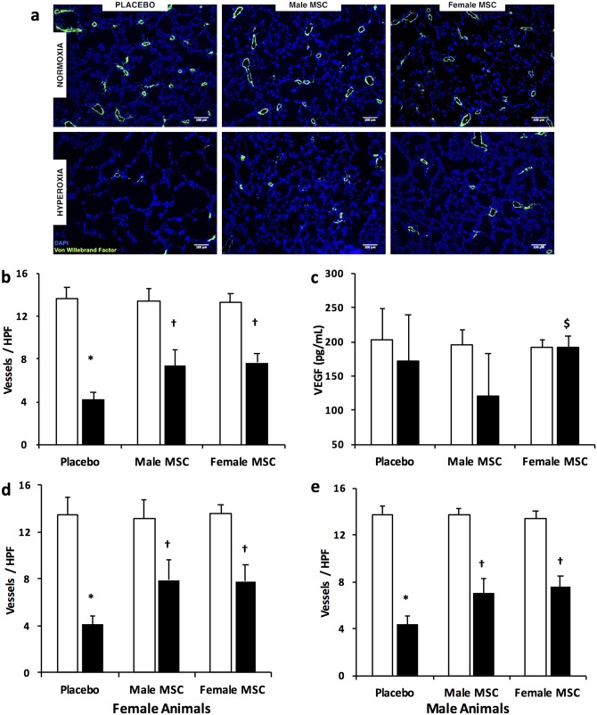
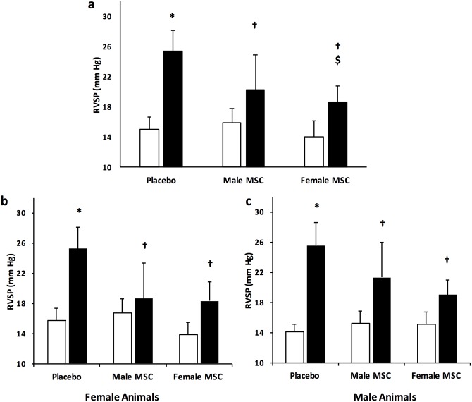
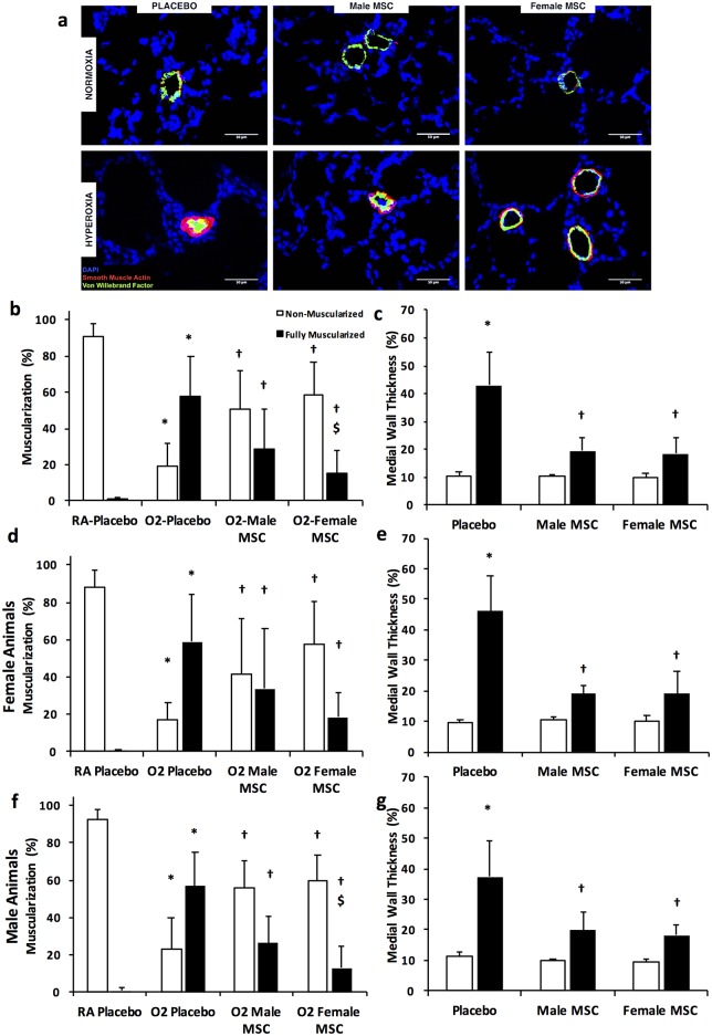
Methods: Newborn Sprague-Dawley rats exposed to normoxia (RA) or hyperoxia (85% O2) from postnatal day (P) 2- P21 were randomly assigned to receive male or female IT bone marrow (BM)-derived green fluorescent protein (GFP+) MSC (1 x 10⁶ cells/50 μl), or Placebo on P7. Pulmonary hypertension (PH), vascular remodeling, alveolarization, and angiogenesis were assessed at P21. PH was determined by measuring right ventricular systolic pressure (RVSP) and pulmonary vascular remodeling was evaluated by quantifying the percentage of muscularized peripheral pulmonary vessels. Alveolarization was evaluated by measuring mean linear intercept (MLI) and radial alveolar count (RAC). Angiogenesis was determined by measuring vascular density. Data are expressed as mean ± SD, and analyzed by ANOVA.

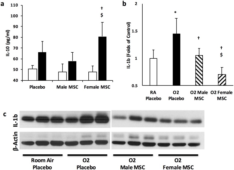
Results: There were no significant differences in the RA groups. Exposure to hyperoxia resulted in a decrease in vascular density and RAC, with a significant increase in MLI, RVSP, and the percentage of partially and fully muscularized pulmonary arterioles. Administration of both male and female MSC significantly improved vascular density, alveolarization, RVSP, percent of muscularized vessels and alveolarization. Interestingly, the improvement in PH and vascular remodeling was more robust in the hyperoxic rodents who received MSC from female donors. In keeping with our hypothesis, male animals receiving female MSC, had a greater improvement in vascular remodeling. This was accompanied by a more significant decrease in lung pro-inflammatory markers and a larger increase in anti-inflammatory and pro-angiogenic markers in male rodents that received female MSC. There were no significant differences in MSC engraftment among groups.

Conclusions: Female BM-derived MSC have greater therapeutic efficacy than male MSC in reducing neonatal hyperoxia-induced lung inflammation and vascular remodeling. Furthermore, the beneficial effects of female MSC were more pronounced in male animals. Together, these findings suggest that female MSC maybe the most potent BM-derived MSC population for lung repair in severe BPD complicated by PH.


Conflict of interest statement

The authors have declared that no competing interests exist.

Figures

Fig 1
Fig 1. Female MSC produce more VEGF and IL-10 after 24 hours in culture.
(a) VEGF concentration as measured by ELISA in male and female MSC (b) IL-10 concentration as measured by ELISA in male and female MSC (* P < 0.05; Male vs Female MSC, N = 3 experiments/group).
Fig 2
Fig 2. Intra-tracheal (IT) administration of male or female MSC similarly improves alveolarization.
(a) Hematoxylin and eosin stained lung sections demonstrating improved alveolar structure in hyperoxia-exposed pups treated with IT male or female MSC. Original magnification X 200. Bars = 100 μm. IT male or female MSC similarly increased mean linear intercept (b) and decreased radial alveolar count (c). IT male or female MSC similarly improve RAC in female (d) and male (e) animals. (*P < 0.05; Room air (RA) vs hyperoxia- placebo (PL), † P <0.05; hyperoxia -PL vs hyperoxia male or female MSC). White bars are RA and black bars are hyperoxia.
Fig 3
Fig 3. IT male or female MSC similarly improve lung angiogenesis.
(a) Lung sections stained with Von Willebrand Factor (green) and 4’6-diamidino-2-phenylindole (DAPI: blue), demonstrating improved vascular density in hyperoxia-exposed pups treated with male and female MSC at P7. Original magnification X 100. Bars = 100 μm. (b) IT male and female MSC similarly increased lung vascular density in hyperoxic pups. (c) Female MSC normalizes lung VEGF concentration in hyperoxia. Female and male MSC exhibit similar efficacy in improving lung angiogenesis in female (d) and male (e) animals. (*P < 0.05: RA vs hyperoxia-PL; † P <0.05: hyperoxia-PL vs hyperoxia male or female MSC; $ P <0.05: hyperoxia male MSC vs hyperoxia female MSC). White bars are RA and black bars are hyperoxia.
Fig 4
Fig 4. Female MSC have more marked effects on pulmonary hypertension.
(a) Female MSC improve right ventricular systolic pressure (RVSP) to a greater degree than male MSC. The efficacy of MSC in improving RVSP in female (b) and male (c) animals is not dependent on the sex of the recipient. (*P < 0.05: RA vs hyperoxia-PL; † P <0.05: hyperoxia-PL vs hyperoxia male or female MSC; $ P <0.05: hyperoxia male MSC vs hyperoxia female MSC). White bars are RA and black bars are hyperoxia.
Fig 5
Fig 5. Female MSC exhibit greater anti-remodeling effects.
(a) Lung sections stained with Von Willebrand Factor (green), α-smooth muscle actin (red), and DAPI (blue), demonstrating improved vascular remodeling in hyperoxia-exposed pups treated with male or female MSC. Original magnification X400. (b) Female MSC are superior to male MSC in reducing the percentage of fully muscularized blood vessels. (c) IT male or female MSC improved medial wall thickness of vessels (20–50 μm). In female pups, IT male or female MSC similarly improve the percentage of muscularized vessels (d), and the medial wall thickness (e). In male pups, IT female MSC are superior to male MSC in improving the percentage of muscularized blood vessels (f), while being as effective in reducing the medial wall thickness (g). (*P < 0.05: RA vs hyperoxia-PL; † hyperoxia-PL vs hyperoxia male or female MSC; $ P <0.05: hyperoxia male MSC vs hyperoxia female MSC). White bars are RA and black bars are hyperoxia unless otherwise specified.
Fig 6
Fig 6. IT MSC and lung inflammation.
(a) Markedly greater lung IL-10 concentration in pups treated with female as compared to male MSC. (b) Female MSC decrease lung IL-1β expression to a greater degree than male MSC (c) Representative Western blot demonstrating a more marked decrease in lung IL-1β expression in hyperoxic pups treated with female MSC. β-actin is utilized as the normalization protein. (*P < 0.05: RA vs hyperoxia-PL; † hyperoxia-PL vs hyperoxia male or female MSC; $ P <0.05: hyperoxia male MSC vs hyperoxia female MSC; N = 5-6/group). White bars are RA and black bars are hyperoxia.

Similar articles

Cited by

References

    1. Northway WH Jr., Rosan RC, Porter DY. Pulmonary disease following respirator therapy of hyaline-membrane disease. Bronchopulmonary dysplasia. N Engl J Med. 1967;276(7):357–68. Epub 1967/02/16. 10.1056/nejm196702162760701 . - DOI - PubMed
    1. Farstad T, Bratlid D, Medbo S, Markestad T. Bronchopulmonary dysplasia—prevalence, severity and predictive factors in a national cohort of extremely premature infants. Acta Paediatr. 2011;100(1):53–8. Epub 2010/07/27. 10.1111/j.1651-2227.2010.01959.x . - DOI - PubMed
    1. Jain D, Bancalari E. Bronchopulmonary dysplasia: clinical perspective. Birth Defects Res A Clin Mol Teratol. 2014;100(3):134–44. Epub 2014/03/01. 10.1002/bdra.23229 . - DOI - PubMed
    1. Jobe AH, Bancalari E. Bronchopulmonary dysplasia. Am J Respir Crit Care Med. 2001;163(7):1723–9. Epub 2001/06/13. 10.1164/ajrccm.163.7.2011060 . - DOI - PubMed
    1. Bhandari A, Bhandari V. “New” Bronchopulmonary Dysplasia: A Clinical Review. Clin Pulm Med. 2011;18(3):137–43 10.1097/CPM.0b013e318218a071 - DOI

MeSH terms

Substances

LinkOut - more resources