. 2017 Apr 14;25(4):523-536.
doi: 10.3727/096504016X14756282819385. Epub 2016 Oct 5.

MicroRNA-221-3p Plays an Oncogenic Role in Gastric Carcinoma by Inhibiting PTEN Expression

Affiliations

MicroRNA-221-3p Plays an Oncogenic Role in Gastric Carcinoma by Inhibiting PTEN Expression

Jianping Shi et al. Oncol Res. .

Retraction in

Abstract

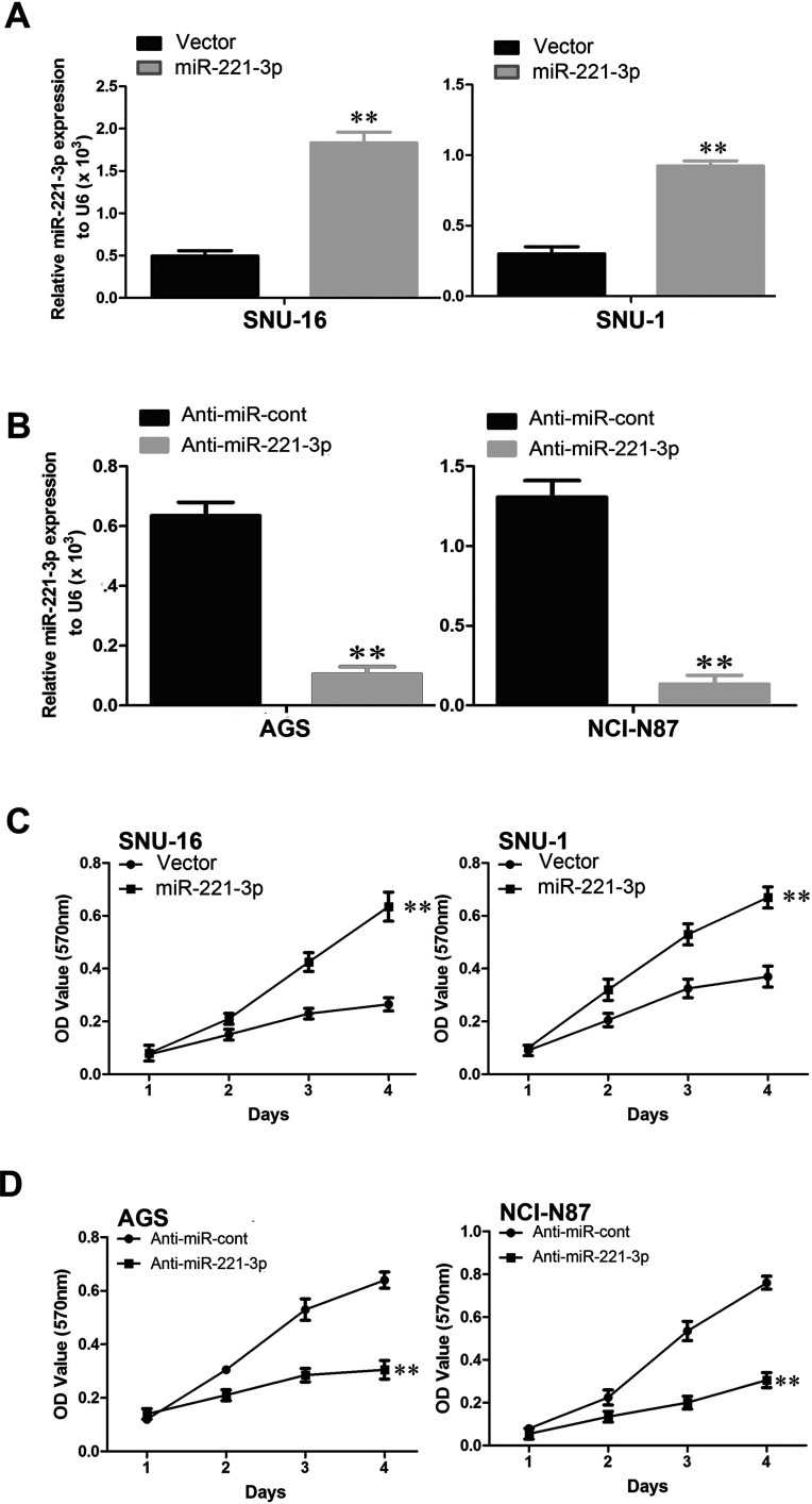
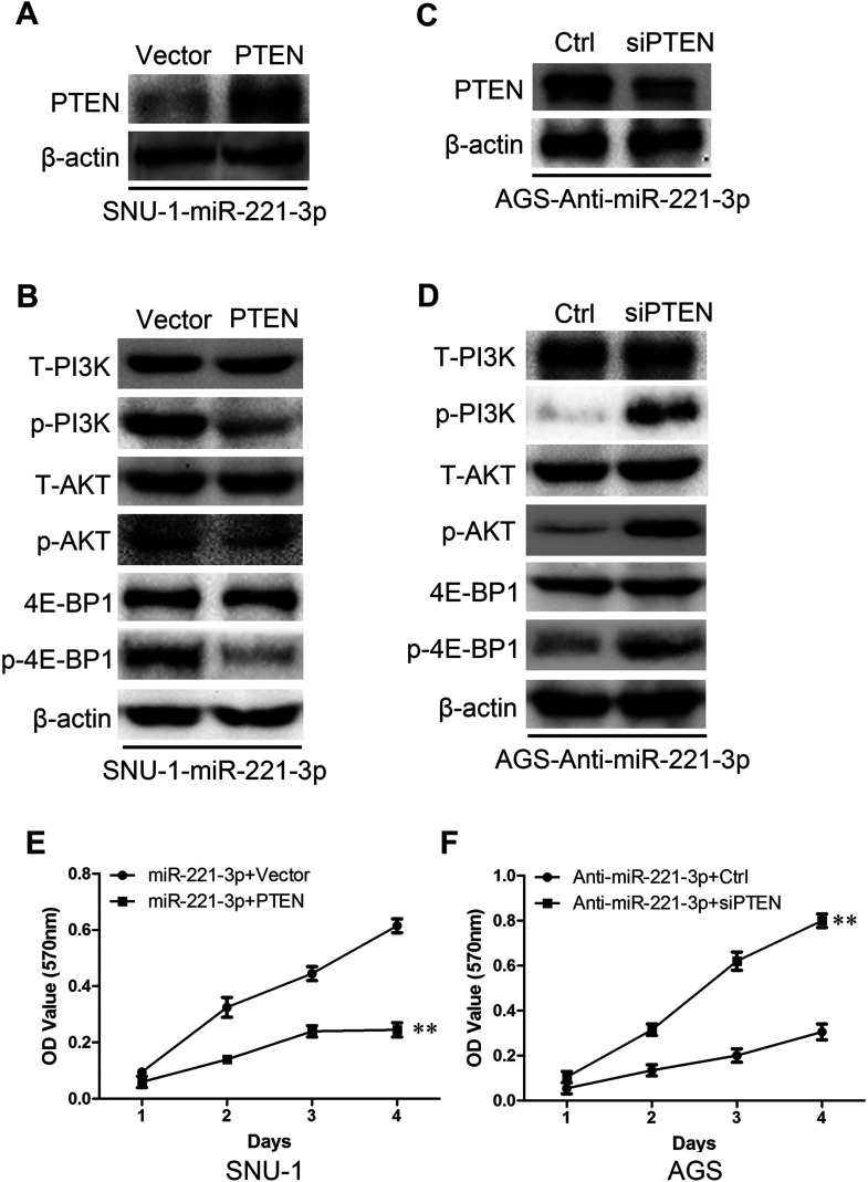
Gastric carcinoma is one of the most common malignancies in men, and microRNA plays a critical role in regulating the signaling networks of gastric carcinoma tumorigenesis and metastasis. We first report the functional characteristics of miR-221-3p in gastric carcinoma. Quantification in gastric carcinoma cell lines and tumor samples reveals significantly increasing miR-221-3p expression. Moreover, a high level of miR-221-3p is correlated with a poor prognosis for gastric carcinoma patients. Ectopic miR-221-3p expression significantly promotes gastric carcinoma cell proliferation, invasion, and sphere formation, while silencing miR-221-3p significantly inhibits these abilities in gastric carcinoma cells. Tests in vivo showed that miR-221-3p significantly promotes tumor growth in xenograft mouse models. In this study, we reveal that miR-221-3p targets PTEN mRNA and downregulates PTEN, which is the possible mechanism of miR-221-3p-induced oncogenic properties. Collectively, we reveal a critical role for miR-221-3p in gastric carcinoma development and progression.


Conflict of interest statement

The authors declare no conflicts of interest.

Figures

Figure 1
Figure 1
miR-221-3p is upregulated in gastric carcinoma tumors and gastric carcinoma cells. (A) Quantification of miR-221-3p in gastric carcinoma cell lines showing higher expression than in normal human stomach epithelial cells (AGS1). (B) Average expression of miR-221-3p in gastric carcinoma cells and control cells. (C) Quantification of miR-221-3p in human gastric carcinoma tumors (T) and normal liver tissue (N). **p < 0.01 based on the Student’s t-test. Error bars, SD.
Figure 2
Figure 2
miR-221-3p is upregulated in gastric carcinoma and inversely correlates with patient survival. (A) Quantification of miR-221-3p in normal liver, stages I–II gastric carcinoma, and stages III–IV gastric carcinoma. (B) Correlation analysis of expression data and patient survival data from TCGA showing that miR-221-3p levels are a risk indicator for survival. **p < 0.01, ##p < 0.01 based on the Student’s t-test. (A) ##Student’s t-test to stages I–II. Error bars, SD.
Figure 3
Figure 3
miR-221-3p accelerates gastric carcinoma cell proliferation in vitro. (A) Quantification of miR-221-3p in SNU-16 and SNU-1 cell lines with overexpressing miR-221-3p. (B) Quantification of miR-221-3p in AGS and NCI-N87 cell lines with silencing miR-221-3p. (C) MTT assay reveals cell growth curves of miR-221-3p overexpressing cells SNU-16 and SNU-1. (D) MTT assay reveals cell growth curves of miR-221-3p silencing cells AGS and NCI-N87. **p < 0.01 based on the Student’s t-test. Error bars, SD.
Figure 4
Figure 4
miR-221-3p accelerates gastric carcinoma cell colony formation. (A) Representative micrographs of crystal violet-stained cell colonies overexpressing miR-221-3p in SNU-16 and SNU-1 cell lines analyzed by colony formation assay for 14 days and quantification of number of clones. (B) Representative micrographs of crystal violet-stained cell colonies silencing miR-221-3p AGS and NCI-N87 cell lines analyzed by colony formation assay for 14 days and quantification of number of clones. **p < 0.01 based on the Student’s t-test. Error bars, SD.
Figure 5
Figure 5
miR-221-3p promotes gastric carcinoma cell tumor growth in vivo. (A) Cells stably overexpressing miR-221-3p in SNU-1 were subcutaneously injected into nude mice. Six weeks later, SNU-1 cells stably overexpressing miR-221-3p had larger tumors than controls. (B) Representative picture of tumors formed. (C) The growth curves of tumor volumes. (D) Tumor weight. **p < 0.01 based on the Student’s t-test. Error bars, SD.
Figure 6
Figure 6
miR-221-3p accelerates invasion of gastric carcinoma cells. (A) Representative results of invasive ability of SNU-1 and SNU-16 cells transfected with miR-221-3p mimics or miR control. (B) Representative results of invasive ability of AGS and NCI-N87 cells transfected with anti-miR-221-3p mimics or anti-miR control. **p < 0.01 based on the Student’s t-test. Error bars, SD.
Figure 7
Figure 7
miR-221-3p inhibits PTEN expression. (A) Correlation of miR-221-3p overexpression with PTEN downregulation in indicated gastric tissues. (B) Western blot of PTEN expression in miR-221-3p-overexpressing cells. (C) Western blot of PTEN expression in miR-221-3p-silencing cells.
Figure 8
Figure 8
PTEN is a direct target of miR-221-3p. (A) Wild-type and mutant-type miR-221-3p target sequences of PTEN 3′-UTR. (B, C) Relative luciferase activity of PTEN in cells after cotransfection with wild-type (WT) or mutant (mut) PTEN 3′-UTR reporter genes and miR-221-3p mimics or control in SNU-1 and SNU-16 cells. (D, E) Relative luciferase activity of PTEN in cells after cotransfection with wild-type (WT) or mutant (mut) PTEN 3′-UTR reporter genes and anti-miR-221-3p mimics or control in AGS and NCI-N87 cells. **p < 0.01 based on the Student’s t-test. Error bars, SD.
Figure 9
Figure 9
miR-221-3p activates both AKT/FOXO3a and AKT/mTOR signaling pathways. (A, B) Western blot analysis of phospho-P13K (p-P13K), total P13K (T-P13K), phospho-AKT (p-AKT), total AKT (T-AKT), phospho-4E-BP1 (p-4E-BP1), and total 4E-BP1 in indicated cells. (C, D) Western blot analysis of protein expression of p21 and cyclin D1 in indicated cells.
Figure 10
Figure 10
AKT/FOXO3a or AKT/mTOR signaling pathway inhibitors restrained the proliferation of miR-221-3p-overexpressing cells. Western blot analysis shows the effect of LY294002 (A), AKT inhibitor III (C), or rapamycin (E). MTT shows the effect of LY294002 (B), AKT inhibitor III (D), or rapamycin (F) on miR-221-3p-overexpressing cells. **p < 0.01 based on the Student’s t-test. Error bars, SD.
Figure 11
Figure 11
Restoration of PTEN inverses miR-221-3p-induced proliferation. (A, C) Western blotting confirmation of PTEN in indicated cells. (B, D) Western blot analysis of indicated phosphorylated proteins in indicated cells. MTT shows the effect on indicated cells after restoration (E) or depletion (F) of PTEN. **p < 0.01 based on the Student’s t-test. Error bars, SD.

Similar articles

Cited by

References

    1. Siegel RL, Miller KD, Jemal A. Cancer statistics, 2015. CA Cancer J Clin. 2015;65:5–29. - PubMed
    1. Huang KH, Lan YT, Fang WL, Chen JH, Lo SS, Li AF, Chiou SH, Wu CW, Shyr YM. The correlation between miRNA and lymph node metastasis in gastric cancer. Biomed Res Int. 2015;2015:543163. - PMC - PubMed
    1. Zhang H, Qu Y, Duan J, Deng T, Liu R, Zhang L, Bai M, Li J, Zhou L, Ning T, Li H, Ge S, Li H, Ying G, Huang D, Ba Y. Integrated analysis of the miRNA, gene and pathway regulatory network in gastric cancer. Oncol Reps. 2016;35:1135–46. - PubMed
    1. Hua HB, Yan TT, Sun QM. miRNA polymorphisms and risk of gastric cancer in Asian population. World J Gastroenterol. 2014;20:5700–7. - PMC - PubMed
    1. Shin VY, Chu KM. MiRNA as potential biomarkers and therapeutic targets for gastric cancer. World J Gastroenterol. 2014;20:10432–9. - PMC - PubMed

Publication types

MeSH terms