An official website of the United States government
The .gov means it’s official.
Federal government websites often end in .gov or .mil. Before
sharing sensitive information, make sure you’re on a federal
government site.
The site is secure.
The https:// ensures that you are connecting to the
official website and that any information you provide is encrypted
and transmitted securely.
1 School of Biological Sciences, University of Reading, Reading, United Kingdom; College of STEM, Texas A&M University, Texarkana, Texarkana, TX, United States. Electronic address: bneuman@tamut.edu.
2 University of California, Irvine, Irvine, CA, United States.
1 School of Biological Sciences, University of Reading, Reading, United Kingdom; College of STEM, Texas A&M University, Texarkana, Texarkana, TX, United States. Electronic address: bneuman@tamut.edu.
2 University of California, Irvine, Irvine, CA, United States.
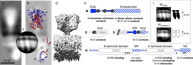
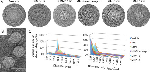
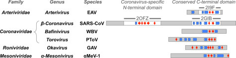
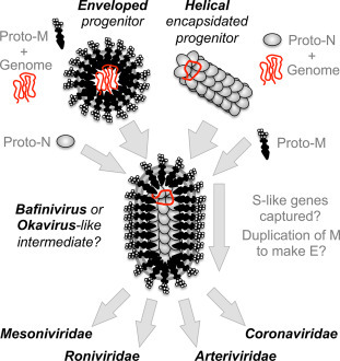
Coronavirus particles serve three fundamentally important functions in infection. The virion provides the means to deliver the viral genome across the plasma membrane of a host cell. The virion is also a means of escape for newly synthesized genomes. Lastly, the virion is a durable vessel that protects the genome on its journey between cells. This review summarizes the available X-ray crystallography, NMR, and cryoelectron microscopy structural data for coronavirus structural proteins, and looks at the role of each of the major structural proteins in virus entry and assembly. The potential wider conservation of the nucleoprotein fold identified in the Arteriviridae and Coronaviridae families and a speculative model for the evolution of corona-like virus architecture are discussed.
Structure and organization of proteins in the virion. Cryo-EM reconstruction of the SARS-CoV…
Fig. 1
Structure and organization of proteins in the virion. Cryo-EM reconstruction of the SARS-CoV structural proteins from virions (A; Neuman et al., 2006) superimposed with solved structures of the MHV S ectodomain (B; 3JCL), SARS-CoV E (C; 2MM4), SARS-CoV C-terminal domain (D; 2GIB), SARS-CoV N-terminal domain (E; 2OFZ), and a cryo-EM reconstruction of the M proteins from MHV VLPs (F; Neuman et al., 2011). An alternative shorter, wider cryo-EM reconstruction of SARS-CoV virion proteins is shown for comparison (G; EMD-1423). Schematics are based on MHV proteins, following the annotation of Kuo et al. (2016). The orientation of N protein domains shown here is hypothetical, and is intended for illustrative purposes only. (H) Domain structure and annotation of MHV S, E, M, and N proteins showing domains outside the virion (solid blue), inside the virion (striped blue), and the position of solved structures. Transmembrane regions (TM), sites of palmitoylation (Acyl), a conserved sequence preceding the transmembrane of S (KW), a serine–arginine-rich unstructured region (SR), phosphorylation sites (stars), and the C-terminal M-interacting domain of N (N3) are marked. Comparison of the appearance of dimeric MLONG and MCOMPACT (I).
Fig. 2
Appearance and characteristics of MHV…
Fig. 2
Appearance and characteristics of MHV virions, MHV VLPs, and copurified exosomal vesicles. Cryo-EM…
Fig. 2
Appearance and characteristics of MHV virions, MHV VLPs, and copurified exosomal vesicles. Cryo-EM images show a vesicle, VLPs produced after coexpression of E and M (EM VLP) or E, M and N (EMN VLP), a virion from tunicamycin-treated cells, and virions from the same preparation that spontaneously formed lacking (− S) or displaying (+ S) visible spikes. A cryoelectron tomography image of MHV is shown to highlight the variation in spike incorporation in virions from the same preparation (B). Sizes (diameter) and shapes (dMAX/dMIN) of virions, VLPs, and vesicles were categorized to illustrate differences in the shape of particles that incorporate different combinations of structural proteins (C).
Fig. 3
Evidence for conservation of the…
Fig. 3
Evidence for conservation of the N protein C-terminal domain fold across the Nidovirales …
Fig. 3
Evidence for conservation of the N protein C-terminal domain fold across the Nidovirales. Proteins are depicted as rectangles with resolved regions of solved protein structures (white regions) and unsolved or unresolved regions (gray regions) indicated. Alpha helix (blue cylinders) and beta strand (red arrows) regions of solved protein secondary structures or JPRED secondary structure predictions are shown manually aligned by secondary structure to facilitate comparison.
Fig. 4
Models of coronavirus virion evolution…
Fig. 4
Models of coronavirus virion evolution by gradual accumulation of structural proteins. Evolution of…
Fig. 4
Models of coronavirus virion evolution by gradual accumulation of structural proteins. Evolution of potential progenitors with enveloped pleomorphic or helical encapsidated virion architecture is shown leading to a filamentous enveloped intermediate stage, superficially resembling virions of the genus Bafinivirus or the family Roniviridae. Structural diversification by capture of attachment and fusion proteins from an unknown source, and partial duplication of M to make E in some lineages then leads to modern nidovirus lineages.
Almazan F., Gonzalez J.M., Penzes Z., Izeta A., Calvo E., Plana-Duran J., Enjuanes L. Engineering the largest RNA virus genome as an infectious bacterial artificial chromosome. Proc. Natl. Acad. Sci. U.S.A. 2000;97(10):5516–5521.
-
PMC
-
PubMed
Arndt A.L., Larson B.J., Hogue B.G. A conserved domain in the coronavirus membrane protein tail is important for virus assembly. J. Virol. 2010;84(21):11418–11428.
-
PMC
-
PubMed
Assiri A., McGeer A., Perl T.M., Price C.S., Al Rabeeah A.A., Cummings D.A. Hospital outbreak of Middle East respiratory syndrome coronavirus. N. Engl. J. Med. 2013;369(5):407–416.
-
PMC
-
PubMed
Barcena M., Oostergetel G.T., Bartelink W., Faas F.G., Verkleij A., Rottier P.J. Cryo-electron tomography of mouse hepatitis virus: insights into the structure of the coronavirion. Proc. Natl. Acad. Sci. U.S.A. 2009;106(2):582–587.
-
PMC
-
PubMed
Belouzard S., Millet J.K., Licitra B.N., Whittaker G.R. Mechanisms of coronavirus cell entry mediated by the viral spike protein. Viruses. 2012;4(6):1011–1033.
-
PMC
-
PubMed