Review
. 2016:96:59-126.
doi: 10.1016/bs.aivir.2016.08.008. Epub 2016 Sep 14.

The Nonstructural Proteins Directing Coronavirus RNA Synthesis and Processing

Affiliations
Review

The Nonstructural Proteins Directing Coronavirus RNA Synthesis and Processing

E J Snijder et al. Adv Virus Res. 2016.

Abstract

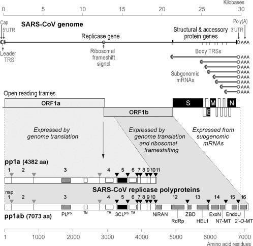
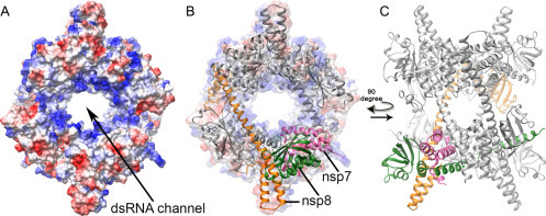
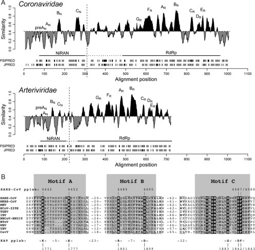
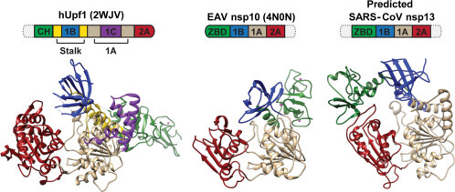
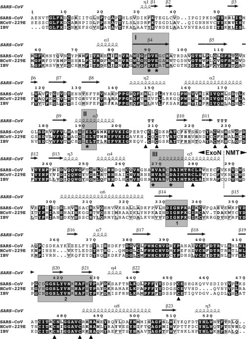
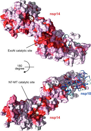
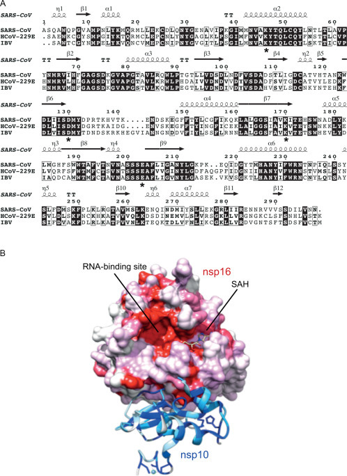
Coronaviruses are animal and human pathogens that can cause lethal zoonotic infections like SARS and MERS. They have polycistronic plus-stranded RNA genomes and belong to the order Nidovirales, a diverse group of viruses for which common ancestry was inferred from the common principles underlying their genome organization and expression, and from the conservation of an array of core replicase domains, including key RNA-synthesizing enzymes. Coronavirus genomes (~26-32 kilobases) are the largest RNA genomes known to date and their expansion was likely enabled by acquiring enzyme functions that counter the commonly high error frequency of viral RNA polymerases. The primary functions that direct coronavirus RNA synthesis and processing reside in nonstructural protein (nsp) 7 to nsp16, which are cleavage products of two large replicase polyproteins translated from the coronavirus genome. Significant progress has now been made regarding their structural and functional characterization, stimulated by technical advances like improved methods for bioinformatics and structural biology, in vitro enzyme characterization, and site-directed mutagenesis of coronavirus genomes. Coronavirus replicase functions include more or less universal activities of plus-stranded RNA viruses, like an RNA polymerase (nsp12) and helicase (nsp13), but also a number of rare or even unique domains involved in mRNA capping (nsp14, nsp16) and fidelity control (nsp14). Several smaller subunits (nsp7-nsp10) act as crucial cofactors of these enzymes and contribute to the emerging "nsp interactome." Understanding the structure, function, and interactions of the RNA-synthesizing machinery of coronaviruses will be key to rationalizing their evolutionary success and the development of improved control strategies.

Keywords: Capping; Coronavirus; Nidovirus; RNA processing; Replication.


Figures

Fig. 1
Fig. 1
Outline of the CoV genome organization and expression strategy, based on SARS-CoV. The top panel depicts the SARS-CoV genome, including various regulatory RNA elements, and the 5′- and 3′-coterminal nested set of subgenomic mRNAs used to express the genes downstream of the replicase gene. UTR, untranslated region; TRS, transcription-regulatory sequence. Below the RNAs, the 14 open reading frames in the genome are indicated, i.e., the replicase ORFs 1a and 1b, the four common CoV structural protein genes (S, E, M, and N) and the ORFs encoding “accessory proteins.” The bottom panel explains the organization and proteolytic processing of the pp1a and pp1ab replicase polyproteins, the latter being produced by -1 ribosomal frameshifting. The nsp3 (PLpro) and nsp5 (3CLpro) proteases and their cleavage sites are indicated in matching colors. The resulting 16 cleavage products (nonstructural proteins (nsps)) are indicated, as are the conserved replicase domains that are relevant for this review. Domain abbreviations and corresponding nsp numbers: PLpro, papain-like proteinase (nsp3); 3CLpro, 3C-like proteinase (nsp5); TM, transmembrane domain (nsp3, nsp4, and nsp6); NiRAN, nidovirus RdRp-associated nucleotidyl transferase (nsp12); RdRp, RNA-dependent RNA polymerase (nsp12); ZBD, zinc-binding domain (nsp13); HEL1, superfamily 1 helicase (nsp13); ExoN, exoribonuclease (nsp14); N7-MT, N7-methyl transferase (nsp14); endoU, uridylate-specific endoribonuclease (nsp15); 2′-O-MT, 2′-O-methyl transferase (nsp16).
Fig. 2
Fig. 2
Crystal structure of the SARS-CoV nsp7–nsp8 hexadecamer (pdb 2AHM) (Zhai et al., 2005). Purified recombinant SARS-CoV nsp7 and nsp8 were found to self-assemble into a supercomplex of which the structure was determined at 2.4 Å resolution. (A) The complex forms a doughnut-shaped hollow structure of which the central channel is lined with positively charged side chains (in blue) and was postulated to mediate double-stranded RNA binding. The outside of the structure is predominantly negatively charged (red) surface shading). (B and C) SARS-CoV nsp8 resembles a “golf club”-like shape that can adopt two conformations, as presented here in orange and green. These nsp8 conformations are integrated into a much larger, hexadecameric structure that is composed of eight nsp8 subunits and eight nsp7 subunits, of which one is shaded pink. In (B), the hexadecamer is depicted against the background of the surface plot presented in (A).
Fig. 3
Fig. 3
Comparison of coronavirus nsp12 and arterivirus nsp9, containing the highly conserved NiRAN and RdRp domains. (A) Similarity density plot derived from a multiple sequence alignment including RdRp subunits from all nidovirus lineages. To highlight local deviations from the average, areas displaying conservation above and below the mean similarity are shaded in black and gray, respectively. Conserved sequence motifs of NiRAN (subscript N; see also B) and RdRp (subscript R) are labeled. Domain boundaries used for bioinformatics analyses and uncertainty with respect to the NiRAN/RdRp domain boundary are indicated with vertical and by dashed horizontal lines, respectively. Below each plot, the predicted secondary structure elements are presented in gray for α-helices and black for β-strands. (B) Multiple sequence alignment showing the three conserved motifs of the NiRAN domain from representative species across the Nidovirales order. Conserved residues in this alignment are shown in white font, while partially conserved residues are boxed. The bottom line depicts residues also conserved in the arterivirus EAV, which was used for a first experimental analysis of the NiRAN domain (Lehmann et al., 2015a). Abbreviations not explained in the main text: NHCoV, night-heron coronavirus HKU19 (genus Deltacoronavirus); BToV, bovine torovirus (family Coronaviridae, subfamily Torovirinae, genus Torovirus); WBV, white bream virus (family Coronaviridae, subfamily Torovirinae, genus Bafinivirus); YHV, yellow head virus (family Roniviridae, genus Okavirus); CavV, Cavally virus (family Mesoniviridae, genus Alphamesonivirus).
Fig. 4
Fig. 4
Three-dimensional models of cellular hUpf1 (the prototype of the Upf1-like family of SF1 helicases), the EAV nsp10 helicase (Deng et al., 2014), and the predicted structure of SARS-CoV nsp13. Based on sequence and structural comparisons, nidovirus helicases are classified into the Upf1-like family. Domain colors in the structures correspond to those used in the domain organization depicted above each structure, in which domain sizes are not drawn to scale. Dashed domains represent parts that could not be modeled. Zn2 + ions bound to the respective N-terminal domains are depicted as pink spheres. The identical coloring of domains other than 1A and 2A does not imply an evolutionary relationship. PDB accession numbers are listed in brackets.
Fig. 5
Fig. 5
Sequence alignment of coronavirus nsp14 homologs representing three genera of the Coronavirinae subfamily: SARS-CoV (genus Betacoronavirus), HCoV-229E (genus Alphacoronavirus), and IBV (genus Gammacoronavirus). The alignment was generated using Clustal Omega (Sievers et al., 2011) and rendered using ESPript version 3.0 (Robert and Gouet, 2014). Conserved ExoN motifs I, II, and III and clusters of residues involved in SAM binding and N7-MTase activity (1 and 2) are highlighted in gray. Catalytic residues of ExoN and residues involved in the formation of zinc fingers are indicated by asterisks and arrowheads, respectively. Also shown are the secondary structure elements of SARS-CoV nsp14 (pdb 5C8S) and the border between the N-terminal ExoN and C-terminal N7-MT (NMT) domains.
Fig. 6
Fig. 6
Surface representation of the three-dimensional structure of the nsp10/nsp14 complex (pdb 5C8S). The nsp10 ribbon structure is shown with conserved residues colored in blue (using a scale from dark to light blue). The coloring of the nsp14 surface is based on the conservation of the respective residues among CoVs (using a scale from dark to light red). The upper panel shows the surface containing the ExoN catalytic site with one Mg2 + ion bound in the active site (green sphere). The lower panel shows the opposite side of the structure with the N7-MTase active site. The cap analog GpppA and SAH are shown in stick representation. The figures were generated using UCSF Chimera (Pettersen et al., 2004). The degree of conservation of specific residues was determined using an alignment of nsp10 and nsp14 sequences of eight coronaviruses representing the four genera of the Coronavirinae subfamily (SARS-CoV, MERS-CoV, MHV, TGEV, FCoV, HCoV-229E, IBV, and bulbul coronavirus HKU11).
Fig. 7
Fig. 7
Coronavirus nsp16 and its interaction with nsp10. (A) Sequence alignment of nsp16 homologs of SARS-CoV (genus Betacoronavirus), HCoV-229E (genus Alphacoronavirus), and IBV (genus Gammacoronavirus). The alignment was generated using Clustal Omega (Sievers et al., 2011) and rendered using ESPript version 3.0 (Robert and Gouet, 2014). Residues of the catalytic tetrad K–D–K–E are indicated by asterisks and secondary structure elements of SARS-CoV nsp16 (pdb 2XYV) are shown. (B) Surface representation of the three-dimensional structure of the nsp10/nsp16 complex (pdb 2XYV). Nsp10 is shown in ribbon representation with conserved residues colored in dark to light blue according to their conservation among CoVs. Zinc molecules are shown as spheres and zinc-coordinating residues are shown in stick representation. The surface of nsp16 is colored in dark to light red according to the conservation of the respective residues among coronaviruses. SAH is depicted in a stick model. The figure was generated using UCSF Chimera (Pettersen et al., 2004). The degree of conservation of specific residues was determined using an alignment of nsp10 and nsp16 sequences of eight coronaviruses (see Fig. 6).
Fig. 8
Fig. 8
Alignment of nidovirus endoU domains and XendoU from Xenopus laevis. Residues involved in catalysis (*) and substrate binding (&) are indicated. Abbreviations not explained in the main text: EToV, Equine torovirus (subfamily Torovirinae, genus Torovirus); WBV, white bream virus (subfamily Torovirinae, genus Bafinivirus).

References

    1. Adedeji A.O., Marchand B., Te Velthuis A.J., Snijder E.J., Weiss S., Eoff R.L. Mechanism of nucleic acid unwinding by SARS-CoV helicase. PLoS One. 2012;7:e36521. - PMC - PubMed
    1. Adedeji A.O., Singh K., Calcaterra N.E., DeDiego M.L., Enjuanes L., Weiss S. Severe acute respiratory syndrome coronavirus replication inhibitor that interferes with the nucleic acid unwinding of the viral helicase. Antimicrob. Agents Chemother. 2012;56:4718–4728. - PMC - PubMed
    1. Adedeji A.O., Singh K., Kassim A., Coleman C.M., Elliott R., Weiss S.R. Evaluation of SSYA10-001 as a replication inhibitor of severe acute respiratory syndrome, mouse hepatitis, and Middle East respiratory syndrome coronaviruses. Antimicrob. Agents Chemother. 2014;58:4894–4898. - PMC - PubMed
    1. Ahn D.G., Choi J.K., Taylor D.R., Oh J.W. Biochemical characterization of a recombinant SARS coronavirus nsp12 RNA-dependent RNA polymerase capable of copying viral RNA templates. Arch. Virol. 2012;157:2095–2104. - PMC - PubMed
    1. Ahola T., Ahlquist P. Putative RNA capping activities encoded by brome mosaic virus: methylation and covalent binding of guanylate by replicase protein 1a. J. Virol. 1999;73:10061–10069. - PMC - PubMed

MeSH terms