Review
. 2016 Nov;24(4):577-591.
doi: 10.1016/j.fsc.2016.06.011.

Microtia Reconstruction

Affiliations
Review

Microtia Reconstruction

Randall A Bly et al. Facial Plast Surg Clin North Am. 2016 Nov.

Abstract

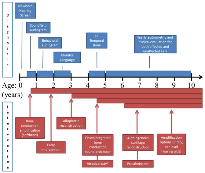
Microtia reconstruction is a challenging endeavor that has seen significant technique evolution. It is important to educate patients and their families to determine the best hearing rehabilitation and ear reconstructive options. Microtia is often associated with aural atresia, hearing loss, and craniofacial syndromes. Optimal care is provided by multiple disciplines, including a reconstructive surgeon, an otologic surgeon, an audiologist, and a craniofacial pediatrician. Microtia management includes observation, prosthetic ear, autologous cartilage reconstruction, or alloplastic implant placement. Hearing management options are observation, bone conduction sound processor, or atresiaplasty with and without hearing aids. Appropriate counseling should be done to manage expectations.

Keywords: Alloplastic reconstruction; Auricular reconstruction; Autologous reconstruction; Cartilage graft; Microtia; Microtia management.


Figures

Fig. 1
Fig. 1
Normal anatomic landmarks of external auricle.
Fig. 2
Fig. 2
Class I, II, III, IV microtia.
Fig. 3
Fig. 3
Timeline of diagnostic and treatment interventions for microtia and atresia. Diagnostic studies are shown in blue and interventions in red. CROS, contralateral routing of signal. * Atresiaplasty is considered if patient has favorable findings on high resolution computed tomography of the temporal bones.
Fig. 4
Fig. 4
Prosthetic ear.
Fig. 5
Fig. 5
(A) Template of normal left ear made from radiograph film. (B) Template placed over right microtic ear and (C) used to help position incisions for graft placement.
Fig. 6
Fig. 6
Two-centimeter incision drawn at inferior aspect of superior limb of synchondrosis to be harvested.
Fig. 7
Fig. 7
(A) Incision made along microtic ear lobule and then onto postauricular scalp to permit microtic remnant removal and lobule transposition. (B) Lobule transposed into desired position. (C) Mastoid pocket elevated with maintenance of subcutaneous pedicle to superior flap (superficial to retractor).
Fig. 8
Fig. 8
(A) Free-floating eighth rib cartilage being dissected from medial tip to lateral bony-cartilaginous junction. (B) Inferior limb being dissected from lateral to medial using hook for retraction. (C) Entire synchondrosis harvested before final superior limb cut. (D) Eighth free floating ribs and synchondrosis completely harvested.
Fig. 9
Fig. 9
(A) Template used to guide carving of auricular framework from rib synchondrosis with excess pieces removed. (B) Framework carved with 15 blade and skin biopsy punches. (C) Rib cartilage pieces shown separately: helical rim, antihelix projection, base framework, and antitragustragus complex. (D) Framework constructed.
Fig. 10
Fig. 10
Completion of stage 1 autologous reconstruction (note that the tragus is native).
Fig. 11
Fig. 11
Bolster in place after completion of stage 1 autologous reconstruction.
Fig. 12
Fig. 12
(A) Incision made around framework. (B) Auricular framework elevated exposing postauricular surface of framework to be grafted and mastoid cortex. (C) Postauricular scalp flap advanced with towel clip in place.
Fig. 13
Fig. 13
(A) Elliptical skin graft designed on upper thigh and harvested with number 20 blade. (B) Additional postauricular skin graft harvested from contralateral ear. (C) Ear elevated after scalp flap advancement and placement of skin grafts.
Fig. 14
Fig. 14
Preoperative and postoperative photographs of left microtia and autologous cartilage reconstruction.
Fig. 15
Fig. 15
Preoperative marking for superficial temporal artery in preparation for harvest of TPF flap (red) and “c”-shaped incision (black).
Fig. 16
Fig. 16
(A) Endoscopic view of TPF harvest, distal TPF flap dissected away from subcutaneous tissue using extended length needle tip cautery. (B) TPF flap elevated.
Fig. 17
Fig. 17
Prepared alloplastic framework.
Fig. 18
Fig. 18
Alloplastic framework in place with skin graft.
Fig. 19
Fig. 19
Preoperative and postoperative photographs of right microtia with alloplastic reconstruction.

References

    1. Luquetti DV, Leoncini E, Mastroiacovo P. Microtia-anotia: a global review of prevalence rates. Birth Defects Res A Clin Mol Teratol. 2011;91(9):813–22. - PMC - PubMed
    1. Luquetti DV, Heike CL, Hing AV, et al. Microtia: epidemiology and genetics. Am J Med Genet A. 2012;158A(1):124–39. - PMC - PubMed
    1. Mastroiacovo P, Corchia C, Botto LD, et al. Epidemiology and genetics of microtia-anotia: a registry based study on over one million births. J Med Genet. 1995;32(6):453–7. - PMC - PubMed
    1. Harris J, Källén B, Robert E. The epidemiology of anotia and microtia. J Med Genet. 1996;33(10):809–13. - PMC - PubMed
    1. Castilla EE, Orioli IM. Prevalence rates of microtia in South America. Int J Epidemiol. 1986;15:364–8. - PubMed

MeSH terms