. 2016 Dec 1;420(1):110-119.
doi: 10.1016/j.ydbio.2016.10.001. Epub 2016 Oct 3.

Bmp4-Msx1 signaling and Osr2 control tooth organogenesis through antagonistic regulation of secreted Wnt antagonists

Affiliations

Bmp4-Msx1 signaling and Osr2 control tooth organogenesis through antagonistic regulation of secreted Wnt antagonists

Shihai Jia et al. Dev Biol. .

Abstract

Mutations in MSX1 cause craniofacial developmental defects, including tooth agenesis, in humans and mice. Previous studies suggest that Msx1 activates Bmp4 expression in the developing tooth mesenchyme to drive early tooth organogenesis. Whereas Msx1-/- mice exhibit developmental arrest of all tooth germs at the bud stage, mice with neural crest-specific inactivation of Bmp4 (Bmp4ncko/ncko), which lack Bmp4 expression in the developing tooth mesenchyme, showed developmental arrest of only mandibular molars. We recently demonstrated that deletion of Osr2, which encodes a zinc finger transcription factor expressed in a lingual-to-buccal gradient in the developing tooth bud mesenchyme, rescued molar tooth morphogenesis in both Msx1-/- and Bmp4ncko/ncko mice. In this study, through RNA-seq analyses of the developing tooth mesenchyme in mutant and wildtype embryos, we found that Msx1 and Osr2 have opposite effects on expression of several secreted Wnt antagonists in the tooth bud mesenchyme. Remarkably, both Dkk2 and Sfrp2 exhibit Osr2-dependent preferential expression on the lingual side of the tooth bud mesenchyme and expression of both genes was up-regulated and expanded into the tooth bud mesenchyme in Msx1-/- and Bmp4ncko/ncko mutant embryos. We show that pharmacological activation of canonical Wnt signaling by either lithium chloride (LiCl) treatment or by inhibition of DKKs in utero was sufficient to rescue mandibular molar tooth morphogenesis in Bmp4ncko/ncko mice. Furthermore, whereas inhibition of DKKs or inactivation of Sfrp2 alone was insufficient to rescue tooth morphogenesis in Msx1-/- mice, pharmacological inhibition of DKKs in combination with genetic inactivation of Sfrp2 and Sfrp3 rescued maxillary molar morphogenesis in Msx1-/- mice. Together, these data reveal a novel mechanism that the Bmp4-Msx1 pathway and Osr2 control tooth organogenesis through antagonistic regulation of expression of secreted Wnt antagonists.

Keywords: Bmp4; Dkk2; Mouse; Msx1; Organogenesis; Osr2; Sfrp2; Tooth development; Wnt signaling.


Figures

Fig. 1
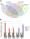
Fig. 1. Opposing effects of Msx1 and Osr2 on expression of Wnt signaling pathway genes in the E13.5 mandibular molar tooth mesenchyme
(A) Venn diagram comparing genes whose expression was changed by more than 2.0-fold in Msx1−/− mutants and 1.5-fold in Osr2−/− mutants compared with their wildtype littermates, respectively, in the RNA-seq datasets from the laser capture microdissected E13.5 mandibular molar tooth mesenchyme. (B) Quantitative real-time RT-PCR analysis of the levels of Dkk2, Sfrp1, Sfrp2, Lef1, and Tcf7 mRNAs in the E13.5 mandibular molar tooth mesenchyme of Msx1−/− and Osr2−/− mutant embryos in comparison with their wildtype littermates. Complementary DNAs were generated from pooled LCM tissue RNA samples from 3 embryos for each genotype. *, p < 0.05. Error bars represent S.E.M.
Fig. 2
Fig. 2. Comparison of patterns of Dkk2 and Sfrp2 mRNA expression in the developing molar tooth mesenchyme in E13.5 Osr2−/−, Msx1−/−, Bmp4ncko/ncko mutant and control embryos
(A–D) Frontal sections showing expression patterns of Dkk2 mRNAs in and around the maxillary (upper) and mandibular (lower) molar tooth mesenchyme in the E13.5 control (A), Osr2−/− (B), Msx1−/− (C), and Bmp4ncko/ncko mutant (D) embryos (n = 3 for each genotype). (E–H) Frontal sections showing expression patterns of Sfrp2 mRNAs in and around the maxillary and mandibular molar tooth mesenchyme in the E13.5 control (A), Osr2−/− (B), Msx1−/− (C), and Bmp4ncko/ncko mutant (D) embryos. A white line separates the pictures for maxillary and mandibular tooth germs in Panels C and G because the two pictures are taken from distinct sections of a series from the same embryo. Arrow points to the lingual side of the mandibular molar tooth germ. Red dashed line marks the boundary between the tooth bud epithelium and mesenchyme.
Fig. 3
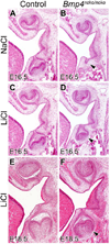
Fig. 3. LiCl treatment rescues mandibular molar tooth development in Bmp4ncko/ncko embryos
(A–F) H-E-stained frontal sections through the maxillary and mandibular first molar tooth germs in control (A, C, E) and Bmp4ncko/ncko (B, D, F) embryos treated with NaCl (A, B), or LiCl (C–F). Arrowhead points to the mandibular first molar tooth germ in Bmp4ncko/ncko mutant embryos in B, D, and F. n=6 for NaCl-treated control; n=6 for LiCl-treated control; n=20 for NaCl-treated Bmp4ncko/ncko.; n=14/30 for LiCl-treated Bmp4ncko/ncko mandibular molar tooth germs.
Fig. 4
Fig. 4. IIIC3a treatment in utero rescues mandibular molar tooth morphogenesis in Bmp4ncko/ncko mutant embryos
(A–D) H-E-stained frontal sections through the maxillary and mandibular first molar tooth germs in E18.5 Control (A, C) and Bmp4ncko/ncko mutant (B, D) embryos treated with DMSO (A, B) or IIIC3a (C, D). Arrowhead points to the mandibular first molar tooth germ in Bmp4ncko/ncko mutant embryos in B and D. n=6 for DMSO or IIIC3a-treated control; n=10 for DMSO-treated Bmp4ncko/ncko; n=8/12 for IIIC3a-treated Bmp4ncko/ncko mandibular molar tooth germs.
Fig. 5
Fig. 5. IIIC3a treatment in utero rescues Wnt signaling activity in and bud-to-cap transition of mandibular molar tooth germs in Bmp4ncko/ncko mutant embryos
(A–P) Comparison of Lef1 (A–D), Axin2 (E–H), Shh (I–L), and p21 (M–P) mRNA expression, respectively, in the maxillary and mandibular molar tooth germs in E14 control (A, C, E, G, I, K, M, O) and Bmp4ncko/ncko (B, D, F, H, J, L, N, P) embryos without (A, B, E, F, I, J, M, N) or with (C, D, G, H, K, L, O, P) IIIC3a treatment. mRNA signals are detected in blue color. A white line separates the pictures for maxillary and mandibular tooth germs in Panels C, D, G, H, K, L, O, and P because the two pictures are taken from distinct sections of a series from the same embryo. Arrowhead points to the mandibular first molar tooth germ in each panel. Red dashed line marks the boundary between the tooth bud epithelium and mesenchyme.
Fig. 6
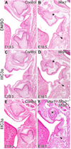
Fig. 6. IIIC3a treatment in utero rescued maxillary molar tooth organogenesis in Msx1−/−Sfrp2−/−Sfrp3−/− embryos
(A–F) H-E-stained frontal sections through the maxillary and mandibular first molar tooth germs at E18.5 in the control (A, C, E), Msx1−/− (B, D), and Msx1−/−Sfrp2−/−Sfrp3−/− mutant (F) embryos treated with DMSO (A, B) or IIIC3a (C–F). A white line separates the pictures for maxillary and mandibular tooth germs in Panel C because the two pictures are taken from distinct sections of a series from the same embryo. Arrowhead points to the mandibular first molar tooth germ and asterisk labels the maxillary first molar tooth mesenchyme in the mutant embryos (B, D, F). n=12 for DMSO-treated control and Msx1−/− samples; n=34 for IIIC3a-treated Msx1−/− and control samples; n=3/10 for IIIC3a-treated Msx1−/−Sfrp2−/−Sfrp3−/− samples.

Similar articles

Cited by

References

    1. Andl T, Reddy ST, Gaddapara T, Millar SE. WNT signals are required for the initiation of hair follicle development. Dev. Cell. 2002;2:643–653. - PubMed
    1. Bei M, Kratochwil K, Maas RL. BMP4 rescues a non-cell-autonomous function of Msx1 in tooth development. Development. 2000;127:4711–4718. - PubMed
    1. Beurel E, Grieco SF, Jope RS. Glycogen synthase kinase-3 (GSK3): Regulation, actions, and diseases. Pharmacol. Ther. 2015;148:114–131. - PMC - PubMed
    1. Brunskill EW, Potter SS. RNA-Seq defines novel genes, RNA processing patterns and enhancer maps for the early stages of nephrogenesis: Hox supergenes. Dev. Biol. 2012;368:4–17. - PMC - PubMed
    1. Chai Y, Jiang X, Ito Y, Bringas P, Jr, Han J, Rowitch DH, Soriano P, McMahon AP, Sucov HM. Fate of the mammalian cranial neural crest during tooth and mandibular morphogenesis. Development. 2000;127:1671–1679. - PubMed

MeSH terms