Comparative Study
. 2016 Nov 29;7(48):78883-78895.
doi: 10.18632/oncotarget.12428.

Selinexor, a Selective Inhibitor of Nuclear Export (SINE) compound, acts through NF-κB deactivation and combines with proteasome inhibitors to synergistically induce tumor cell death

Affiliations
Comparative Study

Selinexor, a Selective Inhibitor of Nuclear Export (SINE) compound, acts through NF-κB deactivation and combines with proteasome inhibitors to synergistically induce tumor cell death

Trinayan Kashyap et al. Oncotarget. .

Abstract

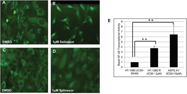
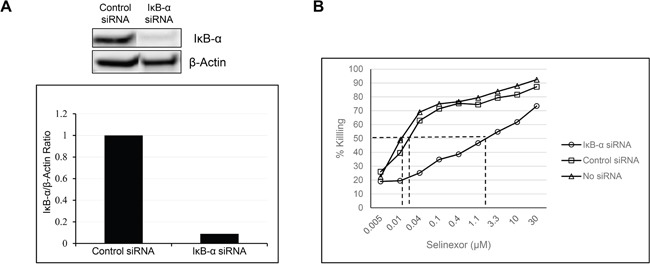
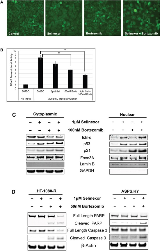
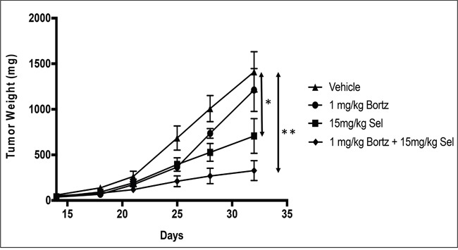
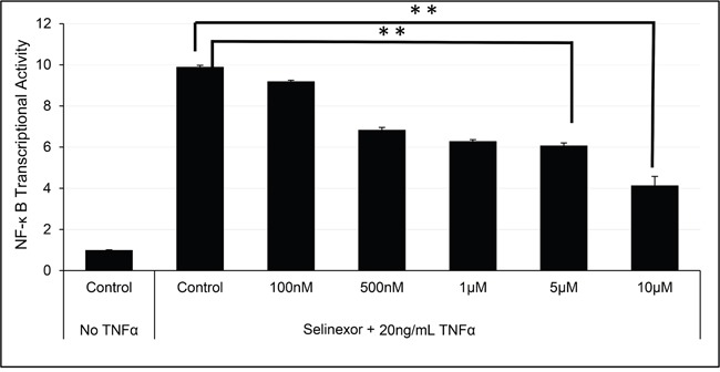
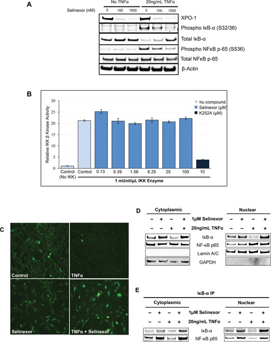
The nuclear export protein, exportin-1 (XPO1/CRM1), is overexpressed in many cancers and correlates with poor prognosis. Selinexor, a first-in-class Selective Inhibitor of Nuclear Export (SINE) compound, binds covalently to XPO1 and blocks its function. Treatment of cancer cells with selinexor results in nuclear retention of major tumor suppressor proteins and cell cycle regulators, leading to growth arrest and apoptosis. Recently, we described the selection of SINE compound resistant cells and reported elevated expression of inflammation-related genes in these cells. Here, we demonstrated that NF-κB transcriptional activity is up-regulated in cells that are naturally resistant or have acquired resistance to SINE compounds. Resistance to SINE compounds was created by knockdown of the cellular NF-κB inhibitor, IκB-α. Combination treatment of selinexor with proteasome inhibitors decreased NF-κB activity, sensitized SINE compound resistant cells and showed synergistic cytotoxicity in vitro and in vivo. Furthermore, we showed that selinexor inhibited NF-κB activity by blocking phosphorylation of the IκB-α and the NF-κB p65 subunits, protecting IκB-α from proteasome degradation and trapping IκB-α in the nucleus to suppress NF-κB activity. Therefore, combination treatment of selinexor with a proteasome inhibitor may be beneficial to patients with resistance to either single-agent.

Keywords: NF-κB; SINE; XPO1; proteasome inhibitors; selinexor.


Conflict of interest statement

CONFLICTS OF INTEREST

TK, CA, TJU, BK, CD, WS, ML, MK, SS and YL are full time Karyopharm, Inc. employees. AA, RMM, IM, ASA are from Wayne State University School of Medicine and they have no conflicts of interest to disclose.

Figures

Figure 1
Figure 1. SINE compound resistant cell lines showed increased basal levels of NF-κB transcriptional activity
SINE compound resistant HT-1080 (fibrosarcoma, HT-1080-R) cells were selected by continued exposure of sensitive parental cells in increasing concentrations of the SINE compound KPT-185. A, B. Parental and C, D. HT-1080-R cells were treated with 1μM of selinexor for 4 hours. SINE compound-induced nuclear retention of IκB-α was evaluated by immunofluorescence microscopy and shown to be impaired in SINE compound resistant cells. E. Parental-sensitive HT-1080, HT-1080-R, and ASPS-KY (Alveolar Soft Part Sarcoma) cells were tested for NF-κB transcriptional activity by ELISA assay. Equal number of cells from the 3 cell lines were lysed with RIPA buffer. The results from 2 independent assays show that higher NF-κB transcriptional activity is correlated with lower sensitivity to the cytotoxic effects of selinexor. The error bars indicate the standard deviation and the Student's t-test was used to calculate p values. **p<0.01.
Figure 2
Figure 2. Reduction in the levels of IκB-α affects the potency of selinexor
A. U-2 OS cells were transfected with either 40nM IκB-α or control siRNA using lipofectamine RNAiMax. At 96 hours post transfection there was a 90% reduction in the protein levels of IκB-α. B. The siRNA transfected and non-transfected U-2 OS cells were treated with increasing concentrations of selinexor and cells were assayed for the cytotoxic effect of selinexor 72 hours post treatment.
Figure 3
Figure 3. Combination with proteasome inhibitors overcomes selinexor resistance in cell lines
A. HT-1080-R cells were treated with 1 μM of selinexor and/or 100 nM bortezomib for 12 hours. The cells were fixed and the cellular localization of IκB-α was evaluated by immunofluorescence microscopy. Selinexor treatment showed minimal nuclear entrapment of IκB-α, but combined treatment with selinexor and bortezomib further increased nuclear retention of IκB-α. B. HT-1080-R cells were pre-treated with 3μM of selinexor (Sel) and/or 100nM bortezomib (Bortz) for 2 hours and then exposed to 20ng/mL TNFα for 4 hours in serum free media followed by evaluation for DNA binding activity. TNFα treatment induced NF-κB transcriptional activity by 8-fold, whereas treatment with the combination of selinexor and bortezomib reduced the activity by 60% compared to 20% and 40% by the single agent respectively. The error bars indicate standard deviation and the Student's t-test was used to calculate p values. *p<0.05. C. Sub-cellular localization of XPO1 cargos in HT-1080-R cells treated with 1 μM of selinexor and/or 100 nM bortezomib for 12 hours was evaluated by cellular fractionation and Western blotting. The combination treatment of selinexor and bortezomib increased nuclear levels of XPO1 cargos compared to either single agent treatment. Lamin B served as a nuclear protein marker; GAPDH as a cytosolic protein marker. D. HT-1080-R and ASPS-KY cells were treated with 1 μM of selinexor and/or 50 nM bortezomib for 24 hours. The combination of selinexor and bortezomib was more cytotoxic than either one of the single agents as indicated by pronounced cleavage of PARP and Caspase 3 with the combination.
Figure 4
Figure 4. In vivo combination of bortezomib with selinexor is more efficacious than either single agent in the SINE compound resistant tumors
HT-1080-R cells (1×10⁶) were subcutaneously injected in the flank of 3 naïve mice. Four weeks post injection, palpable tumors were formed at which time mice were euthanized, and tumors harvested and 50 mg pieces were re-implanted unilaterally into the flank of experimental groups. Bortezomib (Bortz) at 1mg/kg iv; selinexor (Sel) 15mg/kg orally or the combination of bortezomib at 1mg/kg + selinexor at 15mg/kg was administered Monday and Thursday for 2 weeks. Effects on tumor weight were recorded as described in materials and methods. Data shown are the mean for each group. The bortezomib by itself showed no significant tumor reduction. Selinexor as single agent and in combination showed 50% and 76% tumor weight reduction respectively at the end of the study (day 32). The error bars indicate SEM and the Student's t-test was used to calculate p values. *p<0.05, **p<0.01.
Figure 5
Figure 5. Selinexor inhibited NF-κB transcriptional activity
U-2 OS cells were pre-treated with different concentrations of selinexor for 2 hours and then exposed to TNFα for 4 hours in serum free media. TNFα exposure induced NF-κB transcriptional activity by 10-fold. Selinexor inhibited NF-κB transcriptional activity in a dose dependent manner. The error bars indicate standard deviation and the Student's t-test was used to calculate p values. **p<0.01.
Figure 6
Figure 6. Inhibition of NF-κB transcriptional activity by selinexor is associated with nuclear localization of IκB-α and protection from degradation
A. U-2 OS cells were stimulated with or without 20ng/mL TNFα for 2 hours before being treated with vehicle, 100nM or 1μM selinexor for the next 24 hours. Western blot of phospho-IκB-α and phosphor-NF-κB p-65 shows that selinexor reverts the pro-inflammatory effects of TNFα and selinexor also increased cellular levels of IκB-α. Selinexor induces XPO1 degradation. It is mediated by the proteasome degradation pathway (TK and YL, not shown). B. IKKβ kinase activity was analyzed by in vitro kinase assay using recombinant IKKβ, recombinant IκB-α substrate containing serine 32/36 residues and selinexor at different concentrations. IKKβ kinase activity was detected using a phosphorylation specific IκB-α antibody. Selinexor had no inhibitory effects on IKKβ kinase activity. The pan-kinase inhibitor K252A was used as a positive control for the assay. C. Immunofluorescence staining of IκB-α after treatment with 20ng/mL TNFα or/and 1μM selinexor for 24 hours. Selinexor induced nuclear localization of IκB-α in the presence or absence of TNFα. D. Cellular fractionation of U-2 OS cells shows similar increased nuclear levels of IκB-α and NF-κB p65 upon selinexor treatment even in the presence of TNFα. Lamin A/C was used as nuclear protein marker; GAPDH as a cytosolic protein marker. E. IκB-α immunoprecipitation (IP) and Western blotting of cytoplasmic and nuclear fractions of U-2OS cells treated with selinexor and TNFα shows that IκB-α binds to NF-κB p65 subunit both in the nucleus and cytoplasm.

References

    1. Senapedis WT, Baloglu E, Landesman Y. Clinical translation of nuclear export inhibitors in cancer. Semin Cancer Biol. 2014;27:74–86. - PubMed
    1. Chook YM, Süel KE. Nuclear import by karyopherin-βs: recognition and inhibition. Biochim Biophys Acta. 2011;1813:1593–1606. - PMC - PubMed
    1. Fornerod M, Ohno M, Yoshida M, Mattaj IW. CRM1 is an export receptor for leucine-rich nuclear export signals. Cell. 1997;90:1051–1060. - PubMed
    1. Turner JG, Sullivan DM. CRM1-mediated nuclear export of proteins and drug resistance in cancer. Curr Med Chem. 2008;15:2648–2655. - PubMed
    1. Xu D, Farmer A, Collett G, Grishin NV, Chook YM. Sequence and structural analyses of nuclear export signals in the NESdb database. Mol Biol Cell. 2012;23:3677–3693. - PMC - PubMed

Publication types

MeSH terms

LinkOut - more resources