Comparative Study
. 2016 Nov 29;7(48):78320-78330.
doi: 10.18632/oncotarget.12460.

Sulfuretin promotes osteoblastic differentiation in primary cultured osteoblasts and in vivo bone healing

Affiliations
Comparative Study

Sulfuretin promotes osteoblastic differentiation in primary cultured osteoblasts and in vivo bone healing

Q-Schick Auh et al. Oncotarget. .

Abstract

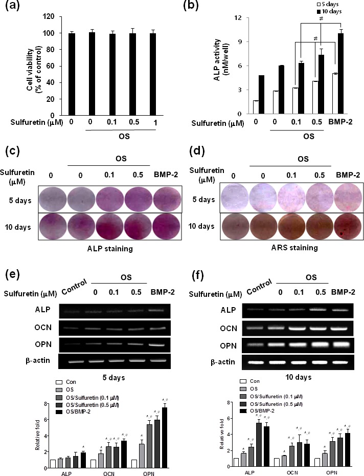
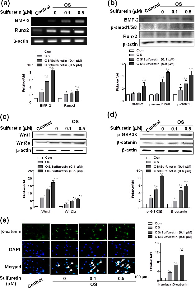
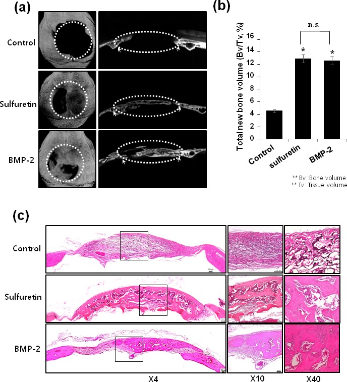
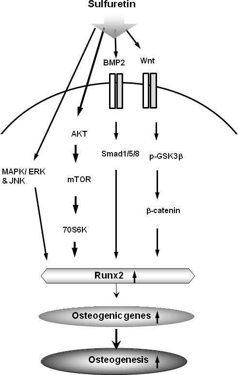
Although sulfuretin, the major flavonoid of Rhus verniciflua Stokes, has a variety of biological actions, its in vitro and in vivo effects on osteogenic potential remain poorly understood. The objective of the present study was to investigate the effects of sulfuretin on in vitro osteoblastic differentiation and the underlying signal pathway mechanisms in primary cultured osteoblasts and on in vivo bone formation using critical-sized calvarial defects in mice. Sulfuretin promoted osteogenic differentiation of primary osteoblasts, with increased ALP activity and mineralization, and upregulated differentiation markers, including ALP, osteocalcin, and osteopontin, in a concentration-dependent manner. The expression levels of Runx2, BMP-2, and phospho-Smad1/5/8 were upregulated by sulfuretin. Moreover, sulfuretin increased phosphorylation of Akt, mTOR, ERK, and JNK. Furthermore, sulfuretin treatment increased mRNA expression of Wnt ligands, phosphorylation of GSK3, and nuclear β-catenin protein expression. In vivo studies with calvarial bone defects revealed that sulfuretin significantly enhanced new bone formation by micro-computed tomography and histologic analysis. Collectively, these data suggest that sulfuretin acts through the activation of BMP, mTOR, Wnt/β-catenin, and Runx2 signaling to promote in vitro osteoblast differentiation and facilitate in vivo bone regeneration, and might be have therapeutic benefits in bone disease and regeneration.

Keywords: Differentiation; In vitro; In vivo; Osteoblasts; Osteogenesis; Signal pathways, Pathology Section.


Conflict of interest statement

CONFLICTS OF INTEREST

The authors declare they have no competing financial interests.

Figures

Figure 1
Figure 1. Effects of sulfuretin on cytotoxicity and osteoblastic differentiation in primary cultured osteoblasts
(A) Cell viability was determined by the MTT assay. Differentiation was assessed by ALP activity (B), ALP or Alizarin red staining (C-D), and mRNA expression (E, F). Cells were cultured in osteogenic supplement medium (OS) containing 50 μg/ml L-ascorbic acid and 10 mM β-glycerophosphate with the indicated concentration of sulfuretin for 5 (A, B, E) or 10 days (B, C, F). Data (A, B) are presented as mean ± standard deviation (SD) (n = 3). *, p < 0.05 vs. control. #, p < 0.05 vs. each group. Graphs in E, F below are presented as mean ± standard deviation (SD) (n = 3). *, p < 0.05 vs. control. #, p < 0.05 vs. OS. The data shown are for n=3 in each category of sample in each experiment.
Figure 2
Figure 2. Effects of sulfuretin on the BMP and Wnt/β-catenin pathway in primary osteoblasts
(A-E) Cells were cultured in osteogenic supplement medium with the indicated concentration of sulfuretin for 3 days (A, B) or 24 h (C-E). mRNA and protein expression were analyzed by RT-PCR (A), western blot (B-D), and immunocytochemistry (E) analysis, respectively. Similar results were obtained from three independent experiments. *, p < 0.05 vs. control. #, p < 0.05 vs. OS. The data shown are for n=3 in each category of sample in each experiment.
Figure 3
Figure 3. Effects of sulfuretin on the activation of Akt (A), mTOR (A, B), and MAP kinase (C) signaling in primary osteoblasts
(A-C) Cells were cultured with osteogenic supplement and the indicated concentration of sulfuretin for 30 min (A, B) and 45 min (C). Where indicated, the cells were pretreated with rapamycin (20 nM) for 1 h and post-treated with 0.5 μM sulfuretin for 30 min (B). Protein expression was assessed by western blot analysis. *, p < 0.05 vs. control. #, p < 0.05 vs. OS. &, p < 0.05 vs. OS/sulfuretin. The data shown are for n=3 in each category of sample in each experiment.
Figure 4
Figure 4. Effects of sulfuretin on in vivo bone regeneration
(A) Representative micro-CT images of the calvarial defect at 6 weeks. (B) Quantitative analysis of new bone formation by 3D μCT. (C) Representative histologic images from H&E-stained calvarial bone defect (left) and higher magnification images (right). Datas (B) are presented as mean ± standard deviation (SD) (n = 6). The data shown are for n = 3 in each category of sample in each experiment. *, p < 0.05, vs. control, NS: non-significant.
Figure 5
Figure 5. Schematic diagram illustrating that sulfuretin can induce osteoblastic differentiation and in vivo osteogenesis via activation of the BMP, mTOR, MAPK, and Wnt/β-catenin signaling pathways.

References

    1. Pietschmann P, Mechtcheriakova D, Meshcheryakova A, Foger-Samwald U, Ellinger I. Immunology of Osteoporosis: A Mini-Review. Gerontology. 2016;62(2):128–137. - PMC - PubMed
    1. Andreopoulou P, Bockman RS. Management of postmenopausal osteoporosis. Annu Rev Med. 2015;66:329–342. - PubMed
    1. Rodan GA, Martin TJ. Therapeutic Approaches to Bone Diseases. Sience. 2000;289(5484):1508–1514. - PubMed
    2. Lindsay R, Hart DM, Clark DM, editors. The minimum effective dose of estrogen for prevention of postmenopausal bone loss. Obstet Gynecol. 1984;63(3):759–763. - PubMed
    1. Nune KC, Kumar A, Misra RDK, Li SJ, Hao YL, Yang R. Osteoblast functions in functionally graded Ti-6Al-4V mesh structures. J Biomater Appl. 2016;30(8):1182–1204. - PubMed
    1. Nune KC, Kumar A, Murr LE, Misra RDK. Interplay between self-assembled structure of bone morphogenetic protein-2 (BMP-2) and osteoblast functions in three-dimensional titanium alloy scaffolds: Stimulation of osteogenic activity. J Biomed Mater Res A. 2016;104(2):517–532. - PubMed

Publication types

MeSH terms