Review
. 2010 Jan 28;3(2):345-368.
doi: 10.3390/ph3020345.

Olesoxime (TRO19622): A Novel Mitochondrial-Targeted Neuroprotective Compound

Affiliations
Review

Olesoxime (TRO19622): A Novel Mitochondrial-Targeted Neuroprotective Compound

Thierry Bordet et al. Pharmaceuticals (Basel). .

Abstract

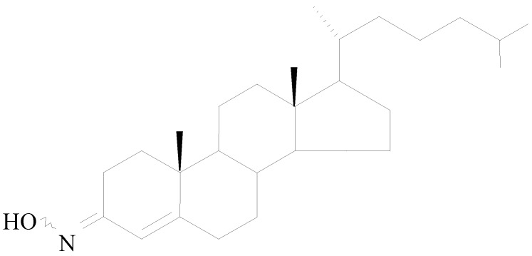
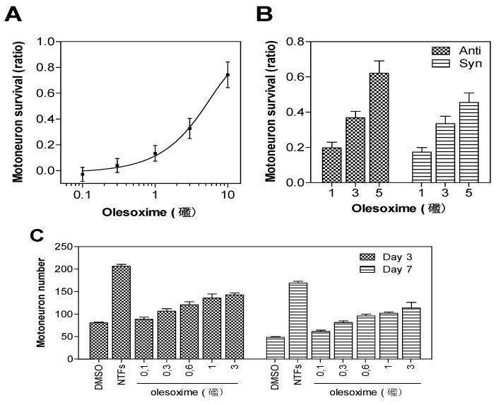
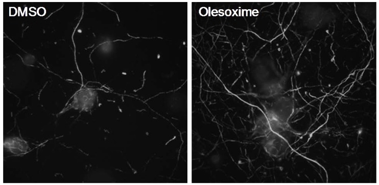
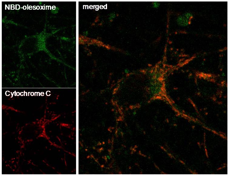
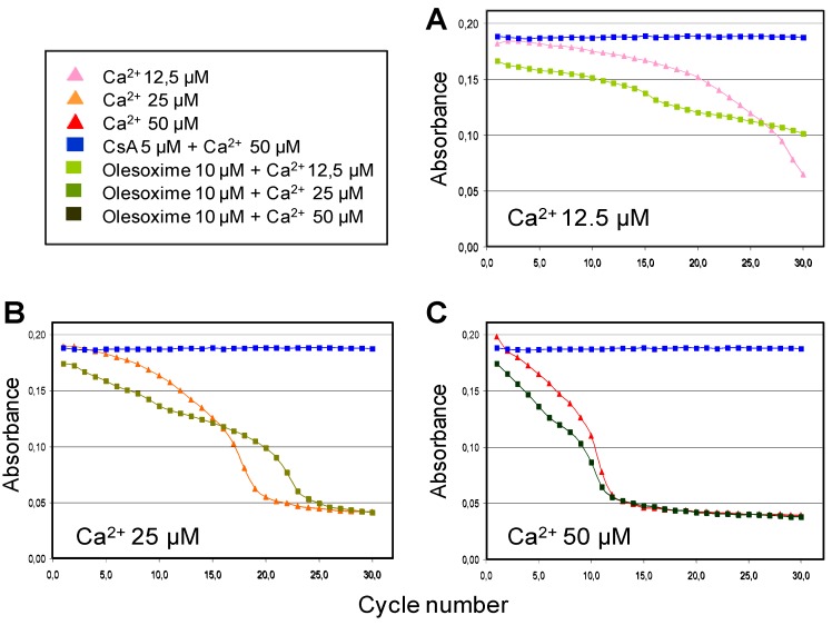
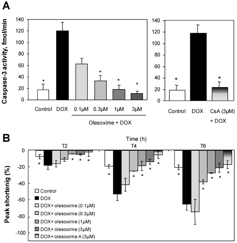
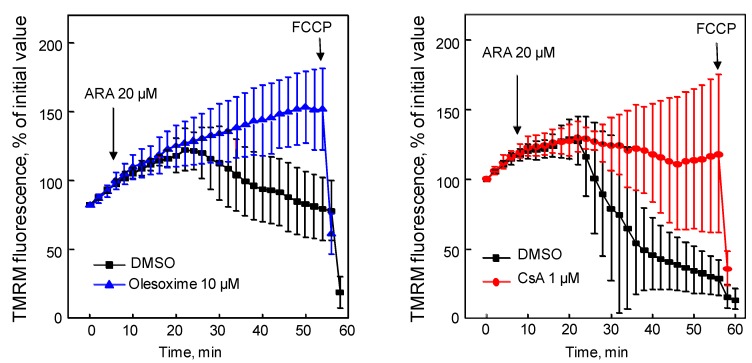
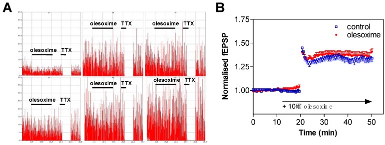
Olesoxime (TRO19622) is a novel mitochondrial-targeted neuroprotective compound undergoing a pivotal clinical efficacy study in Amyotrophic Lateral Sclerosis (ALS) and also in development for Spinal Muscular Atrophy (SMA). It belongs to a new family of cholesterol-oximes identified for its survival-promoting activity on purified motor neurons deprived of neurotrophic factors. Olesoxime targets proteins of the outer mitochondrial membrane, concentrates at the mitochondria and prevents permeability transition pore opening mediated by, among other things, oxidative stress. Olesoxime has been shown to exert a potent neuroprotective effect in various in vitro and in vivo models. In particular olesoxime provided significant protection in experimental animal models of motor neuron disorders and more particularly ALS. Olesoxime is orally active, crosses the blood brain barrier, and is well tolerated. Collectively, its pharmacological properties designate olesoxime as a promising drug candidate for motor neuron diseases.

Keywords: TRO19622; amyotrophic lateral sclerosis; mitochondrial permeability transition pore; motor neuron disease; neuroprotection; olesoxime; spinal muscular atrophies.


Figures

Figure 1
Figure 1
Olesoxime chemical structure.
Figure 2
Figure 2
Neuroprotective dose-effect of olesoxime against motor neuron cell death. Rat embryonic motor neurons were cultured in 96-well plates in the absence of trophic factors and with increasing concentrations of olesoxime (A, C), or with pure syn or anti forms of olesoxime (B). Motor neuron survival was measured after 3 days (A-C) or 7 days (C) in vitro by direct counting of live cells labelled with calcein-AM. Survival is expressed as a ratio of surviving cell (A-B) relative to positive controls treated with a cocktail of neurotrophic factors (NTFs) and negative controls that received DMSO alone (0.1%). Data represent mean ± SEM.
Figure 3
Figure 3
Olesoxime promotes neurite outgrowth of rat primary cortical neurons. E18 cortical neurons were cultured for 6 days in Neurobasal medium, 2% B27 (Invitrogen) and 1% pyruvate. After 24 h treatment with 5µM olesoxime, an increased density in neurite network was observed by immunostaining of neurofilaments.
Figure 4
Figure 4
Mitochondrial localization of NBD-olesoxime in primary cortical neurons. Rat E18 cortical neurons were treated with 10µM NBD-olesoxime (green) for 24 h, then immunostained for cytochrome C (red) and processed for confocal imaging. The orange staining in the merged image demonstrated localization of olesoxime in mitochondria.
Figure 5
Figure 5
Partial effects of olesoxime on calcium-induced mitochondrial swelling. Mitochondria purified from mouse liver were incubated with 10 µM olesoxime or 5 µM CsA for 5 minutes, prior to the induction of swelling by different calcium concentrations (A, 12.5 µM; B, 25 µM; C, 50 µM). Thereafter, mitochondrial swelling was monitored every 2 min by determining the light scattering or decreased absorbance of the samples.
Figure 6
Figure 6
Treatment with olesoxime dose-dependently inhibits doxorubicin-induced apoptosis in adult rabbit cardiomyocytes and maintains contractility with efficacy similar to Cyclosoporin A. (A) Caspase-3 activity was measured in cell lysates by a fluorometric assay (Biomol Reasearch Laboratories, Plymouth Meeting, PA, USA) 8h after 1µM doxorubicin (DOX) treatment with and without olesoxime. (B) Time-dependent inhibition by olesoxime of doxorubicin-induced contractile dysfunction monitored by video microscopy in ventricular cardiomyocytes. Data represent mean ± SEM, n = 5-6 cells per group, * p < 0.05 vs. doxorubicin treated cells as determined by Student’s t-test.
Figure 7
Figure 7
Olesoxime prevented arachidonic acid-induced mPTP opening. Hela cells were loaded with tetramethylrhodamine methyl ester (TMRM) for 5 min then treated with olesoxime or CsA (t0). Arachidonic acid (ARA) induced mitochondrial membrane depolarization measured as a loss of TMRM fluorescence in the mitochondria over time. Fluorescence intensity was measured every 2 minutes from 10 individual mitochondria. FCCP, carbonyl cyanide p-trifluoromethoxyphenylhydrazone. Data represent mean ± SEM.
Figure 8
Figure 8
Olesoxime does not modify neuronal electrophysiological properties. (A) Spontaneous action potential was measured on rat cortical neurons exposed to 10 µM olesoxime for 10 min and then to 100 nM tetrodotoxin (TTX) for 2 min. Spike rate is plotted on a 50 Hz scale. The total session recording was 30 minutes. (B) LTP was recorded on hippocampal slices of neonatal rats in presence of 10 µM olesoxime. Data represent mean ± SEM.

References

    1. Bordet T., Pruss R., Henderson C.E. Screening for ALS drugs. In: Mitsumoto H., Przedborski S., Gordon P.H., editors. Amyotrophic Lateral Sclerosis. Taylor & Francis Group; New York, NY, USA: 2006. pp. 551–582.
    1. Bordet T., Buisson B., Michaud M., Drouot C., Galea P., Delaage P., Akentieva N.P., Evers A.S., Covey D.F., Ostuni M.A., Lacapere J.J., Massaad C., Schumacher M., Steidl E.M., Maux D., Delaage M., Henderson C.E., Pruss R.M. Identification and characterization of cholest-4-en-3-one, oxime (TRO19622), a novel drug candidate for amyotrophic lateral sclerosis. J. Pharmacol. Exp. Ther. 2007;322:709–720. doi: 10.1124/jpet.107.123000. - DOI - PubMed
    1. Henderson C.E., Bloch-Gallego E., Camu W. Purified embryonic motoneurons. In: Cohen J., Wilkin G., editors. Nerve Cell Culture: A practical approach. Oxford UP; London, UK: 1995. pp. 69–81.
    1. Greenlund L.J., Deckwerth T.L., Johnson E.M. Superoxide dismutase delays neuronal apoptosis: A role for reactive oxygen species in programmed neuronal death. Neuron. 1995;14:303–315. - PubMed
    1. Estevez A.G., Spear N., Manuel S.M., Radi R., Henderson C.E., Barbeito L., Beckman J.S. Nitric oxide and superoxide contribute to motor neuron apoptosis induced by trophic factor deprivation. J. Neurosci. 1998;18:923–931. - PMC - PubMed

LinkOut - more resources