

doi: 10.1038/ncomms13052.
Abundant DNA 6mA methylation during early embryogenesis of zebrafish and pig
Affiliations
- PMID: 27713410
- PMCID: PMC5059759
- DOI: 10.1038/ncomms13052
Item in Clipboard
Abundant DNA 6mA methylation during early embryogenesis of zebrafish and pig
Nat Commun.
.
Abstract
DNA N6-methyldeoxyadenosine (6mA) is a well-known prokaryotic DNA modification that has been shown to exist and play epigenetic roles in eukaryotic DNA. Here we report that 6mA accumulates up to ∼0.1-0.2% of total deoxyadenosine during early embryogenesis of vertebrates, but diminishes to the background level with the progression of the embryo development. During this process a large fraction of 6mAs locate in repetitive regions of the genome.
Figures
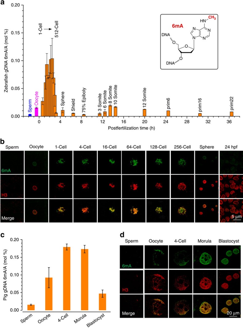
(a,c) Quantification of 6mA modification in isolated gDNA from sperm, oocyte and various embryo stages of zebrafish (a) and pig (c) by UHPLC–QQQ–MS/MS. The mole ratios of 6mA/A are shown, error bars indicate mean±s.d. (n=3). (b,d) Immunofluorescence images of selected embryo stages of zebrafish (b) and pig (d) at single-cell level stained by anti-6mA antibody (green, rabbit polyclonal from SYSY) and anti-Histone 3 (H3) antibody (red, mouse monoclonal from Biodragon Immunotechnologies). Early embryo stages show strong fluorescence indicative of the high abundance of 6mA in gDNA, whereas the signal is diminished with the progression of the embryo development. Full embryo images are presented in Supplementary Fig. 3.
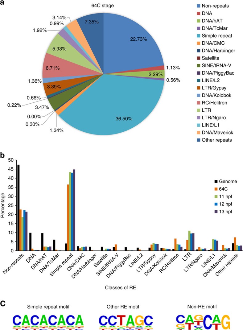
(a) Pie chart showing the distribution of 6mA peaks at 64-cell (64C) stage. The peaks located in REs are classified into subgroups based on RepeatMasker annotation. (b) 6mA peak classification for samples of 64C, 11 hpf, 12 hpf and 13 hpf. The genome background distribution is shown for comparison. The peaks are classified with the same criteria shown in a. (c) Sequence motifs of 6mA peaks in simple repeat, other REs apart from simple repeats (other RE) and non-repetitive regions (non-RE) for the 64C stage sample. Motifs were searched and generated by Homer software. The P-values for simple repeat, RE and non-RE motifs are 5e−1163, 1e−1557 and 1e−390, respectively.
Similar articles
-
Epigenetic marking of the zebrafish developmental program.Curr Top Dev Biol. 2013;104:85-112. doi: 10.1016/B978-0-12-416027-9.00003-6. Curr Top Dev Biol. 2013. PMID: 23587239 Review.
-
Genes for embryo development are packaged in blocks of multivalent chromatin in zebrafish sperm.Genome Res. 2011 Apr;21(4):578-89. doi: 10.1101/gr.113167.110. Epub 2011 Mar 7. Genome Res. 2011. PMID: 21383318 Free PMC article.
-
Embryonic DNA methylation: insights from the genomics era.Brief Funct Genomics. 2014 Mar;13(2):121-30. doi: 10.1093/bfgp/elt039. Epub 2013 Sep 23. Brief Funct Genomics. 2014. PMID: 24064195 Review.
-
Single-Nucleotide-Resolution Sequencing of N6-Methyldeoxyadenosine.Methods Mol Biol. 2021;2198:369-377. doi: 10.1007/978-1-0716-0876-0_28. Methods Mol Biol. 2021. PMID: 32822045
-
The developmental epigenomics toolbox: ChIP-seq and MethylCap-seq profiling of early zebrafish embryos.Methods. 2013 Aug 15;62(3):207-15. doi: 10.1016/j.ymeth.2013.04.011. Epub 2013 Apr 23. Methods. 2013. PMID: 23624103
Cited by
-
The Epigenetics of Gametes and Early Embryos and Potential Long-Range Consequences in Livestock Species-Filling in the Picture With Epigenomic Analyses.Front Genet. 2021 Mar 3;12:557934. doi: 10.3389/fgene.2021.557934. eCollection 2021. Front Genet. 2021. PMID: 33747031 Free PMC article. Review.
-
N6-Methyladenine DNA modification in Xanthomonas oryzae pv. oryzicola genome.Sci Rep. 2018 Nov 2;8(1):16272. doi: 10.1038/s41598-018-34559-5. Sci Rep. 2018. PMID: 30389999 Free PMC article.
-
Environmental Toxicity Assessment of Sodium Fluoride and Platinum-Derived Drugs Co-Exposure on Aquatic Organisms.Toxics. 2022 May 23;10(5):272. doi: 10.3390/toxics10050272. Toxics. 2022. PMID: 35622686 Free PMC article.
-
DNA N6-methyladenine in metazoans: functional epigenetic mark or bystander?Nat Struct Mol Biol. 2017 Jun 6;24(6):503-506. doi: 10.1038/nsmb.3412. Nat Struct Mol Biol. 2017. PMID: 28586322
-
Zebrafish transposable elements show extensive diversification in age, genomic distribution, and developmental expression.Genome Res. 2022 Jul;32(7):1408-1423. doi: 10.1101/gr.275655.121. Epub 2022 Jan 5. Genome Res. 2022. PMID: 34987056 Free PMC article.
References
Publication types
MeSH terms
Substances
Grants and funding
LinkOut - more resources
Full Text Sources
Other Literature Sources
Molecular Biology Databases
