. 2016 Oct 7:6:32833.
doi: 10.1038/srep32833.

In-vivo Dynamics of the Human Hippocampus across the Menstrual Cycle

Affiliations

In-vivo Dynamics of the Human Hippocampus across the Menstrual Cycle

Claudia Barth et al. Sci Rep. .

Abstract

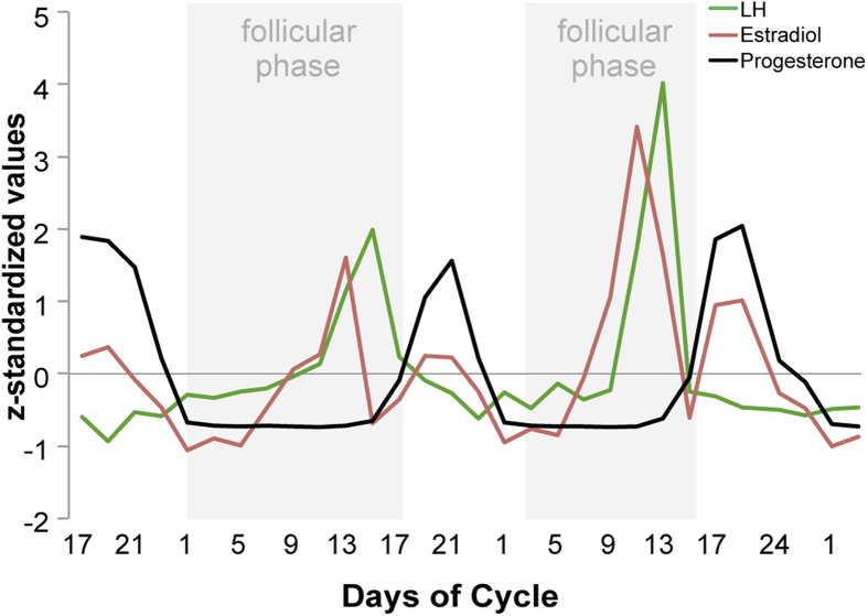
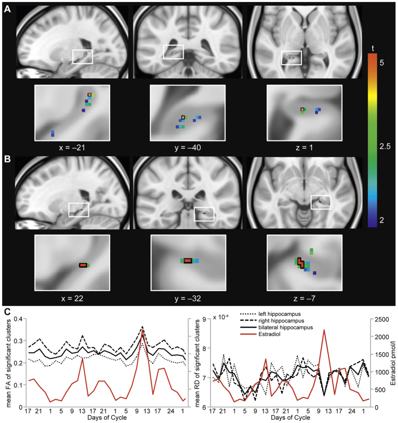
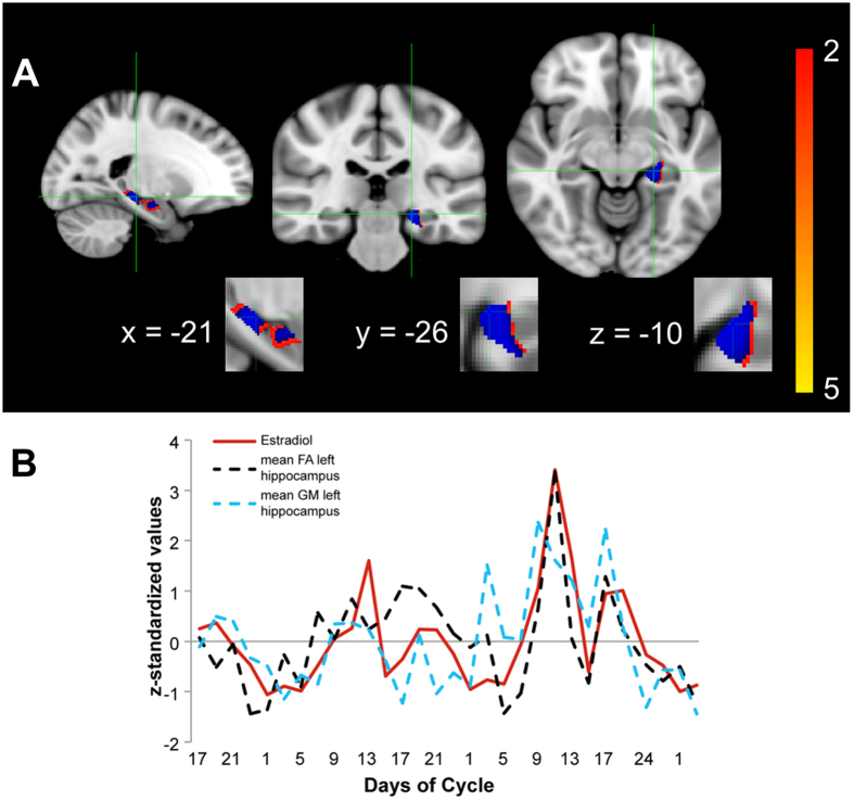
Sex hormones fluctuate during the menstrual cycle. Evidence from animal studies suggests similar subtle fluctuations in hippocampal structure, predominantly linked to estrogen. Hippocampal abnormalities have been observed in several neuropsychiatric pathologies with prominent sexual dimorphism. Yet, the potential impact of subtle sex-hormonal fluctuations on human hippocampal structure in health is unclear. We tested the feasibility of longitudinal neuroimaging in conjunction with rigorous menstrual cycle monitoring to evaluate potential changes in hippocampal microstructure associated with physiological sex-hormonal changes. Thirty longitudinal diffusion weighted imaging scans of a single healthy female subject were acquired across two full menstrual cycles. We calculated hippocampal fractional anisotropy (FA), a measure sensitive to changes in microstructural integrity, and investigated potential correlations with estrogen. We observed a significant positive correlation between FA values and estrogen in the hippocampus bilaterally, revealing a peak in FA closely paralleling ovulation. This exploratory, single-subject study demonstrates the feasibility of a longitudinal DWI scanning protocol across the menstrual cycle and is the first to link subtle endogenous hormonal fluctuations to changes in FA in vivo. In light of recent attempts to neurally phenotype single humans, our findings highlight menstrual cycle monitoring in parallel with highly sampled individual neuroimaging data to address fundamental questions about the dynamics of plasticity in the adult brain.


Figures

Figure 1
Figure 1. Endogenous ovarian sex hormone fluctuation.
Characteristic patterns of serum estradiol (red line), progesterone (black line) and LH (green line) levels are displayed across two menstrual cycles (follicular phase highlighted in grey). The depicted data represent days of cycle with corresponding z-standardized, single hormone values, matching the single scan time points in chronological order. As expected, estradiol shows a first prominent peak in the periovulatory phase, followed by a second smaller peak in the late luteal phase. LH surges after the peak of estrogen, shortly before ovulation. Progesterone levels are low during the follicular phase and high during the luteal phase.
Figure 2
Figure 2. Estrogen-modulated FA differences and time course across the menstrual cycle in the bilateral hippocampus.
Threshold-Free Cluster Enhancement (TFCE) voxel-wise FA correlation with respective estrogen levels in hippocampal ROI-masks (left (panel A) and right (panel B) hippocampus) are displayed. Red voxels outlined with black, superimposed on respective t-values, correspond to significant FWE-corrected results (p < 0.05). In panel C, FA (left) and RD (right) values from significant clusters, respectively peak voxel, are extracted and plotted versus estrogen levels (in red) across the menstrual cycles assessed.
Figure 3
Figure 3. Estrogen-modulated grey matter differences in the left hippocampus.
Threshold-Free Cluster Enhancement (TFCE) voxel-wise grey matter correlation with respective estrogen levels in hippocampal ROI-mask (hair cross on peak voxel; panel A: grey matter changes in left hippocampus) are displayed. Blue voxels, superimposed on respective t-values, correspond to significant FWE-corrected results (p < 0.05). In panel B, bilateral FA and left grey matter values from significant clusters, respectively peak voxel, are extracted and plotted versus estrogen levels (in red) across the menstrual cycles assessed.

References

    1. Boccardi M. et al. Effects of hormone therapy on brain morphology of healthy postmenopausal women: a Voxel-based morphometry study. Menopause 13, 584–591, doi: 10.1097/01.gme.0000196811.88505.10 (2006). - DOI - PubMed
    1. Galea L. A. et al. Endocrine regulation of cognition and neuroplasticity: our pursuit to unveil the complex interaction between hormones, the brain, and behaviour. Can J Exp Psychol 62, 247–260, doi: 10.1037/a0014501 (2008). - DOI - PubMed
    1. Peper J. S., Hulshoff Pol H. E., Crone E. A. & van Honk J. Sex steroids and brain structure in pubertal boys and girls: a mini-review of neuroimaging studies. Neuroscience 191, 28–37, doi: 10.1016/j.neuroscience.2011.02.014 (2011). - DOI - PubMed
    1. Sacher J., Neumann J., Okon-Singer H., Gotowiec S. & Villringer A. Sexual dimorphism in the human brain: evidence from neuroimaging. Magn Reson Imaging 31, 366–375, doi: 10.1016/j.mri.2012.06.007 (2013). - DOI - PubMed
    1. Duarte-Guterman P., Yagi S., Chow C. & Galea L. A. Hippocampal learning, memory, and neurogenesis: Effects of sex and estrogens across the lifespan in adults. Horm Behav, doi: 10.1016/j.yhbeh.2015.05.024 (2015). - DOI - PubMed

Publication types

LinkOut - more resources