. 2017:1495:111-130.
doi: 10.1007/978-1-4939-6451-2_8.

Protein Chemical Modification Inside Living Cells Using Split Inteins

Affiliations

Protein Chemical Modification Inside Living Cells Using Split Inteins

Radhika Borra et al. Methods Mol Biol. 2017.

Abstract

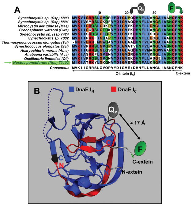
Methods to visualize, track, measure, and perturb or activate proteins in living cells are central to biomedical efforts to characterize and understand the spatial and temporal underpinnings of life inside cells. Although fluorescent proteins have proven to be extremely useful for in vivo studies of protein function, their utility is inherently limited because their spectral and structural characteristics are interdependent. These limitations have spurred the creation of alternative approaches for the chemical labeling of proteins. We describe in this protocol the use of fluorescence resonance emission transfer (FRET)-quenched DnaE split-inteins for the site-specific labeling and concomitant fluorescence activation of proteins in living cells. We have successfully employed this approach for the site-specific in-cell labeling of the DNA binding domain (DBD) of the transcription factor YY1 using several human cell lines. Moreover, we have shown that this approach can be also used for modifying proteins in order to control their cellular localization and potentially alter their biological activity.

Keywords: Fluorescence; Npu intein; Protein labeling; Protein trans-splicing; Split-intein.


Figures

Figure 1
Figure 1
A. Site-specific labeling and fluorescence activation of a protein of interest (POI) by FRET-quenched protein trans-splicing. Key to this approach is the introduction of fluorescence quencher (Q) into the IC polypeptide, which blocks the fluorescence signal of the fluorophore (F) located at the C-terminus of the IC polypeptide before protein trans-splicing happens. When protein trans-splicing occurs the fluorophore is covalently attached to the C-terminus of the POI triggering its fluorescence. The use of this approach for in-cell modification and fluorescence tagging of proteins minimizes the fluorescence background from the unreacted IC polypeptide thus facilitating the optical tracking of the labeled protein inside the cell. B. Scheme showing the approach used for in-cell labeling of a POI with a fluorophore inside a live cell using protein trans-splicing (figure modified from [13]).
Figure 2
Figure 2
Design of FRET-quenched DnaE split inteins. A. Multiple sequence alignment of the DnaE IC for different species indicating the positions used for the introduction of the quencher group in the IC polypeptide. Multiple sequence alignment was performed using T-Coffee and visualized using Jalview [26]. Molecular representations of the DnaE inteins were generated using the PyMol software package. B. Crystal structure of the Npu DnaE intein in the pre-spliced state (PDB code: 2KEQ) [27]. DnaE IC and IN are shown in red and blue respectively. The structural secondary elements are also shown. The position used to place the quencher and fluorophore groups at the IC and C-extein, as well as the distances are indicated (figure modified from [13]).
Figure 3
Figure 3
SDS-PAGE analysis of the in vitro protein trans-splicing/labeling reaction between FRET-quenched DnaE IC (Table 1) and YY1-IN. Protein detection was performed by silver staining (top) and epifluorescence (bottom). TS = trans-splicing.
Figure 4
Figure 4
Expression of protein YY1-IN in U2OS (and HeLa) cells. Cells were collected after 24 h post-transfection, lysed and the soluble fraction analyzed and quantified by western blot using an anti-His antibody.
Figure 5
Figure 5
In-cell site-specific labeling of YY1 DBD with a nuclear localization signal and concomitant fluorescence activation using protein trans-splicing. A. U2OS (and HeLa) cells were first transiently transfected with a plasmid encoding YY1-IN and with DnaE IC polypeptides as described. Cells were then extensively washed and examined by fluorescence microscopy. B. Magnification of cells after 18 h of incubation showing the migration of the labeled-YY1 to the nuclear compartment. C. Quantification of labeling yield for in-cell trans-splicing reaction. Identification of labeled YY1 DBD protein and quantification of in-cell trans-splicing yield was performed by western blot (right panel) and epifluorescence (left panel), respectively. Bar represents 25 μm in panels A and B. TS = trans-splicing.

References

    1. Xie XS, Yu J, Yang WY. Living cells as test tubes. Science. 2006;312:228–230. - PubMed
    1. Chen I, Ting AY. Site-specific labeling of proteins with small molecules in live cells. Curr Opin Biotechnol. 2005;16:35–40. - PubMed
    1. Miller LW, Cornish VW. Selective chemical labeling of proteins in living cells. Curr Opin Chem Biol. 2005;9:56–61. - PubMed
    1. Perler FB. Protein splicing mechanisms and applications. IUBMB Life. 2005;57:469–476. - PubMed
    1. Saleh L, Perler FB. Protein splicing in cis and in trans. Chem Rec. 2006;6:183–193. - PubMed

MeSH terms

Substances