. 2016 Dec;12(12):2311-2325.
doi: 10.1080/15548627.2016.1235122. Epub 2016 Oct 7.

KSHV reduces autophagy in THP-1 cells and in differentiating monocytes by decreasing CAST/calpastatin and ATG5 expression

Affiliations

KSHV reduces autophagy in THP-1 cells and in differentiating monocytes by decreasing CAST/calpastatin and ATG5 expression

R Santarelli et al. Autophagy. 2016 Dec.

Abstract

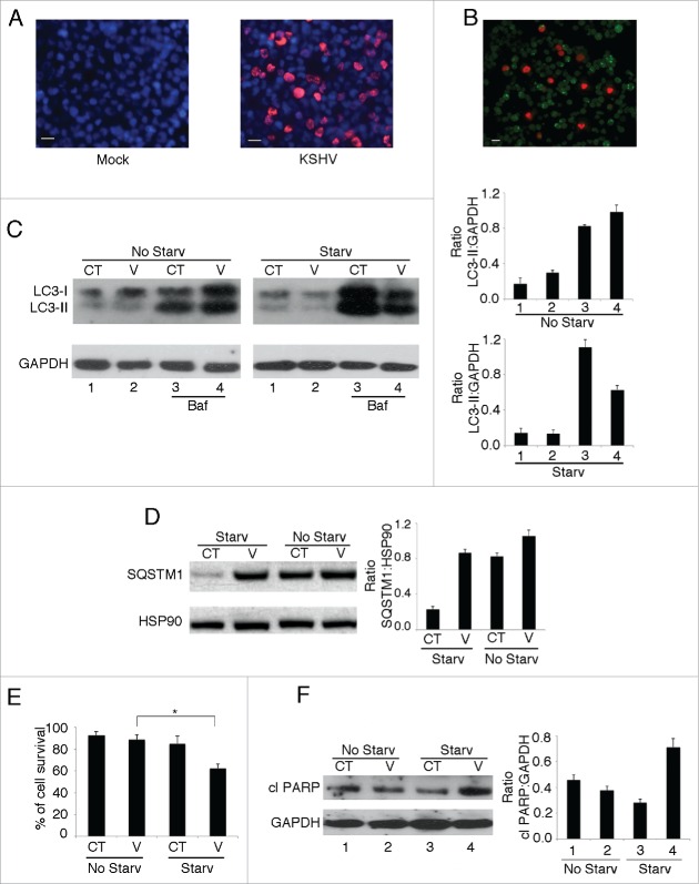
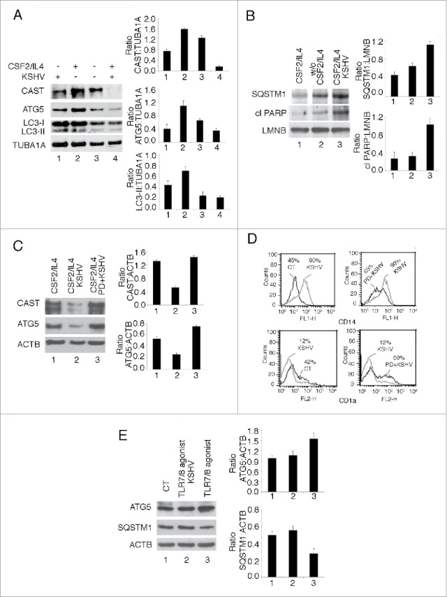
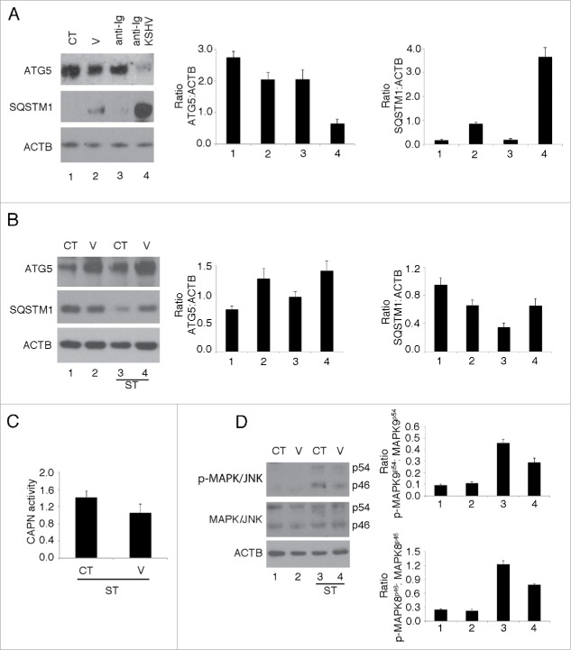
We have previously shown that Kaposi sarcoma-associated herpesvirus (KSHV) impairs monocyte differentiation into dendritic cells (DCs). Macroautophagy/autophagy has been reported to be essential in such a differentiating process. Here we extended these studies and found that the impairment of DC formation by KSHV occurs through autophagy inhibition. KSHV indeed reduces CAST (calpastatin) and consequently decreases ATG5 expression in both THP-1 monocytoid cells and primary monocytes. We unveiled a new mechanism put in place by KSHV to escape from immune control. The discovery of viral immune suppressive strategies that contribute to the onset and progression of viral-associated malignancies is of fundamental importance for finding new therapeutic approaches against them.

Keywords: ATG5; CAPN/calpain; CAST/calpastatin; KSHV; MAPK/JNK; SQSTM1/p62; THP-1; autophagy; dendritic cells; herpesvirus; monocytes.


Figures

Figure 1.
Figure 1.
KSHV infection reduces starvation-induced autophagy in THP-1 cells. (A) KSHV infection of THP-1 cells was revealed by the expression of K-bZIP early viral protein (red) by indirect immunofluorescence assay. After 30 h of infection, 30% of the cells were KSHV infected. Nuclei were stained by DAPI (blue) One experiment out of 3 is shown. (B) THP-1 cells, stably transfected with pEGFP-LC3 plasmid were infected by KSHV and starved for 6 h. Autophagy activation was revealed by the appearance of GFP-LC3 puncta (green) and the KSHV-infected cells by the expression of K-bZIP protein (red). (C) Western blot analysis showing a reduced expression level of the lipidated form of LC3 (LC3-II) in KSHV-infected (V) compared to mock-infected THP-1 cells (CT), after 6 h of starvation (Starv). The same experiment was performed also in the presence of bafilomycin A1 (BAF; 20 nM), added during the last 2 h of starvation. LC3-II expression was also analyzed in KSHV-infected (V) and mock-infected THP-1 (CT) cells, cultured in complete medium (No Starv), in the presence or absence of BAF. GAPDH was used as a loading control and a representative experiment out of 3 is shown. (D) SQSTM1 accumulation in KSHV-infected (V) or mock-infected THP-1 (CT), upon 6 h of starvation (Starv) or in complete medium (No Starv). HSP90 was used as a loading control and a representative experiment out of 3 is shown. (E) A reduction of cell survival, as evaluated by trypan blue exclusion, was observed in KSHV-infected (V) in comparison to mock-infected (CT) THP-1 cells, after 6 h of starvation (Starv). *p=0.03. Mean plus standard deviation (SD) is also reported. (F) Cleavage of PARP (cl PARP) as evaluated by immunoblot assay in KSHV-infected (V) compared to the mock-infected (CT) THP-1 cells, after 6 h of starvation or in complete medium (No Starv). GAPDH was used as a loading control and a representative experiment out of 3 is shown. Densitometric analysis was performed by calculating the ratio of specific proteins to the relative loading control in 3 different experiments. Bar: 10 µm.
Figure 2.
Figure 2.
KSHV infection of THP-1 cells reduces the level of CAST and ATG5 during starvation-induced autophagy. (A) The expression of ATG5 and CAST was analyzed by western blot in KSHV-infected (V) compared to the mock-infected (CT) THP-1 cells, after 6 h of starvation. ACTB was used as a loading control and a representative experiment out of 3 is shown. (B) The expression of BECN1 and ATG7 was analyzed by western blot in KSHV-infected (V) compared to the mock-infected (CT) THP-1 cells, after 6 h of starvation. GAPDH was used as a loading control and a representative experiment out of 3 is shown. (C) CAPN activity was measured in mock and infected THP-1 cells also in the presence of the PD150606 (PD) CAPN inhibitor. *p = 0.03; **p = 0.04. (D) ATG5 mRNA expression in mock and infected THP-1 cells, as measured by real-time RT-PCR. (E) THP-1 cells pretreated or not with the CAPN inhibitor PD150606 were infected with KSHV and starved for 6 h. Immunoblot analysis was performed to determine CAST, ATG5, SQSTM1 and cleaved BID (cl BID) expression levels. ACTB was used as a loading control and a representative experiment out of 3 is shown. Mock-infected THP-1 cells were used as a control. (F) Percentage of cell survival was evaluated by trypan-blue exclusion in mock or KSHV-infected cells in the presence or in the absence of PD150606. Mean plus standard deviation (SD) is reported. p = 0.02; ◊◊p = 0.03. (G) Western blot analysis showing ATG5, cl PARP and LC3-I/-II in THP-1 cells treated with scrambled siRNA (siRNA SC) or ATG5 siRNA (siRNA ATG5), upon 6 h starvation. HSP70 was used as a loading control and a representative experiment out of 3 is shown. Densitometric analysis was performed by calculating the ratio of specific proteins to the relative loading control in 3 different experiments.
Figure 3.
Figure 3.
MAPK9/JNK2 dephosphorylation is involved in CAST reduction mediated by KSHV in THP-1 cells. (A) KSHV-infected (V) or mock-infected (CT) THP-1 cells were starved for 6 h and phospho (p)-MAPK/JNK (p54 and p46 isoforms) and total MAPK/JNK (p54 and p46 isoforms) expression levels were evaluated by western blot analysis. GAPDH was used as a loading control and a representative experiment out of 3 is shown. Histograms represent the mean plus SD of the densitometric analysis of the ratio of p-MAPKp54:MAPKp54 and of p-MAPKp46:MAPKp46 in 3 different experiments. (B) THP-1 cells transfected with siRNA targeting MAPK8/JNK1 or (C) siRNA targeting MAPK9/JNK2 were starved for 6 h and ATG5, CAST, MAPK9/JNK2 and MAPK8/JNK1 levels were evaluated by immunoblot analysis in comparison to the scramble-treated control. GAPDH was used as loading control and a representative experiment out of 3 is shown. Histograms represent the mean plus SD of the densitometric analysis of the ratio of specific proteins to the GAPDH in 3 different experiments.
Figure 4.
Figure 4.
Autophagy and in vitro differentiation induced by TPA are impaired by KSHV infection in THP-1 cells. (A) THP-1 cells were infected with KSHV and treated for 48 h with TPA. LC3-I/-II and SQSTM1 expression was analyzed by western blotting. HSP70 was used as a loading control and a representative experiment out of 3 is shown. (B) THP-1 cells mock infected or infected with KSHV were treated with TPA for 24 or 48 h. CAST, ATG5 and SQSTM1 expression was analyzed by western blotting. GAPDH was used as a loading control and a representative experiment out of 3 is shown. (C) KSHV-infected or mock-infected THP-1 cells were treated with TPA for 48 h to induce cell differentiation. The effect of KSHV-infection on THP-1 differentiation was observed utilizing an optical microscope (40X magnification) or (D) by analyzing the expression level of the ITGAM/CD11b differentiation marker. Histograms represent the mean plus SD of the densitometric analysis of the ratio of specific proteins to the loading controls in 3 different experiments. Bar: 10 µm.
Figure 5.
Figure 5.
KSHV inhibits autophagy in human monocytes and impairs their in vitro differentiation and activation by downregulating ATG5 and reducing autophagy. (A) CAST, ATG5 and LC3-I/-II levels were evaluated by western blot analysis in KSHV- or mock-infected monocytes cultured in the presence or in the absence of CSF2 and IL4 for 48 h. TUBA1A was used as a loading control and a representative experiment out of 3 is shown. (B) Effect on SQSTM1 and PARP cleavage mediated by KSHV infection of monocytes, as determined by western blot analysis. LMNB/Lamin B was used as loading control and a representative experiment out of 3 is shown. (C) Western blot analysis of CAST and ATG5 expression in mock-infected or KSHV-infected differentiating monocytes in the presence or in the absence of PD150606. ACTB was used as a loading control and a representative experiment out of 3 is shown. (D) Monocytes mock-infected or infected with KSHV, in the presence or in the absence of PD150606, were cultured in complete medium containing CSF2 and IL4 for 48 h. The percentage of CD14- and CD1A-positive cells was then evaluated by FACS analysis. A representative experiment out of 3 is shown. (E) KSHV- or mock-infected monocytes were exposed to ORN R-2176-dT TLR7/8 agonist for 24 h. Subsequently, ATG5 and SQSTM1 were analyzed by western blot. Densitometric analysis was performed by calculating the ratio of specific proteins to the relative loading control of 3 different experiments.
Figure 6.
Figure 6.
KSHV reduces autophagy induction in primary B lymphocytes and HUVEC cells. (A) Purified B lymphocytes were mock (CT) or KSHV (V) exposed and treated with anti-human IgM for 24 h to induce BCR stimulation. ATG5 and SQSTM1 expression was analyzed by western blot. (B) HUVEC cells mock (CT) or KSHV-infected (V) were starved for 6 h and ATG5 and SQSTM1 expression was then analyzed by western blot. (C) CAPN activity was measured in mock and KSHV-infected HUVEC cells during starvation. (D) Mock (CT) or KSHV-infected (V) HUVEC cells were starved for 6 h and phosphorylated and total MAPK9/JNK2 expression levels were evaluated by western blot analysis. Densitometric analysis was performed by calculating the ratio of specific proteins to the ACTB loading control in 3 different experiments.

References

    1. Jin M, Klionsky DJ. Regulation of autophagy: modulation of the size and number of autophagosomes. FEBS Lett 2014; 588:2457-63; PMID:24928445; http://dx.doi.org/ 10.1016/j.febslet.2014.06.015 - DOI - PMC - PubMed
    1. Parzych KR, Klionsky DJ. An overview of autophagy: morphology, mechanism, and regulation. Antioxid Redox Signal 2014; 20:460-73; PMID:23725295; http://dx.doi.org/ 10.1089/ars.2013.5371 - DOI - PMC - PubMed
    1. Galluzzi L, Pietrocola F, Levine B, Kroemer G. Metabolic control of autophagy. Cell 2014; 159:1263-76; PMID:25480292; http://dx.doi.org/ 10.1016/j.cell.2014.11.006 - DOI - PMC - PubMed
    1. Feng Y, He D, Yao Z, Klionsky DJ. The machinery of macroautophagy. Cell Res 2014; 24:24-41; PMID:24366339; http://dx.doi.org/ 10.1038/cr.2013.168 - DOI - PMC - PubMed
    1. Levine B, Kroemer G. Autophagy in the pathogenesis of disease. Cell 2008; 132:27-42; PMID:18191218; http://dx.doi.org/ 10.1016/j.cell.2007.12.018 - DOI - PMC - PubMed

Publication types

MeSH terms

LinkOut - more resources