

The impact of autophagy on the development of senescence in primary tubular epithelial cells
- PMID: 27715411
- PMCID: PMC5105933
- DOI: 10.1080/15384101.2016.1234547
The impact of autophagy on the development of senescence in primary tubular epithelial cells
Abstract
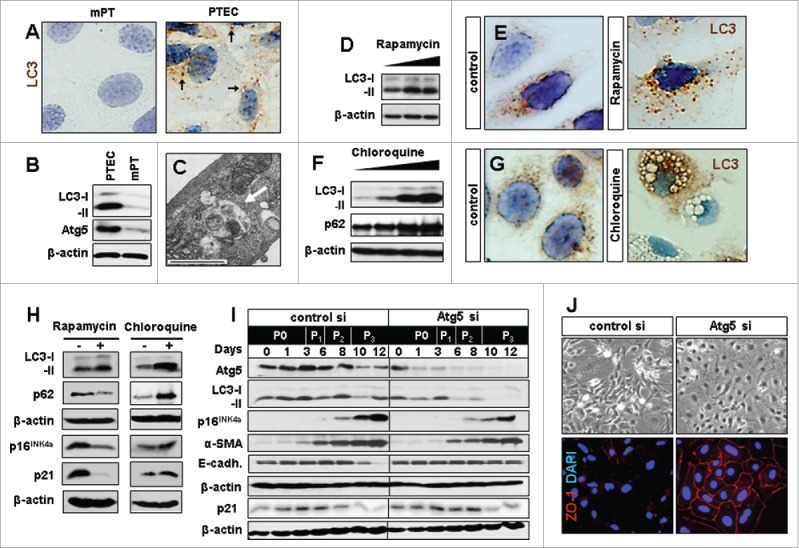
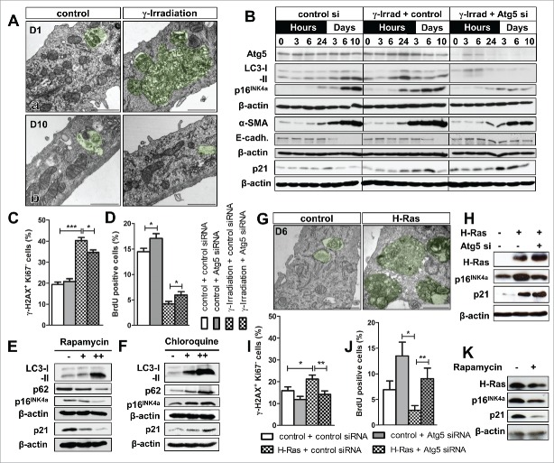
Autophagy and senescence are 2 distinct pathways that are importantly involved in acute kidney injury and renal repair. Recent data indicate that the 2 processes might be interrelated. To investigate the potential link between autophagy and senescence in the kidney we isolated primary tubular epithelial cells (PTEC) from wild-type mice and monitored the occurrence of cellular senescence during autophagy activation and inhibition. We found that the process of cell isolation and transfer into culture was associated with a strong basal autophagic activation in PTEC. Specific inhibition of autophagy by silencing autophagy-related 5 (Atg5) counteracted the occurrence of senescence hallmarks under baseline conditions. Reduced senescent features were also observed in Atg5 silenced PTEC after γ-irradiation and during H-Ras induced oncogenic senescence, but the response was less uniform in these stress models. Senescence inhibition was paralleled by better preservation of a mature epithelial phenotype in PTEC. Interestingly, treatment with rapamycin, which acts as an activator of autophagy, also counteracted the occurrence of senescence features in PTEC. While we interpret the anti-senescent effect of rapamycin as an autophagy-independent effect of mTOR-inhibition, the more specific approach of Atg5 silencing indicates that overactivated autophagy can have pro-senescent effects in PTEC. These results highlight the complex interaction between cell culture dependent stress mechanisms, autophagy and senescence.
Keywords: Autophagy; chloroquine; p16INK4a; rapamycin; senescence; tubular epithelial cells.
Figures
References
-
- Takahashi A, Kimura T, Takabatake Y, Namba T, Kaimori J, Kitamura H, Matsui I, Niimura F, Matsusaka T, Fujita N, et al.. Autophagy guards against cisplatin-induced acute kidney injury. Am J Pathol 2012; 180:517-25; PMID:22265049; http://dx.doi.org/10.1016/j.ajpath.2011.11.001 - DOI - PubMed
-
- Kimura T, Takabatake Y, Takahashi A, Kaimori JY, Matsui I, Namba T, Kitamura H, Niimura F, Matsusaka T, Soga T, et al.. Autophagy protects the proximal tubule from degeneration and acute ischemic injury. J Am Soc Nephrol 2011; 22:902-13; PMID:21493778; http://dx.doi.org/10.1681/ASN.2010070705 - DOI - PMC - PubMed
-
- Kaushal GP, Shah SV. Autophagy in acute kidney injury. Kidney Int 2016; 89:779-91; PMID:26924060; http://dx.doi.org/10.1016/j.kint.2015.11.021 - DOI - PMC - PubMed
-
- Jiang M, Wei Q, Dong G, Komatsu M, Su Y, Dong Z. Autophagy in proximal tubules protects against acute kidney injury. Kidney Int 2012; 82:1271-83; PMID:22854643; http://dx.doi.org/10.1038/ki.2012.261 - DOI - PMC - PubMed
-
- Childs BG, Durik M, Baker DJ, van Deursen JM. Cellular senescence in aging and age-related disease: from mechanisms to therapy. Nat Med 2015; 21:1424-35; PMID:26646499; http://dx.doi.org/10.1038/nm.4000 - DOI - PMC - PubMed
MeSH terms
Substances
LinkOut - more resources
Full Text Sources
Other Literature Sources
Research Materials
Miscellaneous