. 2016 Oct 4;13(1):259.
doi: 10.1186/s12974-016-0728-y.

The antidepressant-like effects of pioglitazone in a chronic mild stress mouse model are associated with PPARγ-mediated alteration of microglial activation phenotypes

Affiliations

The antidepressant-like effects of pioglitazone in a chronic mild stress mouse model are associated with PPARγ-mediated alteration of microglial activation phenotypes

Qiuying Zhao et al. J Neuroinflammation. .

Abstract

Background: Discoveries that microglia-mediated neuroinflammation is involved in the pathological process of depression provided a new strategy for novel antidepressant therapy. Peroxisome proliferator-activated receptor γ (PPARγ) is a nuclear receptor regulating inflammation and microglial polarization and, therefore, a potential target for resolving depressive disorders. Our hypothesis was that antidepressant effects could be achieved through anti-inflammatory and neuroprotective activities by PPARγ-dependent microglia-modulating agents.

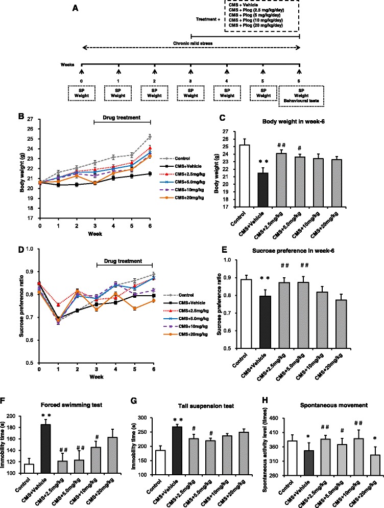
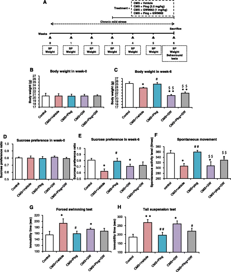
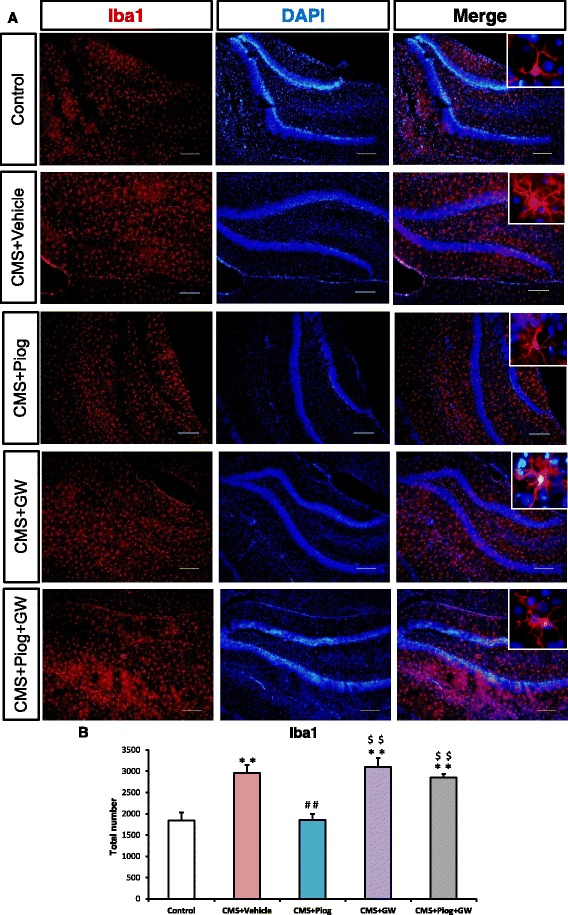
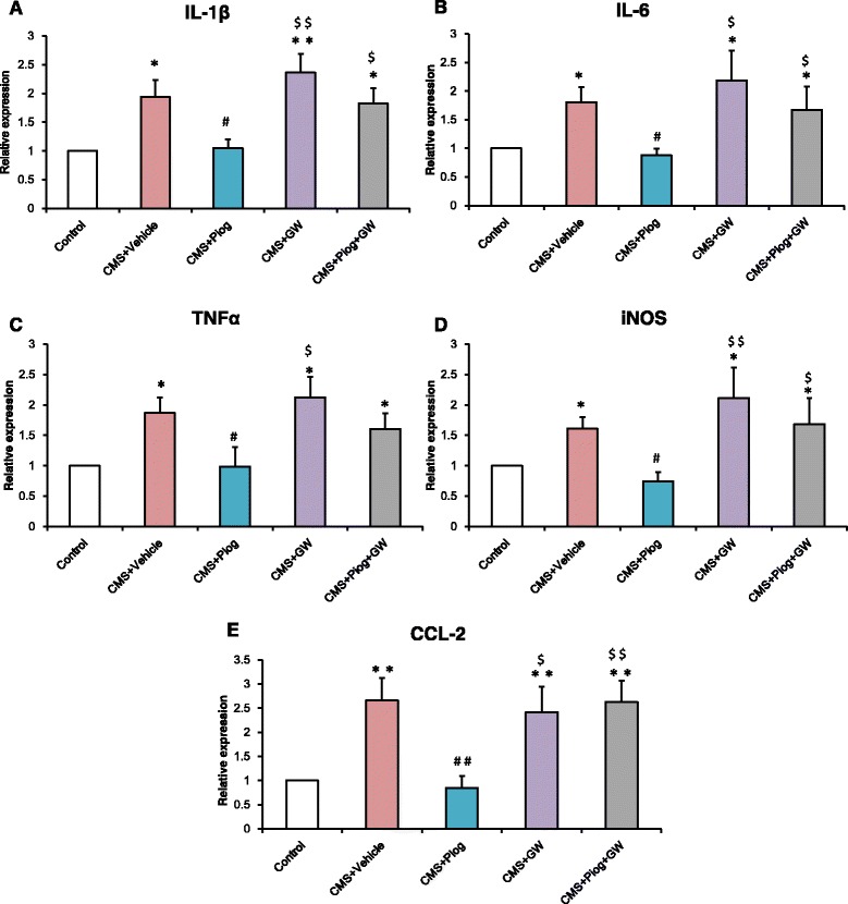
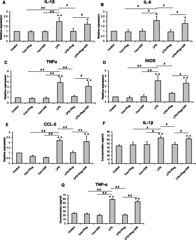
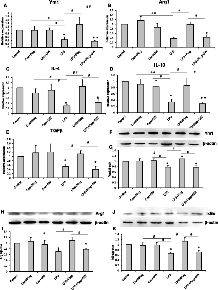
Methods: Chronic mild stress (CMS) treatment was performed on C57BL/6 mice for 6 weeks. After 3 weeks with the CMS procedure, depressive-like behaviors were evaluated by sucrose preference (SP), tail suspension test (TST), forced swimming test (FST), and locomotor activity. Pioglitazone was administered intragastrically once per day for 3 weeks at different doses. Neuroinflammatory cytokines were determined by real time-PCR (RT-PCR), enzyme-linked immunosorbent assay (ELISA), and western blot. The activated microglial state was confirmed by immunohistochemistry. N9 microglial cells were subjected to lipopolysaccharide, pioglitazone, and GW9662 to discuss the phenotype of activated microglia by RT-PCR, ELISA, and western blot.

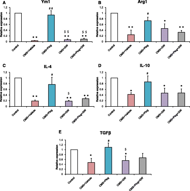
Results: It was demonstrated that the PPARγ agonist pioglitazone (2.5 mg/kg) ameliorated depression-like behaviors in CMS-treated mice, as indicated by body weight (BW), the SP test, the FST, and the TST. The amelioration of the depression was blocked by the PPARγ antagonist GW9662. The expression of M1 markers (IL-1β, IL-6, TNFα, iNOS, and CCL2) increased, and the gene expression of M2 markers (Ym1, Arg1, IL-4, IL-10, and TGFβ) decreased in the hippocampus of the stress-treated mice. Pioglitazone significantly inhibited the increased numbers and morphological alterations of microglia in the hippocampus, reduced the elevated expression of microglial M1 markers, and increased the downgraded expression of microglial M2 markers in C57BL/6 mice exposed to CMS. In an in vitro experiment, pioglitazone reversed the imbalance of M1 and M2 inflammatory cytokines, which is correlated with the inhibition of nuclear factor kB activation and is expressed in LPS-stimulated N9 microglial cells.

Conclusions: We showed that pioglitazone administration induce the neuroprotective phenotype of microglia and ameliorate depression-like behaviors in CMS-treated C57BL/6 mice. These data suggested that the microglia-modulating agent pioglitazone present a beneficial choice for depression.

Keywords: Alternative activation; Antidepressant; CMS; Cytokine; Microglia; PPARγ; Pioglitazone.


Figures

Fig. 1
Fig. 1
Effects of pioglitazone at different doses on CMS mice. a Schematic diagram of the experimental design. BW was measured every week (b). The 2.5 mg/kg dose showed the best result in recovering the BW in the CMS mice (c, n = 10). Anhedonia was detected by SP over the course of the 6-week-long experiment (d, e, n = 10). The duration of immobility in the FST (f) and TST (g) increased after CMS induction at the sixth week (n = 8 in both tests). Pioglitazone (2.5 mg/kg) reduced this increase. In the locomotor activity test, pioglitazone improved the spontaneous activity level after CMS induction (h, n = 8). * p < 0.05, * * p < 0.01 vs. Control; # p < 0.05, ## p < 0.01 vs. CMS + Vehicle. Data are expressed as means ± SEM
Fig. 2
Fig. 2
The activity of PPARγ affected the improvement of depression-like behaviors. a The principle scheme of the experimental project. BW at the baseline level (b) and the sixth week (c) (n = 10). SP showed no differences at week 0 (d). Pioglitazone improved the ratio of the SP in CMS mice but anhedonia was maintained with the antagonist of PPARγ, GW9662, administration (e, n = 10). Spontaneous activity was modulated by pioglitazone and GW9662 (f, n = 8). Pioglitazone decreased the time of immobility, but GW9662 increased the immobility duration in the FST and TST (g, h, n = 8 in both tests). * p < 0.05, ** p < 0.01 vs. Control; # p < 0.05, ## p < 0.01 vs. CMS + Vehicle; $$ p < 0.01 vs. CMS + Piog. Data are expressed as means ± SEM
Fig. 3
Fig. 3
Microglial activated status was influenced by PPARγ in the hippocampus. Pioglitazone decreased the number of Iba1+ microglia compared with the CMS mice. The Iba1+ microglial morphology was amoeboid, and the number of microglia increased in the animals after GW9662 treatment. Administration of pioglitazone and GW9662 had no effect on the microglia (b, n = 5). Enlarged figures indicate typical microglia (a). Scale bars: 10 μm. ** p < 0.01 vs. Control; ## p < 0.01 vs. CMS + Vehicle; $$ p < 0.01 vs. CMS + Piog. Data are expressed as means ± SEM
Fig. 4
Fig. 4
Effect of pioglitazone treatment on microglia-inhibited M1 activation in the DG of the hippocampus. The expression of M1 markers IL-1β (a), IL-6 (b), TNFα (c), iNOS (d), and CCL2 (e) increased; pioglitazone reduced this increase and GW9662 aggravated this increase in the CMS mice. n = 5. * p < 0.05, ** p < 0.01 vs. Control; # p < 0.05, ## p < 0.01 vs. CMS + Vehicle; $ p < 0.05, $$ p < 0.01 vs. CMS + Piog. Data are expressed as means ± SEM
Fig. 5
Fig. 5
Effect of pioglitazone treatment on the microglial shift toward an M2 phenotype. The mRNA expression of M2: Ym1 (a), Arg1 (b), IL-4 (c), IL-10 (d), and TGFβ (e) reduced with a 6-week duration of CMS stress. The decreases were attenuated by pioglitazone administration. GW9662 still reduced the expression of these markers. Pioglitazone and GW9662 treated together had no effect on the M2 level of CMS mice. n = 5. * p < 0.05, ** p < 0.01 vs. Control; # p < 0.05, ## p < 0.01 vs. CMS + Vehicle; $ p < 0.05, $$ p < 0.01 vs. CMS + Piog. Data are expressed as means ± SEM
Fig. 6
Fig. 6
LPS-induced M1 polarization was alleviated by the PPARγ agonist pioglitazone in N9 microglial cells. LPS increased the level of IL-1β (a), IL-6 (b), TNFα (c), iNOS (d), and CCL2 (e), but this was reversed by pioglitazone. The PPARγ antagonist GW9662 inhibited the effect of pioglitazone on the PPARγ of microglia. The protein expression of IL-1β (f) and TNFα (g) was detected by ELISA. * p < 0.05, ** p < 0.01 vs. Control; # p < 0.05, ## p < 0.01. Data are expressed as means ± SEM
Fig. 7
Fig. 7
The M2 molecules: Ym1 (a), Arg1 (b), IL-4 (c), IL-10 (d), and TGFβ (e) decreased with LPS treatment. Pioglitazone increased the expression of the M2 markers. The impact on the LPS+Piog+GW group was similar to that of the LPS microglial cells. The level of Ym1 (f & g) and Arg1 (h & i) was also confirmed by western blot. The protein expression of IκBα decreased in LPS-stimulated cells; pioglitazone significantly ameliorated the expression. GW9662 suppressed the rise after pioglitazone-treated LPS in microglia (j & k).* p < 0.05, ** p < 0.01 vs. Control; # p < 0.05, ## p < 0.01. Data are expressed as means ± SEM

References

    1. Centers for Disease Control and Prevention (CDC). Current depression among adults---United States, 2006 and 2008. MMWR Morb Mortal Wkly Rep. 2010;59:1229–1235. - PubMed
    1. Rush AJ, Trivedi MH, Wisniewski SR, Nierenberg AA, Stewart JW, Warden D, Niederehe G, Thase ME, Lavori PW, Lebowitz BD, et al. Acute and longer-term outcomes in depressed outpatients requiring one or several treatment steps: a STAR*D report. Am J Psychiatry. 2006;163:1905–17. doi: 10.1176/ajp.2006.163.11.1905. - DOI - PubMed
    1. Anderson HD, Pace WD, Libby AM, West DR, Valuck RJ. Rates of 5 common antidepressant side effects among new adult and adolescent cases of depression: a retrospective US claims study. Clin Ther. 2012;34:113–23. doi: 10.1016/j.clinthera.2011.11.024. - DOI - PubMed
    1. Fonseka TM, McIntyre RS, Soczynska JK, Kennedy SH. Novel investigational drugs targeting IL-6 signaling for the treatment of depression. Expert Opin Investig Drugs. 2015;24:459–75. doi: 10.1517/13543784.2014.998334. - DOI - PubMed
    1. Raison CL, Capuron L, Miller AH. Cytokines sing the blues: inflammation and the pathogenesis of depression. Trends Immunol. 2006;27:24–31. doi: 10.1016/j.it.2005.11.006. - DOI - PMC - PubMed

MeSH terms