. 2016 Oct 4;11(1):132.
doi: 10.1186/s13014-016-0709-4.

Precise film dosimetry for stereotactic radiosurgery and stereotactic body radiotherapy quality assurance using Gafchromic™ EBT3 films

Affiliations

Precise film dosimetry for stereotactic radiosurgery and stereotactic body radiotherapy quality assurance using Gafchromic™ EBT3 films

Ning Wen et al. Radiat Oncol. .

Abstract

Purpose: The purpose of this study is to evaluate the dosimetric uncertainty associated with Gafchromic™ (EBT3) films and establish a practical and efficient film dosimetry protocol for Stereotactic Radiosurgery (SRS) and Stereotactic Body Radiotherapy (SBRT).

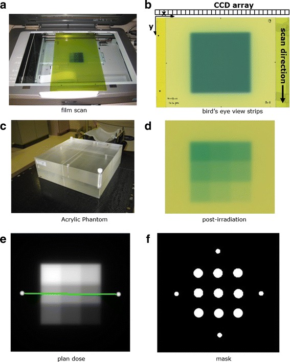
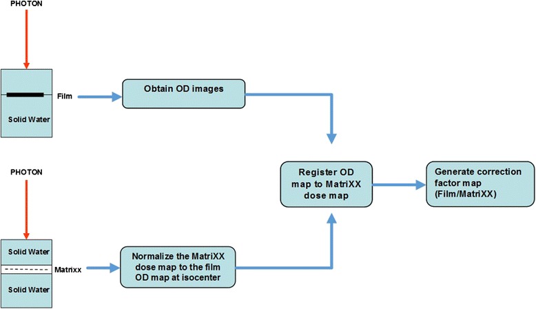
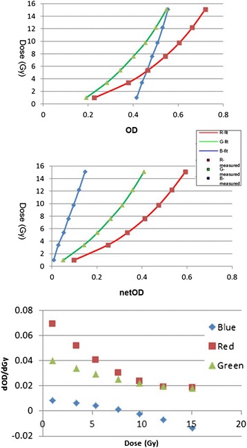
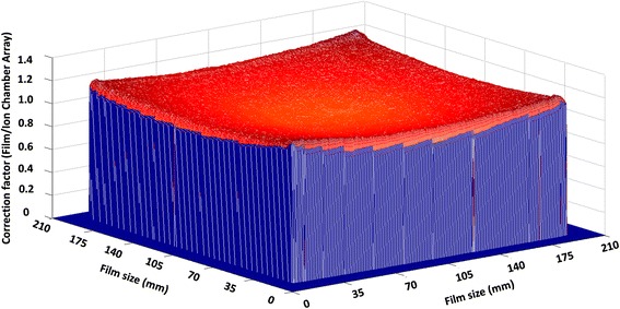
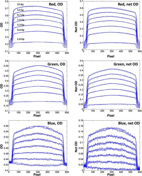
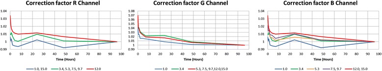
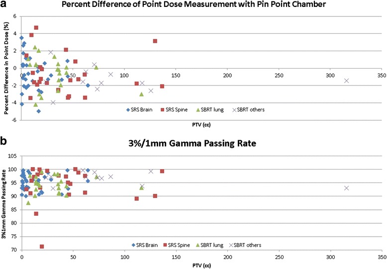
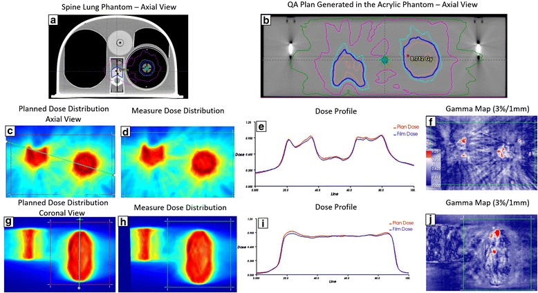
Method and materials: EBT3 films were irradiated at each of seven different dose levels between 1 and 15 Gy with open fields and standard deviations of dose maps were calculated at each color channel for evaluation. A scanner non-uniform response correction map was built by registering and comparing film doses to the reference ion chamber array-based dose map delivered with the same doses. To determine the temporal dependence of EBT3 films, the average correction factors of different dose levels as a function of time were evaluated up to 4 days after irradiation. An integrated film dosimetry protocol was developed for dose calibration, calibration curve fitting, dose mapping, and profile/gamma analysis. Patient specific quality assurance (PSQA) was performed for 83 SRS/SBRT treatment plans, and analysis of the measurements and calculations are presented here.

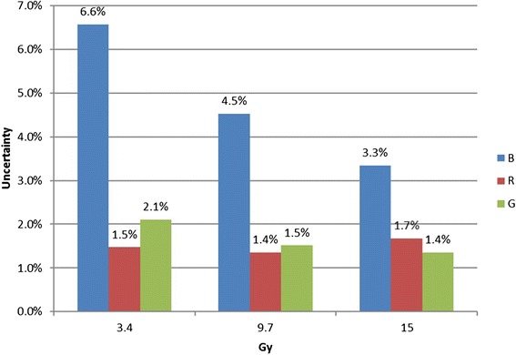
Results: The scanner response varied within 1 % for the field sizes less than 5 × 5 cm2, and up to 5 % for the field sizes of 10 × 10 cm2 for all color channels. The scanner correction method was able to remove visually evident, irregular detector responses for larger field sizes. The dose response of the film changed rapidly (~10 %) in the first two hours and became smooth plateaued afterwards, ~3 % change between 2 and 24 h. The uncertainties were approximately 1.5, 1.7 and 4.8 % over the dose range of 3~15 Gy for the red, green and blue channels. The green channel showed very high sensitivity and low uncertainty in the dose range between 10 and 15 Gy, which is suitable for SRS/SBRT commissioning and PSQA. The difference between the calculated dose and measured dose of ion chamber measurement at isocenter was -0.64 ± 2.02 for all plans, corresponding to a 95 % confidence interval of (-1.09, -0.26). The percentage of points passing the 3 %/1 mm gamma criteria in absolute dose, averaged over all tests was 95.0 ± 4.2.

Conclusion: We have developed the EBT3 films based dosimetry protocol to obtain absolute dose values. The overall uncertainty has been established to be 1.5 % for SRS and SBRT PSQA.

Keywords: Gafchromic films; Quality assurance; Stereotactic body radiation therapy; Stereotactic radiosurgery.


Figures

Fig. 1
Fig. 1
a film scanning orientation with the longer side of film aligned to the scanner detector array. b A bird’s eye view of film scanning orientation with film strips attached for positioning accuracy. c Acrylic phantom for film dose calibration. d A calibration film irradiated with a nine 2 × 2 cm2 square dose pattern. e The corresponding planar isodose distribution. f A mask pattern used to sample optical density values to establish the calibration curve
Fig. 2
Fig. 2
Scanner non-uniform response correction flowchart. The films were irradiated in a solid water phantom at 5 cm depth and the dose distribution was measured with MatriXX at the same setup. The OD images obtained from the films were registered to the normalized MatriXX dose map. The scanner correction map was generated by taking the ratio of the film OD map to MatriXX dose map
Fig. 3
Fig. 3
Dose response curves of OD (upper) and netOD methods (middle) and the sensitivity as a function of dose (lower). Cubic B-spline was employed as interpolant (R2 = 1 for all the curves)
Fig. 4
Fig. 4
The 3D scanner non-uniform correction map of the 20 × 20 cm2 field size at the 12 Gy dose level for the green channel. The vertical scale is normalized to 12 Gy
Fig. 5
Fig. 5
Horizontal (orthogonal to scan direction) OD and netOD profiles for dose levels of 1.0, 3.4, 5.3, 7.5, 9.7 12.0 and 15.0 Gy. The rows from top correspond respectively to the red, green, and blue channels. Each line represents a profile from one of the four film images
Fig. 6
Fig. 6
The uncertainty at the three dose levels for the red, green and blue channel
Fig. 7
Fig. 7
The average correction factors of different dose levels as a function of time, normalized at 4 days after exposure. The starting time of each scanning was recorded in hours
Fig. 8
Fig. 8
a Percent point dose difference between measured (pin point chamber) and calculated dose (Eclipse); b The percentage of points passing the gamma criteria of 3 %/1 mm using films based on absolute dose comparison
Fig. 9
Fig. 9
The upper row: a the spine and lung phantom from IROC for single ISO multiple target irradiation for NRG BR001 and BR002 protocol; b Planned axial dose distribution at central level of 6XFFF Hard C-Shape plan using VMAT technique on the acrylic phantom; The middle row: (c-d) Planned and measured dose distribution in the axial plane; e The dose profile comparison along the oblique line across both spine and lung targets shown in (c); f film gamma analysis results with 3 %/1 mm criteria (99.4 % of pixels passed within region of interest shown in (c); The lower row: The same comparisons in the coronal view. The film gamma pass rate with 3 %/1 mm criteria was 98.9 %

References

    1. Kragl G, af Wetterstedt S, Knausl B, Lind M, McCavana P, Knoos T, et al. Dosimetric characteristics of 6 and 10MV unflattened photon beams. Radiother Oncol. 2009;93:141–146. doi: 10.1016/j.radonc.2009.06.008. - DOI - PubMed
    1. Vassiliev ON, Titt U, Ponisch F, Kry SF, Mohan R, Gillin MT. Dosimetric properties of photon beams from a flattening filter free clinical accelerator. Phys Med Biol. 2006;51:1907–1917. doi: 10.1088/0031-9155/51/7/019. - DOI - PubMed
    1. Ponisch F, Titt U, Vassiliev ON, Kry SF, Mohan R. Properties of unflattened photon beams shaped by a multileaf collimator. Med Phys. 2006;33:1738–1746. doi: 10.1118/1.2201149. - DOI - PubMed
    1. Poppe B, Djouguela A, Blechschmidt A, Willborn K, Ruhmann A, Harder D. Spatial resolution of 2D ionization chamber arrays for IMRT dose verification: single-detector size and sampling step width. Phys Med Biol. 2007;52:2921–2935. doi: 10.1088/0031-9155/52/10/019. - DOI - PubMed
    1. Spezi E, Angelini AL, Romani F, Ferri A. Characterization of a 2D ion chamber array for the verification of radiotherapy treatments. Phys Med Biol. 2005;50:3361–3373. doi: 10.1088/0031-9155/50/14/012. - DOI - PubMed

LinkOut - more resources