Review
. 2017 Jan;31(1):10-25.
doi: 10.1038/eye.2016.216. Epub 2016 Oct 7.

Structural and molecular changes in the aging choroid: implications for age-related macular degeneration

Affiliations
Review

Structural and molecular changes in the aging choroid: implications for age-related macular degeneration

K R Chirco et al. Eye (Lond). 2017 Jan.

Abstract

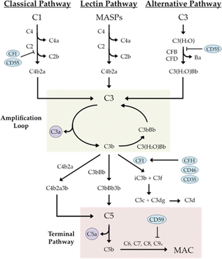
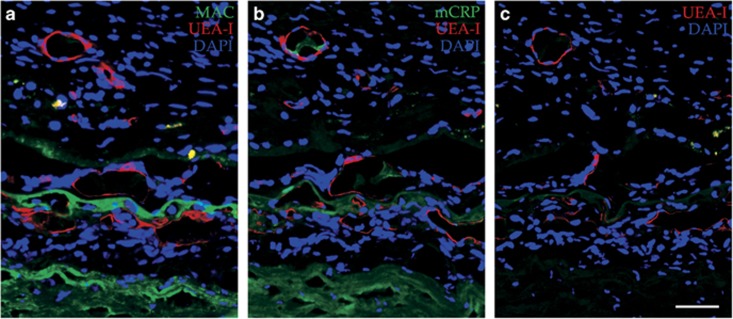
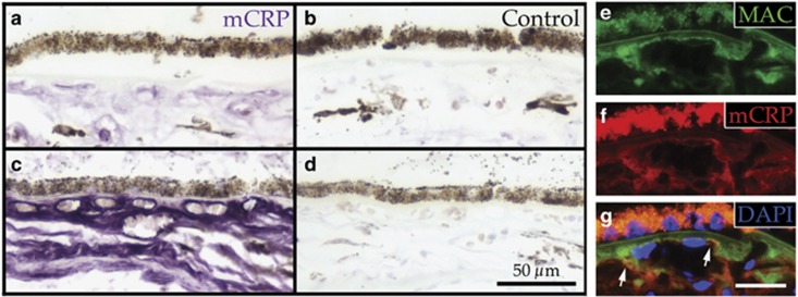
Age-related macular degeneration (AMD) is a devastating disease-causing vision loss in millions of people around the world. In advanced stages of disease, death of photoreceptor cells, retinal pigment epithelial cells, and choroidal endothelial cells (CECs) are common. Loss of endothelial cells of the choriocapillaris is one of the earliest detectable events in AMD, and, because the outer retina relies on the choriocapillaris for metabolic support, this loss may be the trigger for progression to more advanced stages. Here we highlight evidence for loss of CECs, including changes to vascular density within the choriocapillaris, altered abundance of CEC markers, and changes to overall thickness of the choroid. Furthermore, we review the key components and functions of the choroid, as well as Bruch's membrane, both of which are vital for healthy vision. We discuss changes to the structure and molecular composition of these tissues, many of which develop with age and may contribute to AMD pathogenesis. For example, a crucial event that occurs in the aging choriocapillaris is accumulation of the membrane attack complex, which may result in complement-mediated CEC lysis, and may be a primary cause for AMD-associated choriocapillaris degeneration. The actions of elevated monomeric C-reactive protein in the choriocapillaris in at-risk individuals may also contribute to the inflammatory environment in the choroid and promote disease progression. Finally, we discuss the progress that has been made in the development of AMD therapies, with a focus on cell replacement.


Figures

Figure 1
Figure 1
Layers of the neural retina, RPE, and choroid. H&E (a), immunofluorescence (b), and TEM (c) images showing the layers of the neural retina (cones labeled with calbindin in green), retinal pigment epithelium (yellow), and choroid (vessels labeled with CD34 in red). (b) Immunofluorescence was carried out using anti-calbindin (green) and anti-CD34 (red) antibodies to visualize cone photoreceptor cells and endothelial cells, respectively. Section was counterstained with 4',6-diamidino-2-phenylindole (DAPI; blue). GCL, ganglion cell layer; IPL, inner plexiform layer; INL, inner nuclear layer; OPL, outer plexiform layer; ONL, outer nuclear layer; IS, inner segment; OS, outer segment; RPE, retinal pigment epithelium; BrM, Bruch's membrane; CC, choriocapillaris; CH, choroid. Scale bars in a and b=50 μm, scale bar in c=5 μm.
Figure 2
Figure 2
Ultrastructure of BrM. TEM image shows the basal side of the retinal pigment epithelium, all layers of BrM (1=retinal pigment epithelium basement membrane; 2=inner collagenous layer; 3=elastic layer, asterisks; 4=outer collagenous layer; 5=choriocapillary endothelial cell basement membrane), and the apical side of the choriocapillaris with fenestrations (arrowheads). RPE=retinal pigment epithelium; BrM=Bruch's membrane; CC=choriocapillaris. Scale bar=1 μm.
Figure 3
Figure 3
Drusen as a site for CNV invasion. Immunofluorescence image from a human donor with wet AMD exhibits vessels (Ulex europaeus agglutinin I, UEA-I; red; asterisks) present within the druse deposit, who also contains high levels of the MAC (green). Section was counterstained with DAPI (blue). Scale bar=50 μm.
Figure 4
Figure 4
Ghost vessels in the choriocapillaris. Choroidal endothelial cell loss (ghost vessels; asterisks) in a 96-year-old donor with atrophic AMD is indicated by loss of UEA-I lectin labeling (red). The MAC (green) is present throughout the choriocapillaris. The section was counterstained with DAPI (blue). ONL, outer nuclear layer; RPE, retinal pigment epithelium; CC, choriocapillaris. Scale bar=50 μm.
Figure 5
Figure 5
Overview of the complement system. Complement regulators are depicted as blue ovals and anaphylatoxins are presented as purple circles. The alternative pathway amplification loop is highlighted in green and the common terminal pathway is highlighted in red.
Figure 6
Figure 6
Membrane attack complex and monomeric C-reactive protein accumulation in wet AMD. An anti-C5b-9 antibody directed against the MAC (a, green), and an anti-mCRP antibody (b, green) were utilized in sections from a wet AMD donor. A secondary control shows UEA-I lectin only (c, red). All sections were counterstained with DAPI. Scale bar=50 μm.
Figure 7
Figure 7
Monomeric C-reactive protein is elevated in the choriocapillaris of high-risk donor eyes and colocalizes with the membrane attack complex. mCRP is more abundant in the choriocapillaris of a donor homozygous for the CFH Y402H polymorphism (c; purple), compared with a control donor (a; purple). Secondary controls for each donor are shown in b and d. An anti-C5b-9 antibody (e and g; green) and an anti-mCRP antibody (f and g; red) display regions of co-labeling at the edge of the vessels within the choriocapillaris (g; arrows). DAPI was used as a counterstain in g. Scale bar in d=50 μm, scale bar in g=25 μm.

References

    1. Friedman DS, O'Colmain BJ, Muñoz B, Tomany SC, McCarty C, de Jong PTVM et al. Prevalence of age-related macular degeneration in the United States. Arch Ophthalmol 2004; 122(4): 564–572. - PubMed
    1. Wong WL, Su X, Li X, Cheung CMG, Klein R, Cheng C-Y et al. Global prevalence of age-related macular degeneration and disease burden projection for 2020 and 2040: a systematic review and meta-analysis. Lancet Global Health 2014; 2(2): 106–116. - PubMed
    1. Folk JC, Stone EM. Ranibizumab therapy for neovascular age-related macular degeneration. N Engl J Med 2010; 363(17): 1648–1655. - PubMed
    1. Wangsa-Wirawan ND, Linsenmeier RA. Retinal oxygen: fundamental and clinical aspects. Arch Ophthalmol 2003; 121(4): 547–557. - PubMed
    1. Feeney-Burns L, Burns RP, Gao C-L. Age-related macular changes in humans over 90 years old. Am J Ophthalmol 1990; 109(3): 265–278. - PubMed