. 2016 Oct 7;11(10):e0164179.
doi: 10.1371/journal.pone.0164179. eCollection 2016.

Signaling Properties of Chemerin Receptors CMKLR1, GPR1 and CCRL2

Affiliations

Signaling Properties of Chemerin Receptors CMKLR1, GPR1 and CCRL2

Olivier De Henau et al. PLoS One. .

Abstract

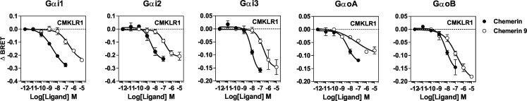
Chemerin is a small chemotactic protein originally identified as the natural ligand of CMKLR1. More recently, two other receptors, GPR1 and CCRL2, have been reported to bind chemerin but their functional relevance remains poorly understood. In this study, we compared the binding and signaling properties of the three human chemerin receptors and showed differences in mode of chemerin binding and receptor signaling. Chemerin binds to all three receptors with low nanomolar affinities. However, the contribution of the chemerin C-terminus to binding efficiency varies greatly amongst receptors. By using BRET-based biosensors monitoring the activation of various G proteins, we showed that binding of chemerin and the chemerin 9 nonapeptide (149YFPGQFAFS157) to CMKLR1 activates the three Gαi subtypes (Gαi1, Gαi2 and Gαi3) and the two Gαo isoforms (Gαoa and Gαob) with potencies correlated to binding affinities. In contrast, no significant activation of G proteins was detected upon binding of chemerin to GPR1 or CCRL2. Binding of chemerin and the chemerin 9 peptide also induced the recruitment of β-arrestin1 and 2 to CMKLR1 and GPR1, though to various degree, but not to CCRL2. However, the propensity of chemerin 9 to activate β-arrestins relative to chemerin is higher when bound to GPR1. Finally, we showed that binding of chemerin to CMKLR1 and GPR1 promotes also the internalization of the two receptors and the phosphorylation of ERK1/2 MAP kinases, although with a different efficiency, and that phosphorylation of ERK1/2 requires both Gαi/o and β-arrestin2 activation but not β-arrestin1. Collectively, these data support a model in which each chemerin receptor displays selective signaling properties.


Conflict of interest statement

The authors have declared that no competing interests exist.

Figures

Fig 1
Fig 1. Chemerin binding assay.
A-C. Saturation binding assay using CHO-K1 cells expressing CMKLR1 (A), GPR1 (B) or CCRL2 (C) that were incubated with increasing concentrations of [125I]-chemerin (total binding, ●). Non-specific binding (◯) was determined in the presence of a 100-fold excess of unlabeled chemerin and specific binding (■) was calculated as the difference. D-F. Competition binding assay using CHO-K1 cells expressing CMKLR1 (D), GPR1 (E) or CCRL2 (F) that were incubated with 0.1 nM [125I]-chemerin as tracers and increasing concentrations of unlabelled chemerin (●) or the nonapeptide chemerin 9 (◯) as competitors. The data were normalized for nonspecific binding (0%) in the presence of 300 nM chemerin, and specific binding in the absence of competitor (100%). The displayed data represent the mean ± S.E.M. of three independent experiments.
Fig 2
Fig 2. Peptide binding assay.
A CHO-K1 cells expressing CMKLR1, GPR1 or CCRL2 were incubated with 0.1 nM [125I]-[145–157]-chemerin only (TOTAL) or 0.1 nM [125I]-[145–157]-chemerin in combination with an excess of chemerin or chemerin 9 as competitors. B. CHO-K1 cells expressing CMKLR1 or GPR1 were incubated with 0.1 nM [125I]-[145–157]-chemerin as tracer and increasing concentrations of unlabelled chemerin (●) or chemerin 9 (◯) as competitors. The data were normalized for nonspecific binding (0%) in the presence of 300 nM chemerin, and specific binding in the absence of competitor (100%). The displayed data represent the mean ± S.E.M. of three independent experiments.
Fig 3
Fig 3. Chemokine binding to CCRL2.
CHO-K1 cells expressing CCRL2 were incubated with 0.1 nM [125I]-chemerin (A) or 0.1 nM [125I]-CCL19 (B) in combination or not with an excess of unlabeled chemerin, CCL5, CCL19 or CCL21 (300nM). All points were run in triplicates (error bars indicate S.E.M.).
Fig 4
Fig 4. Determination of the range of G proteins activated by chemerin receptors.
Real-time measurement of BRET signal in HEK293T cells coexpressing G protein biosensors and CMKLR1, GPR1, CCRL2, GPR44, GPR33 or FPR1, following stimulation for 1 minute by 100 nM chemerin. Results are expressed as the difference in BRET signals measured in the presence and absence of chemerin. As controls, cells expressing the α2c adrenergic receptor (Gαi/o) were stimulated with UK14304; cells expressing the AT1 angiotensin receptor (Gα11/q) were stimulated with angiotensin II; cells expressing the β2 adrenergic receptor (Gαs) were stimulated with isoproterenol; cells expressing thromboxane A2 receptor (Gα12/13) were stimulated with U46619. Data represent the mean ± S.E.M. of three to six independent experiments. Statistical significance was assessed using Tukey's test (***P < 0.0001).
Fig 5
Fig 5. Activation of Gαi and Gαo by CMKLR1.
Real-time measurement of BRET signal in HEK293T cells expressing G protein biosensors and CMKLR1, following stimulation for 1 minute with increasing concentrations of chemerin (●) or the chemerin-9 nonapeptide (◯). Results are expressed as the difference in BRET signals measured in the presence and absence of chemerin. Data represent the mean ± S.E.M. of three independent experiments.
Fig 6
Fig 6. Recruitment of β-arrestins by CMKLR1 and GPR1.
A. Real-time measurement of BRET signal in HEK293T cells expressing either β-arrestin1-RLuc only (✳) or together with CMKLR1-Venus (●), GPR1-Venus (◯) or CCRL2-Venus (△), following stimulation by 100 nM chemerin. B. Real-time measurement of BRET signal in HEK293T cells expressing either β-arrestin2-RLuc only (✳) or together with CMKLR1-Venus (●), GPR1-Venus (◯) or CCRL2-Venus (△), following stimulation by 100 nM chemerin. C-D Localization of β-arrestin in cells coexpressing β-arrestin1-EYFP (C) or β-arrestin2-GFP and CMKLR1 or GPR1, before (NS) and 5 minutes after stimulation with 100 nM chemerin. E-F. Real-time measurement of BRET signal in HEK293T cells expressing β-arrestin1-Rluc and CMKLR1-Venus or GPR1-Venus following stimulation with increasing concentrations of chemerin (●) or the chemerin 9 nonapeptide (◯). G-H Real-time measurement of BRET signal in HEK293T cells expressing β-arrestin2-Rluc and CMKLR1-Venus or GPR1-Venus following stimulation with increasing concentrations of chemerin (●) or the chemerin 9 nonapeptide. Results of BRET experiments are expressed as the difference in BRET signals measured in the presence and absence of chemerin. Data represent the mean ± S.E.M. of three independent experiments.
Fig 7
Fig 7. Impact of Pertussis toxin on arrestins recruitment to CMKLR1 and GPR1.
Real-time measurement of BRET signal in HEK293T cells expressing β-arrestin1-Rluc (A-B) or β-arrestin2-Rluc (C-D) in combination with CMKLR1-Venus or GPR1-Venus, following stimulation with 100 nM chemerin in the absence (●) or the presence of Pertussis toxin (PTX, ◯). Results of BRET experiments are expressed as the difference in BRET signals measured in the presence and absence of chemerin. Data represent the mean ± S.E.M. of three independent experiments.
Fig 8
Fig 8
Down-regulation of CMKLR1 and GPR1 A. CHO-K1 cells expressing chemerin receptors were incubated with 100 nM chemerin for various periods of time. Cell surface receptor was detected by flow cytometry using a saturating concentration of antibodies specific for CMKLR1 (●), GPR1 (◯) or CCRL2 (△). Results were normalized for the fluorescence of unstimulated cells (100%) and for background fluorescence (0%). Data represent the mean ± S.E.M. of three independent experiments. B. CHO-K1 cells expressing chemerin receptors were first incubated with 125I-chemerin at 4°C and washed with binding buffer containing 500 mM NaCl to eliminate the unbound tracer. Then, cells were either left at 4°C or shifted to 28°C to allow receptor internalization. After 90 minutes, cells were acid-washed and the amount of radioactivity remaining associated with the cells was measured. Data represent the mean ± S.E.M. of three independent experiments.
Fig 9
Fig 9. Functional response of chemerin receptors.
A. Calcium mobilization was measured in CHO-K1 cells using the aequorin-based functional assay. Cells expressing chemerin receptors were stimulated with increasing concentrations of chemerin and luminescence was recorded for 30 s. The results were normalized for the basal luminescence of the cells in absence of agonist (0%) and the maximal response obtained for each cell line with 10 μM ATP (100%). B. Enlarged panel derived from Fig 9 A showing that GPR1 signal triggered by 3 μM chemerin accounts for about 15% of the CMKLR1 signal. Data represent the mean ± S.E.M. of three independent experiments C. Immunoblot detection of phosphorylated ERK1/2 MAP kinases revealed with anti-phospho ERK1/2 (upper panel). CHO-K1 cells expressing chemerin receptors were stimulated with 300 nM chemerin for various times. Detection of total ERK1/2 by Western blotting was used to ascertain that an equal amount of material was loaded in each lane (lower panel). A typical experiment out of three performed independently is shown.
Fig 10
Fig 10. Contribution of Gi/o proteins and arrestins to CMKLR1- and GPR1-mediated ERK1/2 phosphorylation.
Mouse embryonic fibroblasts (MEF) expressing CMKLR1 or GPR1 were stimulated with 300 nM chemerin (wlack bars) or buffer only (white bars) for two minutes and phosphorylation of ERK1/2 estimated by Western blotting. Results are expressed as the ratio between the amounts of phospho-ERK1/2 and total ERK1/2 following quantification. Data represent the mean ± S.E.M. of three independent experiments. MEF cells were derived form β-Arr1 KO and WT1 or β-Arr2 KO and WT2 siblings. Data represent the mean ± S.E.M. of three independent experiments. Statistical significance was assessed using Tukey's test (***P < 0.0001; ** and ## P< 0.001; * and # P< 0.01).
Fig 11
Fig 11. Overview of the three chemerin receptors.
Binding of chemerin to CMKLR1 leads to the activation of Gi/o proteins and arrestins as well as to calcium mobilization, Erk1/2 phosphorylation and internalization of the chemerin–receptor complex. Binding of chemerin to GPR1 leads mainly to the activation arrestins, although we cannot exclude contribution of G protein to GPR1 signaling. A weak calcium mobilization and Erk1/2 phosphorylation is also detected in response chemerin. GPR1 also internalizes efficiently in response to chemerin. In contrast, CCRL2 binds efficiently chemerin but does not signal nor internalize. The current hypotheses is that CCRL2 might present chemerin C-terminus (in red) to nearby cells expressing functional receptors or play the role of chemerin scavenger.

References

    1. Wittamer V, Franssen JD, Vulcano M, Mirjolet JF, Le Poul E, Migeotte I et al. (2003) Specific recruitment of antigen-presenting cells by chemerin, a novel processed ligand from human inflammatory fluids. Journal of Experimental Medicine 198: 977–985. 10.1084/jem.20030382 - DOI - PMC - PubMed
    1. Meder W, Wendland M, Busmann A, Kutzleb C, Spodsberg N, John H, Richter R et al. (2003) Characterization of human circulating TIG2 as a ligand for the orphan receptor ChemR23. FEBS Lett 555: 495–499. [pii]. 10.1016/s0014-5793(03)01312-7 - DOI - PubMed
    1. Zabel BA, Silverio AM, Butcher EC (2005) Chemokine-like receptor 1 expression and chemerin-directed chemotaxis distinguish plasmacytoid from myeloid dendritic cells in human blood. J Immunol 174: 244–251. 174/1/244 [pii]. - PubMed
    1. Wittamer V, Gregoire F, Robberecht P, Vassart G, Communi D, Parmentier M (2004) The C-terminal nonapeptide of mature chemerin activates the chemerin receptor with low nanomolar potency. Journal of Biological Chemistry 279: 9956–9962. 10.1074/jbc.M313016200 - DOI - PubMed
    1. Zabel BA, Allen SJ, Kulig P, Allen JA, Cichy J, Handel TM et al. (2005) Chemerin activation by serine proteases of the coagulation, fibrinolytic, and inflammatory cascades. J Biol Chem 280: 34661–34666. [pii]; 10.1074/jbc.M504868200 - DOI - PubMed