. 2016 Oct 7;12(10):e1006358.
doi: 10.1371/journal.pgen.1006358. eCollection 2016 Oct.

Round Spermatid Injection Rescues Female Lethality of a Paternally Inherited Xist Deletion in Mouse

Affiliations

Round Spermatid Injection Rescues Female Lethality of a Paternally Inherited Xist Deletion in Mouse

Federica Federici et al. PLoS Genet. .

Abstract

In mouse female preimplantation embryos, the paternal X chromosome (Xp) is silenced by imprinted X chromosome inactivation (iXCI). This requires production of the noncoding Xist RNA in cis, from the Xp. The Xist locus on the maternally inherited X chromosome (Xm) is refractory to activation due to the presence of an imprint. Paternal inheritance of an Xist deletion (XpΔXist) is embryonic lethal to female embryos, due to iXCI abolishment. Here, we circumvented the histone-to-protamine and protamine-to-histone transitions of the paternal genome, by fertilization of oocytes via injection of round spermatids (ROSI). This did not affect initiation of XCI in wild type female embryos. Surprisingly, ROSI using ΔXist round spermatids allowed survival of female embryos. This was accompanied by activation of the intact maternal Xist gene, initiated with delayed kinetics, around the morula stage, resulting in Xm silencing. Maternal Xist gene activation was not observed in ROSI-derived males. In addition, no Xist expression was detected in male and female morulas that developed from oocytes fertilized with mature ΔXist sperm. Finally, the expression of the X-encoded XCI-activator RNF12 was enhanced in both male (wild type) and female (wild type as well as XpΔXist) ROSI derived embryos, compared to in vivo fertilized embryos. Thus, high RNF12 levels may contribute to the specific activation of maternal Xist in XpΔXist female ROSI embryos, but upregulation of additional Xp derived factors and/or the specific epigenetic constitution of the round spermatid-derived Xp are expected to be more critical. These results illustrate the profound impact of a dysregulated paternal epigenome on embryo development, and we propose that mouse ROSI can be used as a model to study the effects of intergenerational inheritance of epigenetic marks.


Conflict of interest statement

The authors have declared that no competing interests exist.

Figures

Fig 1
Fig 1. Chromatin remodeling and Xist expression in ROSI-derived embryos.
A) Left panel: Representative image of a decondensed nucleus of a mature mouse spermatozoon stained with H3.1/2 (green) and anti-centromere antibody (ACA) (red), to illustrate the limited presence of histones in association with pericentromeric chromatin in mature mouse sperm, as shown previously by others [24]. Right panels: Representative image of immunolocalization of H3K9me3 (green) in chromosome spreads of prometaphase-arrested mouse zygotes obtained by ICSI (as represented by the drawing, n = 3 zygotes). The condensed chromosomes of paternal and maternal origin are cutouts from the whole zygote images (paternal chromosome set on top; maternal below). DNA is counterstained with DAPI (blue). B) Left panel: Representative image of a nucleus of a mouse round spermatid immunostained for H3K9me3 (green), to illustrate the enrichment of H3K9me3 on the chomocenter (encircled) and adjacent sex chromosome (arrowhead) as described previously [4]. Right panels: Representative image of immunolocalization of H3K9me3 (green) in chromosome spreads of prometaphase-arrested zygotes obtained by ROSI (as represented by the drawing, n = 20 zygotes). The condensed chromosomes of paternal and maternal origin are cutouts from the whole zygote images (paternal chromosome set on top; maternal below). The X chromosome is indicated by a white dashed square box, whose identity is inferred from the known enrichment of the round spermatid X chromosome for this marker [5]. C) Representative image of immunofluorescence analysis for H3K9me3 (green) on chromosome spreads of a representative prometaphase-arrested 2-cell stage embryo obtained by ROSI (as indicated by the drawing on the left, n = 2). Each blastomere from the same embryo is cutout into separate images (1st blastomere on top; 2nd blastomere below). The X chromosome is indicated by a white dashed square box. Xist DNA FISH (left panel) was performed on the same chromosome spreads represented on the left. Square boxes on the right are blowups of each corresponding boxed area containing one X chromosome (1st blastomere, one X is not visible) or two X chromosomes (2nd blastomere). D) Representative images of Xist RNA FISH on a ROSI-derived 4-cell stage embryo on the left (n = 5 ROSI embryos), and a 4-cell stage embryo derived by in vivo fertilization (n = 4). Dashed red square boxes are blowups of each corresponding boxed area. E) Representative image of Xist RNA FISH on a ROSI-derived 8-cell stage embryo on the left (n = 5 ROSI embryos), and an 8-cell stage embryo derived by in vivo fertilization. Dashed red square boxes are blowups of each corresponding boxed area.
Fig 2
Fig 2. ROSI rescues female-specific lethality of a paternally inherited Xist deletion (XpΔXist).
A) Representative E15 XpΔXist ROSI-derived female embryo (E) and attached placenta (P). Genotypes on DNA isolated from embryo and placenta of 4 ROSI-derived XpΔXist E15 female embryos and the single ROSI-derived XO embryo are shown below. Genotype was determined by the presence of a PCR product for the wt Xist allele (431bp band) and deleted allele (513 bp band). Water negative control and 100 bp marker were also loaded. B) Bar graph showing X:A ratios determined for different tissues in E15 embryos as indicated below the X-axis. To determine the X:A ratios, qPCR on genomic DNA isolated from E15 embryos, placentas and gonads was performed for Xist (chromosome X) and Rex1 (chromosome 8). Results for each gene in each tissue were normalized to the values obtained in one reference wild type female. The X:A ratio was then determined for each individual tissue in 4 wild type females, 4 wild type males and in the 4 XpΔXist ROSI-derived female embryos. Results for each individual tissue are shown as dots, the average values (black horizontal line) and standard deviations (error bars) are also indicated. C) Image of 5 newborn pups with normal appearance derived with ROSI using XpΔXist round spermatids (2 females on the left and 3 males on the right). Genotypes for the wt and deleted Xist alleles are shown below. As expected, both females were heterozygotes, while the males only had the wt Xist allele inherited from the mother.
Fig 3
Fig 3. ΔXist female survival is mediated by a shift to inactivation of the maternal X (Xm).
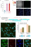
A) Average Xist gene expression levels ± s.d. on RNA isolated from E15 placentas of 3 control males (light blue bars), 3 control females (pink bars) and 3 ROSI-derived XpΔXist females (red bars). The data were normalized to actin. B) Representative image of the Xist RNA FISH (in green) on cryosections from a E15 ROSI-derived XpΔXist female placenta (n = 2). From the phase contrast image on top (right), the Reichert’s membrane on the embryonic side of the placenta can be visualized (marked by asterisk). DNA is counterstained with DAPI. C) Top: Schematic drawing of the RNA/DNA FISH probes used to detect the wild type (wt) Xist and the ΔXist gene and/or RNA. Exon numbers are indicated. The green probe localizes to sequences present in both the wild type Xist and ΔXist gene, whereas the red probe localizes to sequences that are only present in the wild type Xist gene (for further details see the Materials and Methods section). Left: representative merged image of the Xist RNA/DNA FISH using a probe recognizing the RNA produced by the wild type maternal allele and the DNA of both wild type Xist (signal is hidden under Xist RNA FISH cloud) and ΔXist alleles (green), and a probe recognizing only the DNA of the wild type Xist allele (hidden under Xist RNA FISH cloud) and RNA produced by the wild type maternal X chromosome (red) on cryosections from a E15 ROSI-derived XpΔXist female placenta (n = 2). DNA (Dapi) is shown in blue. Examples of nuclei are shown with an RNA FISH cloud signal with both probes, and a separate DNA FISH pinpoint in green (type 1), or with only an RNA FISH cloud signal with both probes (type 2). Cells lacking a cloud (some nuclear sections may not include the Xi) or with complicated staining patterns (due to the presence of polyploid cells, as was documented previously [53]) were not counted. Examples of such nuclei are indicated by asterisks. Scale bar represents 10 μm. Separate images of the nuclei in the boxed area are shown below, and the wild type and mutant chromosome are indicated with arrows for one nucleus. Scale bar represents 5 μm. Right: Quantification of type 1 and type 2 nuclei in two E15 ROSI-derived XpΔXist female placentas. Cells with a single green/red RNA FISH cloud and a separate red DNA FISH pinpoint were never observed.
Fig 4
Fig 4. Maternal Xist expression in ROSI-derived XpΔXist females is variable and delayed compared to paternal Xist expression during iXCI.
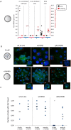
A) Dot plot showing Xist (black) and Eif2s3y (red) mRNA expression levels for RNA isolated from individual in vivo fertilized wt and XpΔXist male (blue) and female (pink) mouse morulas, and from ROSI-derived XpΔXist male and female morulas. Expression levels were normalized to Actin. Grey lines indicate the average values, n values are indicated below the graph. B) Representative images of Xist clouds in wild type in vivo fertilized female embryos, and wild type and XpΔXist ROSI-derived female embryos, analysed at the 8-cell and morula stages. Scale bars in whole embryo and single cell images represent 10, and 5 μm, respectively. C) Quantification of Xist cloud formation in the embryo types described in B. Individual measurements are indicated, horizontal bars represent the average value. Asterisk indicates significantly different from the corresponding stage in wild type ROSI female embryos (P = 0,00025). Numbers of analysed embryos are indicated at the bottom, and followed by total numbers of nuclei that could be scored (some nuclei were lost during procedures, and some embryos that were in the “8-cell” group contained 9 or 10 cells) in parenthesis.
Fig 5
Fig 5. H3K27me3 marks the inactive Xm in ROSI-derived XpΔXist female blastocysts.
H3K27me3 (green) marks the inactive Xp in wild type in vivo fertilized female blastocyst and the inactive Xm in ROSI-derived XpΔXist female blastocysts. Dapi is shown in blue. Size bars in whole embryo and single cell images represent 20, and 5 μm, respectively. H3K27me3 domain is indicated by arrows in the enlargements.
Fig 6
Fig 6. Enhanced expression of RNF12 in ROSI-derived compared to in vivo fertilized 8-cell embryos.
A) Quantification of RNF12 protein levels ± s.d. per male 8-cell embryo as measured from immunocytochemical staining using Image J (see Materials and Methods for details). Representative images of each analysed condition are shown on the right. Size bars represent 10 μm. Asterisk indicates significant difference (P = 0,05) with in vivo wild type derived embryos. B) As in A, for female embryos. Asterisk indicates significant difference (P≤0,05) with in vivo fertilized embryos. The following P values were obtained per comparison: wt in vivo versus wt ROSI derived embryos P = 0,016, wt in vivo versus XpΔXist ROSI derived embryos P = 0,0005, in vivo XpΔXist versus wt ROSI derived embryos P = 0,0098 and in vivo XpΔXist versus XpΔXist ROSI derived embryos P = 0,004.
Fig 7
Fig 7. Schematic representation of critical factors during iXCI in wild type and parthenogenetic females, compared to XpΔXist females arising from in vivo fertilization or ROSI.
Xp (blue) and Xm (pink) are schematically drawn, and the Xist and Rnf12 loci are indicated where relevant. RNF12 protein levels are represented by the size of the orange circle. A green Xist locus indicates that it is primed for expression, whereas a red locus indicates that it is repressed. An open Xist locus represents the ΔXist allele. During normal iXCI, that initiates around the four cell-stage, high maternally regulated RNF12 levels ensure Xist expression from Xp, establishing iXCI by the morula stage (green signal on Xp representing the Xist cloud). In parthenogenic embryos, the imprint on Xm is lost by the morula stage and Xist can be activated in a manner that appears to be regulated by dosage dependent XCI activators such as RNF12 (effect symbolized by two black arrows coming from the Rnf12 loci), but this is inefficient. In contrast, when Xist is deleted from Xp, iXCI cannot be induced, most likely because the presence of a paternal genome helps to maintain repression of the maternal Xist gene (red arrow). Upon ROSI, round spermatid-specific epigenetic regulation (green arrow), possibly in combination with the double dosage of X chromosomes, allows activation of Xm by the morula stage. In addition, RNF12 levels are relatively high and may also aid in this process.

References

    1. Puschendorf M, Terranova R, Boutsma E, Mao X, Isono K, et al. (2008) PRC1 and Suv39h specify parental asymmetry at constitutive heterochromatin in early mouse embryos. Nat Genet 40: 411–420. 10.1038/ng.99 - DOI - PubMed
    1. van der Heijden GW, Dieker JW, Derijck AA, Muller S, Berden JH, et al. (2005) Asymmetry in histone H3 variants and lysine methylation between paternal and maternal chromatin of early mouse zygotes. Mech Dev 122: 1008 10.1016/j.mod.2005.04.009 - DOI - PubMed
    1. Turner JM (2007) Meiotic sex chromosome inactivation. Development 134: 1823–1831. 10.1002/9780470015902.a0022109 - DOI - PubMed
    1. van der Heijden GW, Derijck AA, Posfai E, Giele M, Pelczar P, et al. (2007) Chromosome-wide nucleosome replacement and H3.3 incorporation during mammalian meiotic sex chromosome inactivation. Nat Genet 39: 251–258. 10.1038/ng1949 - DOI - PubMed
    1. Namekawa SH, Park PJ, Zhang LF, Shima JE, McCarrey JR, et al. (2006) Postmeiotic sex chromatin in the male germline of mice. Curr Biol 16: 660–667. 10.1016/j.cub.2006.01.066 - DOI - PubMed

LinkOut - more resources