. 2016 Nov;40(11):2339-2350.
doi: 10.1111/acer.13218. Epub 2016 Sep 26.

Effects of Ethanol on Cellular Composition and Network Excitability of Human Pluripotent Stem Cell-Derived Neurons

Affiliations

Effects of Ethanol on Cellular Composition and Network Excitability of Human Pluripotent Stem Cell-Derived Neurons

Zoe H Larsen et al. Alcohol Clin Exp Res. 2016 Nov.

Abstract

Background: Prenatal alcohol exposure (PAE) in animal models results in excitatory-inhibitory (E/I) imbalance in neocortex due to alterations in the GABAergic interneuron (IN) differentiation and migration. Thus, E/I imbalance is a potential cause for intellectual disability in individuals with fetal alcohol spectrum disorder (FASD), but whether ethanol (EtOH) changes glutamatergic and GABAergic IN specification during human development remains unknown. Here, we created a human cellular model of PAE/FASD and tested the hypothesis that EtOH exposure during differentiation of human pluripotent stem cell-derived neurons (hPSNs) would cause the aberrant production of glutamatergic and GABAergic neurons, resulting in E/I imbalance.

Methods: We applied 50 mM EtOH daily to differentiating hPSNs for 50 days to model chronic first-trimester exposure. We used quantitative polymerase chain reaction, immunocytochemical, and electrophysiological analysis to examine the effects of EtOH on hPSN specification and functional E/I balance.

Results: We found that EtOH did not alter neural induction nor general forebrain patterning and had no effect on the expression of markers of excitatory cortical pyramidal neurons. In contrast, our data revealed highly significant changes to levels of transcripts involved with IN precursor development (e.g., GSX2, DLX1/2/5/6, NR2F2) as well as mature IN specification (e.g., SST, NPY). Interestingly, EtOH did not affect the number of GABAergic neurons generated nor the frequency or amplitude of miniature excitatory and inhibitory postsynaptic currents.

Conclusions: Similar to in vivo rodent studies, EtOH significantly and specifically altered the expression of genes involved with IN specification from hPSNs, but did not cause imbalances of synaptic excitation-inhibition. Thus, our findings corroborate previous studies pointing to aberrant neuronal differentiation as an underlying mechanism of intellectual disability in FASD. However, in contrast to rodent binge models, our chronic exposure model suggests possible compensatory mechanisms that may cause more subtle defects of network processing rather than gross alterations in total E/I balance.

Keywords: Cortical Neurons; Excitatory-Inhibitory Balance; Fetal Alcohol Spectrum Disorder; Interneurons; Somatostatin.


Conflict of interest statement

Disclosure of Potential Conflicts of Interest: The authors indicate no potential conflict of interest.

Figures

Figure 1
Figure 1. Ethanol does not affect neural induction
(A) Diagram illustrating the methods and time course of neuronal differentiation used in the study. Representative images show cellular morphology at various time points during differentiation; image labeled “immature neurons” was taken from neurons at day 30 and shows βIII-Tubulin while the image labeled “functional neurons” was taken at day 50 and shows βIII-Tubulin (red) and Synapsin-1 (green). Scale bars indicate 100μm. (B) Paired confocal images of hPSCs at day 0 (upper panels) and neuroepithelia that were untreated (middle panels) or treated with 50mM EtOH (lower panels). Representative images illustrate the presence of Oct4 (red) and absence of Pax6 (green) at day 0. Cells at day 10 showed robust Pax6 (green) with little Oct4 (red) present. Nuclei are labeled with DAPI (blue). Scale bars indicate 50μm. (C) Fluorescence-activated cell sorting data confirmed that treated and untreated cells at day 10 lost Oct4 expression (red; lower left panels) but showed strong Pax6 expression (green; lower right panels), while cells at day 0 showed the opposite pattern of expression (upper panels). Plots illustrated in black indicate independent IgG controls for each antibody.
Figure 2
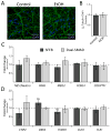
Figure 2. Ethanol does not affect neuroepithelial nor forebrain cortical neuron differentiation
(A) Representative confocal micrographs of control (left) and EtOH-treated (right) hPSN cultures labeled with the neuronal marker βIII-Tubulin (green) and DAPI (blue). Scale bar indicates 50μm. (B) Pooled data reveled no significant difference in the proportion of βIII-Tubulin+/DAPI+ cells between groups. N = 6; p>0.05. (C) Pooled qPCR data shows that mean expression of neuroepithelial markers (NES, PAX6, MEIS2) and forebrain markers (FOXG1, COUPTF1) in EtOH-treated cells did not significantly differ from controls (normalized expression illustrated by dashed line) using either SFEB- or Dual-SMAD inhibition methods. (D) Expression of superficial layer (CTIP2, DKK3, FOXP2), and deep layer (CUX1, TLE1) cortical markers was not significantly different between EtOH-treated and control cells at day 50. N = 3; p>0.05.
Figure 3
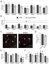
Figure 3. Ethanol specifically and significantly affects GABAergic gene expression without altering the proportion of GABAergic neurons
(A) Pooled qPCR data shows that mean expression levels of markers for post-synaptic NMDA receptors (GRIN1, 2B), AMPA receptors (GRIA1-3), VGLUT2 and PSD95, were not significantly altered in EtOH-treated cells compared to controls (normalized expression illustrated by dashed line). (B) In EtOH-treated cells, multiple transcripts involved with GABAergic IN specification were significantly reduced using either SFEB- or Dual-SMAD inhibition methods compared to untreated-controls (* indicates significance for all markers shown). (C) Representative confocal micrographs of control (left) and EtOH-treated (right) hPSN cultures labeled with GABA (green), βIII-Tubulin (red), and DAPI (blue). Arrows indicate GABA+III-Tubulin+ cells while arrowheads indicate GABA-III-Tubulin+ cells. Scale bar indicates 50μm. (D) Pooled data reveled no significant difference in the proportion of GABA+III-Tubulin+ cells between groups. N = 6; p>0.05. (E) Pooled data showing mRNA expression for postsynaptic GABA receptors (α2, β2, γ2) remained unchanged, while EtOH significantly reduced expression of the presynaptic marker VGAT. N = 3; *p<0.05. (F) Pooled data indicating expression of IN progenitor markers ID4 and COUPTFII (NR2F2) were significantly elevated by EtOH treatment (N = 3; *p<0.05), while CALB2 and PARV were not significantly changed. N = 3; p>0.05.
Figure 4
Figure 4. Ethanol does not affect voltage-gated currents in hPSNs
(A-B) Representative traces from voltage-clamped neurons during voltage steps revealed the presence of robust transient inward and sustained outward currents in EtOH-exposed and controls. (C) Expanded timescale of inward currents. (D-F) Current-voltage relationships for peak inward (D), peak outward (E), and sustained outward (F) currents were not significantly different between untreated hPSNs and those that received 50mM EtOH. N = 6; p>0.05.
Figure 5
Figure 5. Ethanol does not affect spontaneous post-synaptic currents in hPSNs
(A-B) Representative traces from neurons in voltage-clamp mode and held at -40mV revealed the presence of robust EPSCs (downward deflections) and IPSCs (upward deflections) in EtOH-exposed and control hPSNs. (C-D) Pooled data demonstrate that mean frequency of EPSCs (C) and IPSCs (D) were not significantly different between untreated hPSNs and those that received 50mM EtOH for the duration of differentiation. N = 6; p>0.05. (E-F) Pooled data demonstrate no difference in amplitude of EPSCs (E) and IPSCs (F) between controls and hPSNs that received EtOH. N = 6; p>0.05.

References

    1. Ayoola AB, Stommel M, Nettleman MD. Late recognition of pregnancy as a predictor of adverse birth outcomes. American journal of obstetrics and gynecology. 2009;201:156, e151–156. - PubMed
    1. Bell SH, Stade B, Reynolds JN, Rasmussen C, Andrew G, Hwang PA, Carlen PL. The remarkably high prevalence of epilepsy and seizure history in fetal alcohol spectrum disorders. Alcoholism, clinical and experimental research. 2010;34:1084–1089. - PubMed
    1. Ben-Ari Y. Excitatory actions of gaba during development: the nature of the nurture. Nature reviews Neuroscience. 2002;3:728–739. - PubMed
    1. Borodinsky LN, Root CM, Cronin JA, Sann SB, Gu X, Spitzer NC. Activity-dependent homeostatic specification of transmitter expression in embryonic neurons. Nature. 2004;429:523–530. - PubMed
    1. Boy F, Evans CJ, Edden RA, Singh KD, Husain M, Sumner P. Individual differences in subconscious motor control predicted by GABA concentration in SMA. Current biology : CB. 2010;20:1779–1785. - PMC - PubMed

Publication types

MeSH terms