Comparative Study
. 2017 Jan;140(2):216-230.
doi: 10.1111/jnc.13861. Epub 2016 Dec 15.

Trafficking of adeno-associated virus vectors across a model of the blood-brain barrier; a comparative study of transcytosis and transduction using primary human brain endothelial cells

Affiliations
Comparative Study

Trafficking of adeno-associated virus vectors across a model of the blood-brain barrier; a comparative study of transcytosis and transduction using primary human brain endothelial cells

Steven F Merkel et al. J Neurochem. 2017 Jan.

Abstract

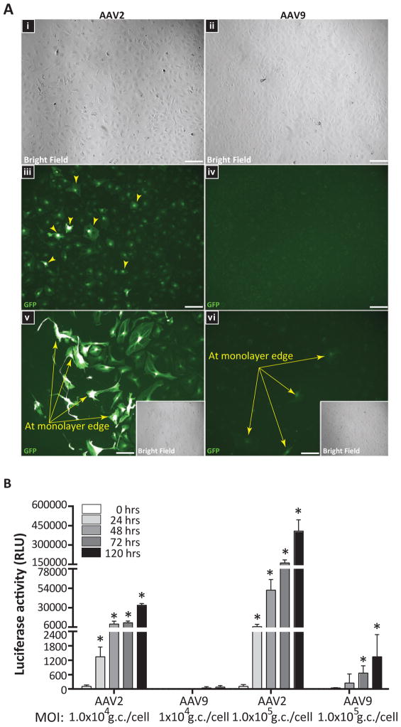
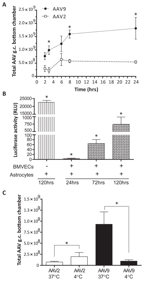
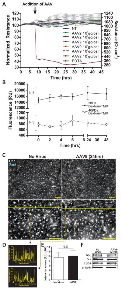
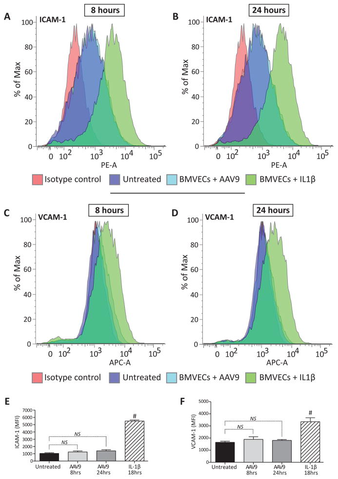
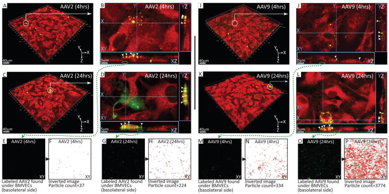
Developing therapies for central nervous system (CNS) diseases is exceedingly difficult because of the blood-brain barrier (BBB). Notably, emerging technologies may provide promising new options for the treatment of CNS disorders. Adeno-associated virus serotype 9 (AAV9) has been shown to transduce cells in the CNS following intravascular administration in rodents, cats, pigs, and non-human primates. These results suggest that AAV9 is capable of crossing the BBB. However, mechanisms that govern AAV9 transendothelial trafficking at the BBB remain unknown. Furthermore, possibilities that AAV9 may transduce brain endothelial cells or affect BBB integrity still require investigation. Using primary human brain microvascular endothelial cells as a model of the human BBB, we performed transduction and transendothelial trafficking assays comparing AAV9 to AAV2, a serotype that does not cross the BBB or transduce endothelial cells effectively in vivo. Results of our in vitro studies indicate that AAV9 penetrates brain microvascular endothelial cells barriers more effectively than AAV2, but has reduced transduction efficiency. In addition, our data suggest that (i) AAV9 penetrates endothelial barriers through an active, cell-mediated process, and (ii) AAV9 fails to disrupt indicators of BBB integrity such as transendothelial electrical resistance, tight junction protein expression/localization, and inflammatory activation status. Overall, this report shows how human brain endothelial cells configured in BBB models can be utilized for evaluating transendothelial movement and transduction kinetics of various AAV capsids. Importantly, the use of a human in vitro BBB model can provide import insight into the possible effects that candidate AVV gene therapy vectors may have on the status of BBB integrity. Read the Editorial Highlight for this article on page 192.

Keywords: adeno-associated virus; blood-brain barrier; gene therapy; neurological disorders.


Conflict of interest statement

The authors declare no competing financial interests.

Figures

Figure 1
Figure 1. AAV2 outperforms AAV9 at transgene expression in primary human BMVEC cultures
(A) Confluent BMVEC monolayers from two donors were incubated with sc-AAV2-eGFP or sc-AAV9-eGFP vectors for 4 hours at 1×10⁴ gc/cell. Cells were then washed to remove unbound virus and incubated for 5 days to allow for eGFP expression. Fluorescent images were acquired using equal capture parameters between serotypes. Representative images acquired from one of the BMVEC donors evaluated are presented. (i–iv) Bright field and GFP images obtained from confluent BMVEC monolayers incubated with either AAV2 or AAV9; 20X objective magnification. (v–vi) GFP filter images from under-confluent BMVEC cultures incubated with either AAV2 or AAV9 with bright field inserts; 40X objective magnification. Arrows indicate expression of eGFP in BMVEC cultures. (B) Additional experiments with a firefly luciferase reporter. BMVEC cultures from a third donor were incubated with either AAV2 or AAV9 expressing firefly luciferase (Fluc) at two different concentrations (1×10⁴ gc/cell and 1×10⁵ gc/cell) for 24, 48, 72, and 120 hours. At the indicated time points, viral-mediated transgene expression was assessed using a luciferase assay system. Data were normalized to the 0-hour treatments for each group and are presented as mean luciferase activity in relative light units (RLU) for treatments performed in quadruplicate + SEM. Statistical significance was analyzed as a function of the 24-hour read-outs for each group. Asterisks denote p < 0.05.
Figure 2
Figure 2. Trafficking of AAV9 across an in vitro BBB model composed of primary human BMVECs is more efficient when compared to AAV2
(A) BMVEC cultures from two different donors exhibiting monolayer formation indicative of barrier properties were prepared on collagen-coated cell culture inserts, creating a two-compartment design to model the BBB. Cultures were incubated with either AAV2 or AAV9 vectors (2×10⁵ gc/cell) for 2, 3, 6, 8, or 24 hours. At the indicated time points, media was collected from the bottom chamber (below the cell culture insert) and analyzed for the presence of vector DNA by qPCR. All treatments were performed in quadruplicate for both donors. Data from a single donor are presented as the mean of total AAV genomes retrieved from the bottom chamber ± SEM. Asterisks denote p < 0.05. (B) Proof-of-concept experiments were performed to show that AAV9 vectors are able to transduce parenchymal brain cells after trafficking across human BMVECs. Primary human astrocytes were seeded in the lower compartment of our BBB model, and AAV9-Fluc vectors (2×10⁵ gc/cell) were applied to the upper compartment and incubated for 24, 72, and 120 hours. Primary astrocyte cultures incubated directly with AAV9-Fluc (no BMVECs) for 120 hours served as a positive control. At the indicated time points, AAV9-mediated transgene expression in primary human astrocytes was evaluated using a luciferase assay. Data were normalized to 0-hour controls and are presented as mean luciferase activity in relative light units (RLU) for treatments performed in quadruplicate + SEM. Statistical significance was analyzed as a function of the 24-hour read-outs. Asterisks denote p < 0.05. (C) Additional experiments were performed as described above in (A); however, BMVEC cultures from one of the donors were incubated with AAV vectors at either 37°C (physiological temperature) or 4°C, to reduce active, cell-mediated transport of AAV particles. After 3 hours, media was collected from the bottom chamber and analyzed for the presence of vector DNA by qPCR. All treatments were performed in quadruplicate. Data are presented as the mean of total AAV genomes retrieved from the bottom chamber + SEM. Asterisks denote p < 0.05.
Figure 3
Figure 3. AAV9 does not affect in vitro indicators of BMVEC barrier integrity
(A) BMVEC cultures from a single donor were prepared on collagen-coated electrode arrays designed for use with an Electric Cell-substrate Impedance System. Transendothelial electrical resistance (TEER) readings were acquired continuously in 30-minute intervals for one week. After BMVEC cultures established stable TEER readings, cells were exposed to either AAV2 or AAV9 at three different concentrations (1×10⁴ gc/cell, 1×10⁵ gc/cell, and 1×10⁶ gc/cell) for 48 hours. All treatments were performed in triplicate. Cultures never exposed to virus were used as stable baseline controls. EDTA was used as a control for the reduction of baseline TEER values. Data are presented as the normalized change in resistance from baseline TEER + SEM over a 45-hour period. Right-hand y-axis shows absolute TEER values (Ohms • cm2). Arrow indicates addition of AAV vectors to BMVEC cultures with stable TEER readings (last 10 hours shown). Statistical analysis revealed no significant changes in TEER between AAV treated cultures and controls. (B) BMVECs exhibiting monolayer formation indicative of barrier properties were prepared on collagen-coated cell culture inserts and incubated with AAV9 vectors (2×10⁵ gc/cell) for 0, 2, 4, 6, 24 and 48 hours. At the indicated times, permeability measures were determined using two tetramethylrhodamine (TMR)-labeled dextrans (3 kDa and 40 kDa) applied to the upper compartment for 20 minutes. Media from the lower compartment was then analyzed for the presence of dextran-TMR tracers. Data are presented as mean fluorescence (RU) + SEM for treatments performed in quadruplicate. Statistical analyses revealed no significant difference between cultures incubated with AAV9 and 0-hour controls. NS denotes p > 0.05. (C-E) Additional BMVEC cultures were incubated with AAV9 (2.5×10⁵ gc/cell) for 24 hours in order to determine the cellular localization and relative expression of key tight junction (TJ) proteins that help to stabilize BBB integrity. BMVEC cultures not exposed to AAV were used as controls. (C) Immunocytochemistry reveals cellular localization of occludin (Occ) at intercellular borders consistent with TJ formation both in control cultures and in the presence of AAV9. (D) The histogram represent inserts values from intensity/pixel from intensity line profile drawn across the images. (E) On the basis of the intensity line profiles from sets of N=5, the graph of the average ± SEM shows no statistical significance observed between no virus and AAV9 exposed cultures. (F) Western blots depict similar expression levels of ZO-1, Occ, and claudin-5 (CLD-5) in BMVEC control cultures and those incubated with AAV9. β-actin is presented as a loading control for equal protein.
Figure 4
Figure 4. BMVECs maintain baseline activation status after AAV9 exposure
BMVEC cultures from a single donor were incubated with AAV9 (2.5×10⁵ gc/cell) for either 8 or 24 hours. At the indicated time points, BMVECs were harvested to measure the surface expression of cellular adhesion molecules (ICAM-1 and VCAM-1) by flow cytometry. All treatments were run in triplicate. Cultures never exposed to virus were used as controls for the baseline expression of ICAM-1 and VCAM-1. BMVECs treated with recombinant IL-1β for 18 hours were used as positive controls. (A–D) Representative histograms depicting the shift in ICAM-1 and VCAM-1 expression in BMVEC cultures treated with AAV9 compared to controls after 8 or 24 hours as indicated. (E–F) Data are presented as the average mean fluorescence intensity (MFI) + SEM of ICAM-1 and VCAM-1 in BMVEC cultures exposed to AAV9 for either 8 or 24 hours compared to controls. NS denotes p > 0.05. Hashtags denote p < 0.05.
Figure 5
Figure 5. Cellular localization of AAV9 and AAV2 in primary human BMVEC cultures
BMVECs from a single donor were seeded in MatTek dishes outfitted with a 14 mm glass-bottomed microwell. Next, cultures were expanded and maintained for one week as previously described to remove heparin. BMVEC cultures were then incubated with DyLight-488 (green signal) conjugated AAV2 or AAV9 particles (2.5×10⁶ gc/cell) for either 4 or 24 hours. Following incubation, cells were washed to remove unbound virus, stained with a viability tracker dye, CMTPX (red signal), and imaged using multiphoton live cell microscopy. (A, C, I, K) Representative fields of view acquired from BMVEC cultures exposed to fluorescently labeled AAV2 or AAV9 for 4 or 24 hours as indicated. White circles with arrows identify areas under close observation. (B, D, J, L) High-resolution images of fluorescently labeled AAV capsid clusters visualized in XZ and YZ planes. White arrowheads point to intracellular accumulations of either AAV2 or AAV9 as indicated. Green circles with arrows identify fluorescently labeled AAV capsid clusters at the basolateral aspect of the XZ plane (below BMVEC monolayers). (E, G, M, O) Extracellular, DyLight-488 conjugated AAV capsid clusters at the basolateral side of BMVEC cultures incubated with either AAV2 or AAV9 for 4 or 24 hours as indicated. Using NIH Image J software, AAV capsid clusters were quantified by converting the fluorescent images to binary and performing particle counting based on area and circularity. (F, H, N, P) Inverted images derived from binary conversion of the fluorescent AAV capsid signal used for particle counting in NIH Image J software.

References

    1. Abbott NJ, Ronnback L, Hansson E. Astrocyte-endothelial interactions at the blood-brain barrier. Nature reviews. Neuroscience. 2006;7:41–53. - PubMed
    1. Banks WA, Akerstrom V, Kastin AJ. Adsorptive endocytosis mediates the passage of HIV-1 across the blood-brain barrier: evidence for a post-internalization coreceptor. Journal of cell science. 1998;111(Pt 4):533–540. - PubMed
    1. Banks WA, Freed EO, Wolf KM, Robinson SM, Franko M, Kumar VB. Transport of human immunodeficiency virus type 1 pseudoviruses across the blood-brain barrier: role of envelope proteins and adsorptive endocytosis. Journal of virology. 2001;75:4681–4691. - PMC - PubMed
    1. Bell CL, Gurda BL, Van Vliet K, Agbandje-McKenna M, Wilson JM. Identification of the galactose binding domain of the adeno-associated virus serotype 9 capsid. Journal of virology. 2012;86:7326–7333. - PMC - PubMed
    1. Bell CL, Vandenberghe LH, Bell P, Limberis MP, Gao GP, Van Vliet K, Agbandje-McKenna M, Wilson JM. The AAV9 receptor and its modification to improve in vivo lung gene transfer in mice. The Journal of clinical investigation. 2011;121:2427–2435. - PMC - PubMed

Publication types