Review
. 2016 Nov;54(6):1033-1046.
doi: 10.1016/j.rcl.2016.05.013. Epub 2016 Aug 11.

Imaging of Hypersensitivity Pneumonitis

Affiliations
Review

Imaging of Hypersensitivity Pneumonitis

Andrea L Magee et al. Radiol Clin North Am. 2016 Nov.

Abstract

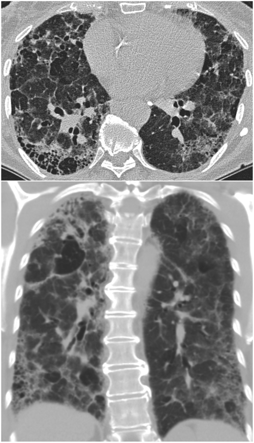
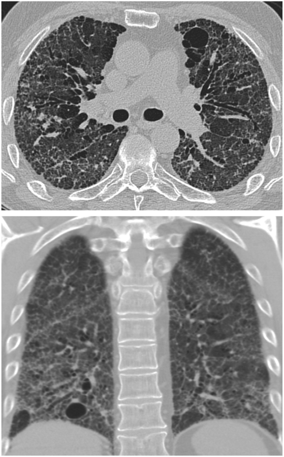
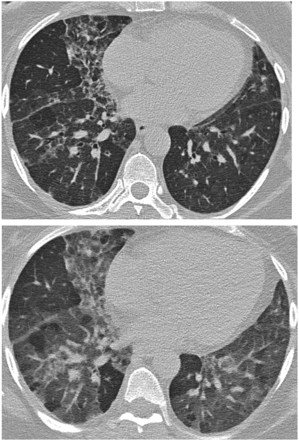
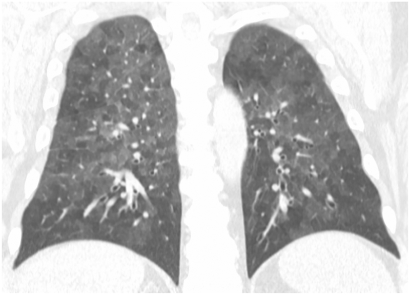
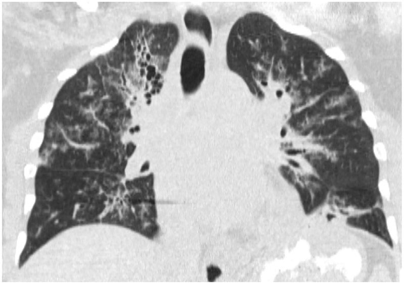
The management of hypersensitivity pneumonitis (HP) depends on early identification of the disease process, which is complicated by its nonspecific clinical presentation in addition to variable and diverse laboratory and radiologic findings. HP is the result of exposure and sensitization to myriad aerosolized antigens. HP develops in the minority of antigenic exposures, and conversely has been documented in patients with no identifiable exposure, complicating the diagnostic algorithm significantly. Prompt diagnosis and early intervention are critical in slowing the progression of irreversible parenchymal damage, and additionally in preserving the quality of life of affected patients.

Keywords: High-resolution computed tomography; Hypersensitivity pneumonitis; Interstitial lung disease; Parenchymal lung disease.


Figures

Figure 1
Figure 1
Lung biopsy from a patient with acute exacerbation of HP shows a chronic inflammatory infiltrate composed mainly of lymphocytes in both the interstitium and the alveoli. Several multinucleated giant cells (arrows) are also present.
Figure 2
Figure 2
Medium power photomicrograph shows an area of bronchiolar metaplasia (left) and a lymphoid aggregate (center) in the same patient.
Figure 3
Figure 3
Medium power photomicrograph shows a large area of fibrosis within which there are two fibroblastic foci (arrows) containing bluish myxoid stroma.
Figure 4
Figure 4
Medium power micrograph shows poorly-formed granuloma in a perivascular location.
Figure 5
Figure 5
Low power photomicrograph of lung showing extensive interstitial fibrosis (solid white arrow) and honeycomb change (short black arrows) in a patient with chronic HP. Complete destruction of alveoli is seen, with only mucus-filled, dilated airspaces lined by bronchial epithelial cells. There is also secondary pulmonary hypertension, as demonstrated by the presence of a thick-walled pulmonary arterial branch (dashed black arrow). Pleural surface is indicated by red arrows.
Figure 6
Figure 6
Medium power photomicrograph of lung biopsy in a patient with subacute HP showing non-necrotizing granuloma (black arrow) with multinucleated giant cells (white arrow).
Figure 7
Figure 7
Coronal CT images (a and b) from two separate patients with HP show upper lung zone-predominant reticular fibrosis, subpleural honeycombing and traction bronchiectasis. The 2nd image (b) shows mosaic attenuation consistent with concomitant air-trapping.
Figure 8
Figure 8
Axial CT in a patient with Bird Fancier’s Disease shows diffuse centrilobular ground glass opacities within the lungs.
Figure 9
Figure 9
Axial (9a) and coronal (9b) expiratory phase CT showing fibrotic changes of HP with superimposed finding of air-trapping in this patient with known HP.
Figure 10
Figure 10
Axial (10a) and coronal (10b) CT show predominantly subpleural fibrotic changes including honeycombing and traction bronchiectasis typical of fibrotic HP with superimposed focal cystic regions.
Figure 11
Figure 11
Inspiratory phase imaging (11a) in this patient with HP demonstrates mosaic attenuation and patchy pulmonary fibrosis. Expiratory phase imaging (11b) demonstrates superimposed air trapping.
Figure 12
Figure 12
Coronal CT reformatted image shows the headcheese sign, featuring sharp geographic margination along the edges of the secondary pulmonary lobules with three distinct levels of attenuation representing normal, ground glass and hyperinflated regions of lung. This sign in the subacute or chronic setting is highly suggesting of HP.
Figure 13
Figure 13
Axial (13a) CT in a case of UIP shows peripheral reticular abnormality, traction bronchiectasis and honeycombing. Coronal (13b) CT reformat shows honeycombing and reticular opacities in a subpleural and basilar distribution.
Figure 14
Figure 14
Axial CT in a patient with NSIP shows bilateral lower lung predominant fine reticular abnormality and ground glass opacity with subpleural sparing.
Figure 15
Figure 15
Axial CT in a case of NSIP shows symmetric lower lung predominant reticular abnormality, exuberant traction bronchiectasis and ground glass opacity as well as a dilated esophagus in this patient with underlying collagen vascular disease.
Figure 16
Figure 16
Axial (16a) and Coronal (16b) CT in a patient with sarcoidosis show bilateral upper and middle lung zone reticulonodular opacities and changes of pulmonary fibrosis.
Figure 16
Figure 16
Axial (16a) and Coronal (16b) CT in a patient with sarcoidosis show bilateral upper and middle lung zone reticulonodular opacities and changes of pulmonary fibrosis.

References

    1. Demedts M, et al., Interstitial lung diseases: an epidemiological overview. Eur Respir J Suppl, 2001. 32: p. 2s–16s. - PubMed
    1. Keskinen P, et al., Regulation of HLA class I and II expression by interferons and influenza A virus in human peripheral blood mononuclear cells. Immunology, 1997. 91(3): p. 421–9. - PMC - PubMed
    1. Dakhama A, et al., Common respiratory viruses in lower airways of patients with acute hypersensitivity pneumonitis. Am J Respir Crit Care Med, 1999. 159(4 Pt 1): p. 1316–22. - PubMed
    1. Wang SZ, et al., Adhesion molecule expression on epithelial cells infected with respiratory syncytial virus. Eur Respir J, 2000. 15(2): p. 358–66. - PubMed
    1. Cormier Y and Israel-Assayag E, The role of viruses in the pathogenesis of hypersensitivity pneumonitis. Curr Opin Pulm Med, 2000. 6(5): p. 420–3. - PubMed

MeSH terms