Review
. 2017 Jan;33(1):65-73.
doi: 10.1016/j.pt.2016.09.003. Epub 2016 Oct 4.

Disease Centered Around Calcified Taenia solium Granuloma

Affiliations
Review

Disease Centered Around Calcified Taenia solium Granuloma

Theodore E Nash et al. Trends Parasitol. 2017 Jan.

Abstract

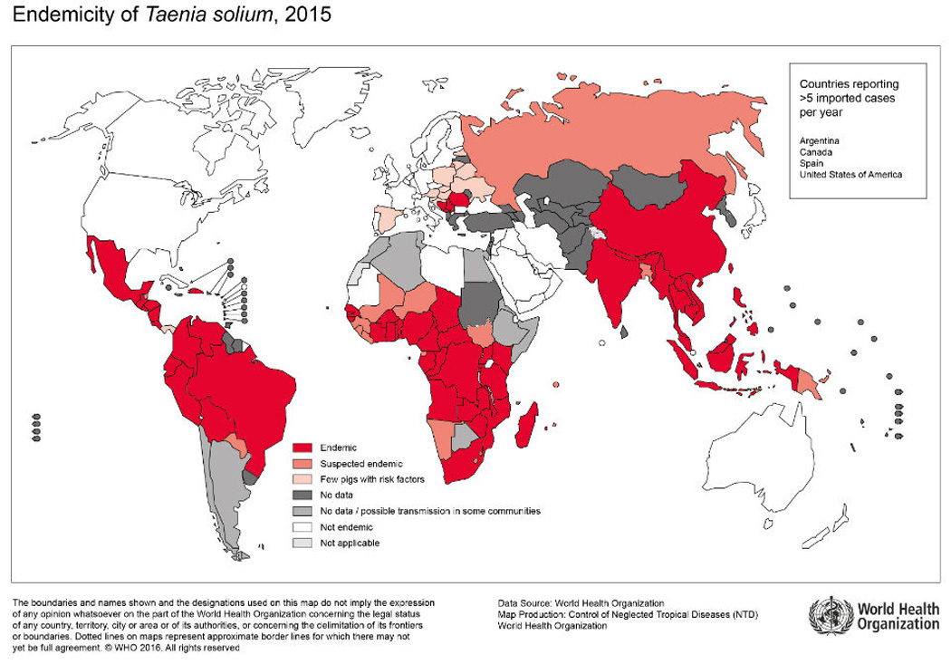
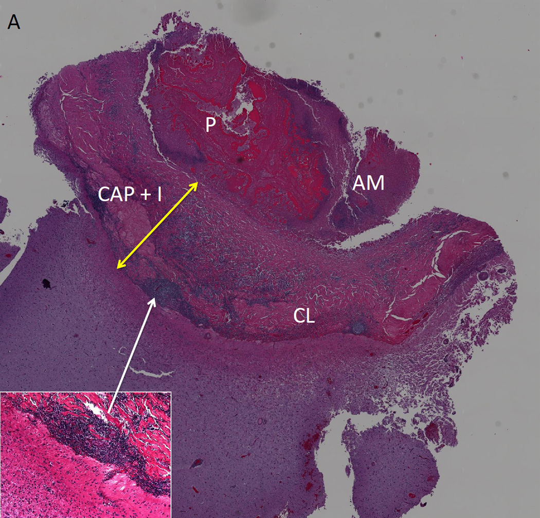
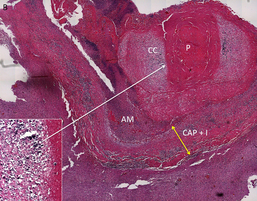
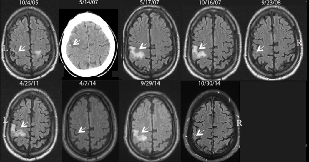
Taenia solium (the pork tapeworm) is present in most developing countries, where it is a frequent cause of seizures and other neurological disease. Parasitic larvae invade the human brain, establish, and eventually resolve, leaving a calcified scar. While these lesions are common in endemic regions, and most of these are clinically silent, a proportion of individuals with calcified cysticerci develop seizures from these lesions, and 30-65% of these cases are associated with perilesional edema (PE), likely due to host inflammation. This manuscript summarizes the importance, characteristics, natural history, and potential prevention and treatments of symptomatic calcified neurocysticercosis (NCC).

Keywords: calcified granuloma; epilepsy; neurocysticercosis; perilesional edema; seizures.


Figures

Figure 1
Figure 1. Endemicity of Taenia solium
Reproduced, with permission, from http://www.who.int/mediacentre/factsheets/Endemicity_Taenia_Solium_2015-1000x706.jpg?ua=1. Accessed September 7th, 2016.
Figure 2
Figure 2. Histopathology of a Calcified Taenia solium Lesion
Two histological sections of excised calcified lesion that was causing multiple episodes of perilesional edema. In A and B, parasite remnants (P) are surrounded by a capsule that contains inflammation (CAP+I, yellow arrow) and in A, an increase in collagen (CL). Amorphous material (AM) is closely associated with P. The adjacent brain has increased glial fibrillary acid protein presence (not shown), perivascular infiltrates and eosinophilic material. In B, aggregates of calcareous corpuscles (CC) surround P, and are likely responsible for the calcification. The inset in (A) shows a higher magnification of one area of inflammation; the inset in B shows a higher magnification of a dense aggregation of calcareous corpuses. Adapted from [25], with permission.
Figure 3
Figure 3. Patient with Multiple Episodes of Perilesional Edema Around Calcifications
Serial fluid-attenuated inversion-recovery (FLAIR) images over time from left to right demonstrating appearance and resolution of perilesional edema around a Taenia solium calcification in the right frontal lobe. The patient was diagnosed and treated on 10/4/05; a viable cyst is noted by the arrow that subsequently calcifies as documented by a computed tomography examination a few days before (5/14/07) the first episode on 5/17/07. Perilesional edema develops on 10/16/07, 4/25/11, and 9/29/14 with interim resolution on 9/23/08, 4/7/14 and 10/30 14. The associated edema of a left sided cyst is partially seen on 10/4/05. R and L denote right and left sides of the brain. Arrows point to the site of the lesion with or without the presence of edema. The date the images were taken is shown.

References

    1. Garcia HH, Del Brutto OH. Neurocysticercosis: updated concepts about an old disease. The Lancet. Neurology. 2005;4:653–661. - PubMed
    1. Newton CR, Garcia HH. Epilepsy in poor regions of the world. Lancet. 2012;380:1193–1201. - PubMed
    1. Schantz PM. Taenia solium cysticercosis: an overview of global distribution and transmission. In: Singh G, Prabhakar S, editors. Taenia solium cysticercosis : from basic to clinical science. CABI Pub; 2002. pp. 63–73.
    1. O'Neal SE, Flecker RH. Hospitalization frequency and charges for neurocysticercosis, United States, 2003–2012. Emerging infectious diseases. 2015;21:969–976. - PMC - PubMed
    1. Ndimubanzi PC, et al. A systematic review of the frequency of neurocyticercosis with a focus on people with epilepsy. PLoS neglected tropical diseases. 2010;4:e870. - PMC - PubMed

Publication types