

Intestinal barrier dysfunction in human necrotizing enterocolitis
- PMID: 27720222
- PMCID: PMC5245981
- DOI: 10.1016/j.jpedsurg.2016.09.011
Intestinal barrier dysfunction in human necrotizing enterocolitis
Abstract
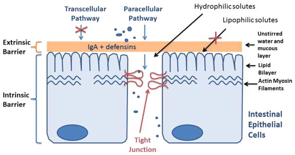
Background: Intestinal barrier dysfunction has been implicated in necrotizing enterocolitis (NEC), but has not been directly measured in human NEC.
Methods: Small intestines removed during surgery were immediately mounted in an Ussing chamber. mRNA expression of tight junction (TJ) proteins was measured with RT-PCR.
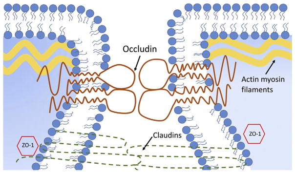
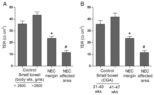
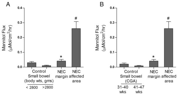
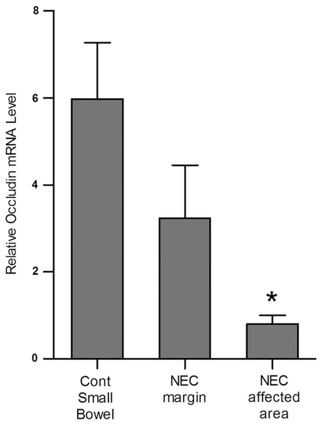
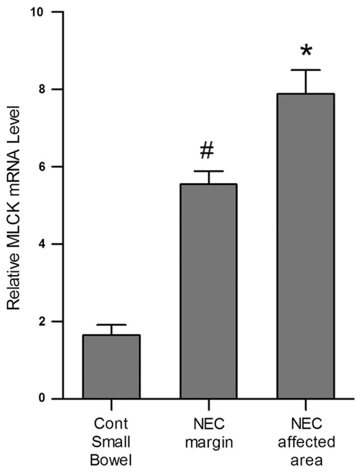
Results: Fifteen infants were included, 5 with NEC and 10 with other diagnoses. Average transepithelial resistance (TER) was 11.61±1.65Ω/cm2 in NEC specimens, 23.36±1.48Ω/cm2 at resection margin, and 46.48±5.65Ω/cm2 in controls. Average flux of permeability marker mannitol was 0.23±0.06μMol/cm2 per h in NEC, 0.04±0.01 μMol/cm2 per h at resection margin, and 0.017±0.004 μMol/cm2 per h in control tissue (p<0.05). RT-PCR analysis showed marked decrease in mRNA expression of a TJ protein occludin in NEC affected tissue (p<0.03 vs. control). Additionally, mRNA expression of myosin light chain kinase (MLCK), an important regulator of TJ permeability, was increased in NEC specimens.
Conclusion: These studies show for the first time that NEC intestinal tissue have increased intestinal permeability, even at grossly healthy-appearing resection areas. The increase in intestinal permeability in NEC appeared to be related in part to a decrease in occludin and an increase in MLCK expression.
Level of evidence: Level 2.
Keywords: Intestinal barrier function; Necrotizing enterocolitis; Occludin; Tight junction.
Published by Elsevier Inc.
Figures
Similar articles
-
Bifidobacteria stabilize claudins at tight junctions and prevent intestinal barrier dysfunction in mouse necrotizing enterocolitis.Am J Pathol. 2013 May;182(5):1595-606. doi: 10.1016/j.ajpath.2013.01.013. Epub 2013 Mar 5. Am J Pathol. 2013. PMID: 23470164 Free PMC article.
-
Rho kinase inhibition maintains intestinal and vascular barrier function by upregulation of occludin in experimental necrotizing enterocolitis.Am J Physiol Gastrointest Liver Physiol. 2018 Oct 1;315(4):G514-G528. doi: 10.1152/ajpgi.00357.2017. Epub 2018 Jun 21. Am J Physiol Gastrointest Liver Physiol. 2018. PMID: 29927318 Free PMC article.
-
Protective Effects of Bifidobacterium on Intestinal Barrier Function in LPS-Induced Enterocyte Barrier Injury of Caco-2 Monolayers and in a Rat NEC Model.PLoS One. 2016 Aug 23;11(8):e0161635. doi: 10.1371/journal.pone.0161635. eCollection 2016. PLoS One. 2016. PMID: 27551722 Free PMC article.
-
IL-1β and the Intestinal Epithelial Tight Junction Barrier.Front Immunol. 2021 Oct 25;12:767456. doi: 10.3389/fimmu.2021.767456. eCollection 2021. Front Immunol. 2021. PMID: 34759934 Free PMC article. Review.
-
Mechanisms of nitric oxide-mediated intestinal barrier failure in necrotizing enterocolitis.Semin Pediatr Surg. 2005 Aug;14(3):159-66. doi: 10.1053/j.sempedsurg.2005.05.004. Semin Pediatr Surg. 2005. PMID: 16084403 Review.
Cited by
-
Neurodevelopmental outcome of infants who develop necrotizing enterocolitis: The gut-brain axis.Semin Perinatol. 2023 Feb;47(1):151694. doi: 10.1016/j.semperi.2022.151694. Epub 2022 Dec 20. Semin Perinatol. 2023. PMID: 36572620 Free PMC article.
-
Direct Implementation of Intestinal Permeability Test in NMR Metabolomics for Simultaneous Biomarker Discovery-A Feasibility Study in a Preterm Piglet Model.Metabolites. 2020 Jan 1;10(1):22. doi: 10.3390/metabo10010022. Metabolites. 2020. PMID: 31906404 Free PMC article.
-
The Role of Glycosaminoglycans in Protection from Neonatal Necrotizing Enterocolitis: A Narrative Review.Nutrients. 2020 Feb 20;12(2):546. doi: 10.3390/nu12020546. Nutrients. 2020. PMID: 32093194 Free PMC article. Review.
-
Intrauterine Inflammation, Epigenetics, and Microbiome Influences on Preterm Infant Health.Curr Pathobiol Rep. 2018;6(1):15-21. doi: 10.1007/s40139-018-0159-9. Epub 2018 Jan 20. Curr Pathobiol Rep. 2018. PMID: 29938128 Free PMC article. Review.
-
Alterations in Intestinal Permeability: The Role of the "Leaky Gut" in Health and Disease.J Equine Vet Sci. 2017 May;52:10-22. doi: 10.1016/j.jevs.2017.02.009. Epub 2017 Mar 7. J Equine Vet Sci. 2017. PMID: 31000910 Free PMC article.
References
-
- Lemons JA, Bauer CR, Oh W, et al. Very low birth weight outcomes of the National Institute of Child Health and Human Development Neonatal Research Network, January through December 1996. Pediatrics. 2001;107:E1. - PubMed
-
- Claud EC, Walker WA. Bacterial colonization, probiotic and necrotizing enterocolitis. J Clin Gastroenterol. 2008;42:S46–52. - PubMed
-
- Guthrie SO, Gordon PV, Thomas V, et al. Necrotizing enterocolitis among infants in the United States. Perinatology. 2003;23:278–85. - PubMed
-
- Bolisetty S, Lui K, Oei J, et al. A regional study of underlying congenital diseases in term infants with necrotizing enterocolitis. Acta Pediatr. 2007;89:1226–30. - PubMed
-
- McElhinney DB, Hindrick HL, Bush DM, et al. Necrotizing enterocolitis in neonates with congenital heart disease: risk factors and outcomes. Pediatrics. 2000;106:1080–7. - PubMed
MeSH terms
Substances
Grants and funding
LinkOut - more resources
Full Text Sources
Other Literature Sources
Research Materials
