. 2016 Oct 24;39(2):155-168.
doi: 10.1016/j.devcel.2016.09.002. Epub 2016 Oct 6.

Adaptation to Stressors by Systemic Protein Amyloidogenesis

Affiliations

Adaptation to Stressors by Systemic Protein Amyloidogenesis

Timothy E Audas et al. Dev Cell. .

Abstract

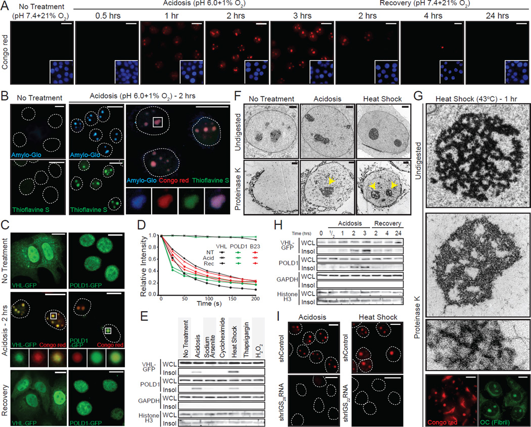
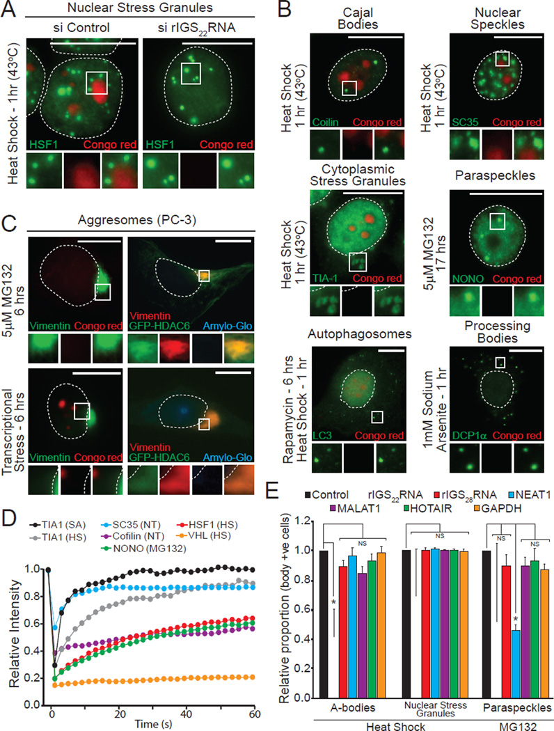
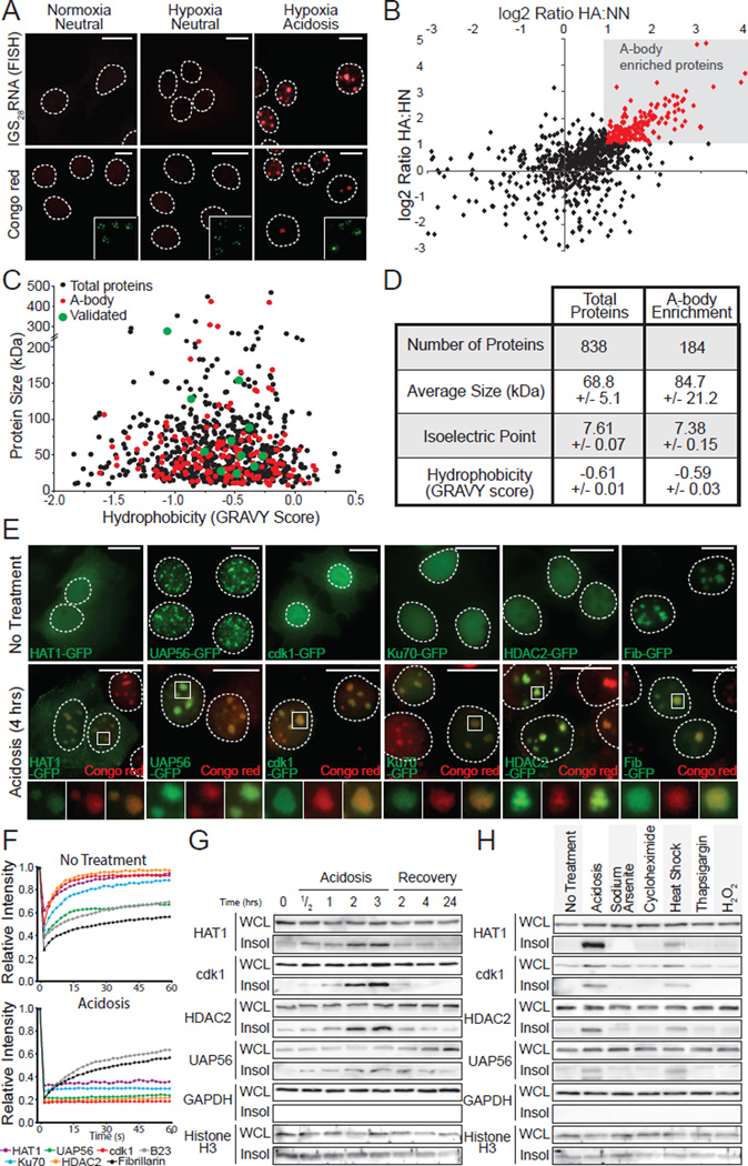
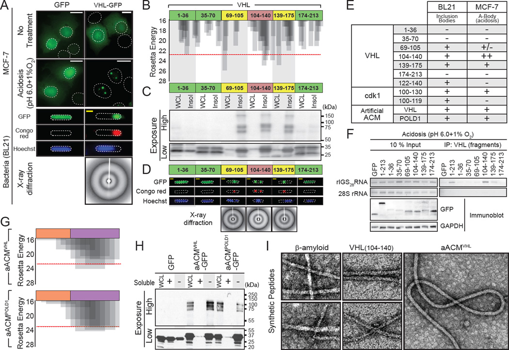
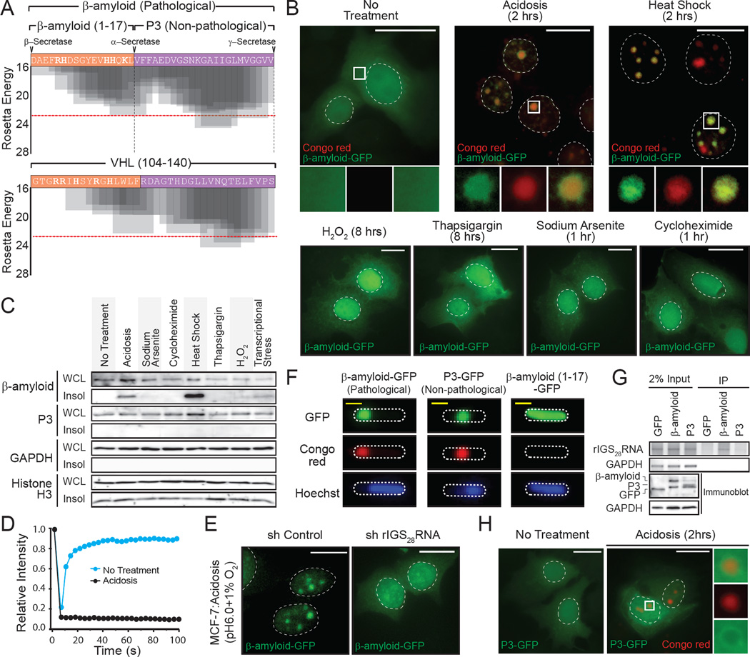
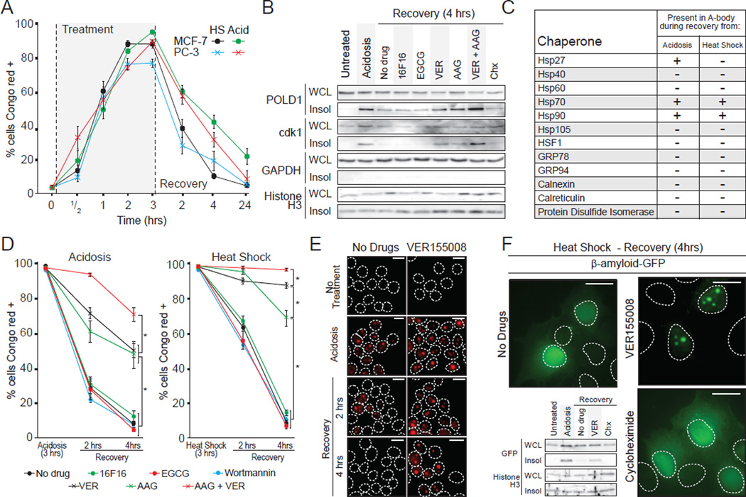
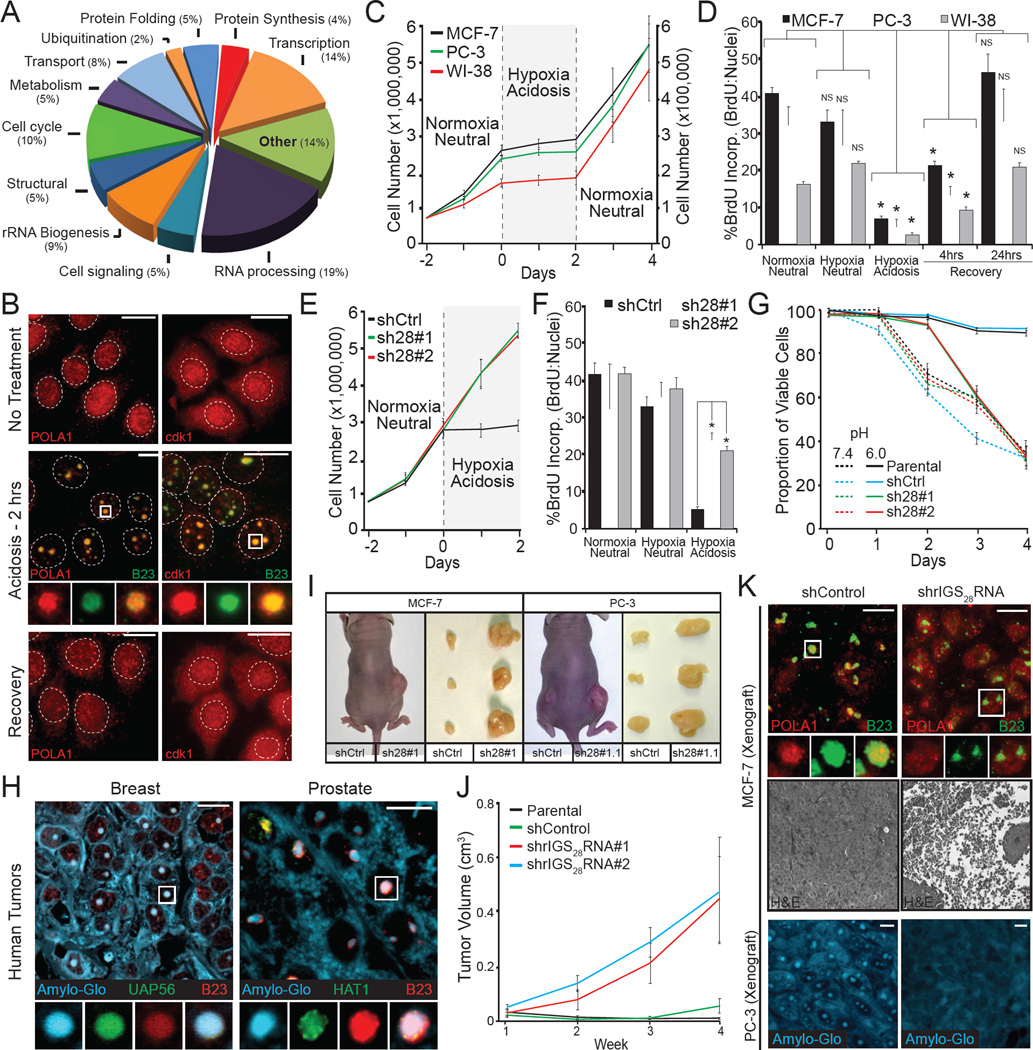
The amyloid state of protein organization is typically associated with debilitating human neuropathies and is seldom observed in physiology. Here, we uncover a systemic program that leverages the amyloidogenic propensity of proteins to regulate cell adaptation to stressors. On stimulus, cells assemble the amyloid bodies (A-bodies), nuclear foci containing heterogeneous proteins with amyloid-like biophysical properties. A discrete peptidic sequence, termed the amyloid-converting motif (ACM), is capable of targeting proteins to the A-bodies by interacting with ribosomal intergenic noncoding RNA (rIGSRNA). The pathological β-amyloid peptide, involved in Alzheimer's disease, displays ACM-like activity and undergoes stimuli-mediated amyloidogenesis in vivo. Upon signal termination, elements of the heat-shock chaperone pathway disaggregate the A-bodies. Physiological amyloidogenesis enables cells to store large quantities of proteins and enter a dormant state in response to stressors. We suggest that cells have evolved a post-translational pathway that rapidly and reversibly converts native-fold proteins to an amyloid-like solid phase.

Keywords: Hsp70; amyloid-bodies (A-bodies); dormancy; extracellular stress; heat shock chaperones; long noncoding RNA (lncRNA); physiological amyloidogenesis; β-amyloid.


Figures

Figure 1
Figure 1. Uncovering the distinct biophysical properties of the cellular A-bodies
(A) Physiological amyloidogenesis is rapid and reversible. MCF-7 cells exposed to extracellular acidosis and returned to standard growth conditions, for the indicated times, were stained with Congo red and Hoechst (blue inset). (B) Nuclear foci stain positively with amyloid-specific dyes. Untreated or acidotic MCF-7 cells were stained with Amylo-Glo (blue), Thioflavin S (green) and/or Congo red (red). Selected regions (white box) were expanded below with merged image included (far right panel). Dashed circles represent nuclei. (C) Established proteins are targeted to the A-bodies. MCF-7 cells expressing VHL-GFP or POLD1-GFP were grown under standard, hypoxic/acidotic conditions or recovered for 24 hours post-acidosis treatment. Acidotic cells were stained with Congo red. Selected regions (white box) were expanded (below). (D) A-body targets are reversibly immobilized. VHL-GFP, POLD1-GFP or GFP-B23 transfected MCF-7 cells were treated as above and bleached repeatedly for fluorescent loss in photobleaching. Quantification is presented as the mean relative intensity of at least 5 data sets. (E) Stimuli-specific insolubilization of A-body components. Insoluble proteins (Insol) were extracted from whole cell lysates (WCL) of untreated, acidotic (2 hrs), heat shocked (2 hrs), sodium arsenite (1 hr), cycloheximide (1hr), thapsigargin (8 hrs) or H2O2 (8 hrs) treated cells. A-body components; VHL-GFP and endogenous POLD1 or the GAPDH and Histone H3 control proteins were detected by western blot. (F) Stimuli-induced A-bodies are proteinase K resistant. Transmission electron microscopy (TEM) of untreated, heat shocked (1hr) and acidotic (1hr) MCF-7 cells were left undigested or exposed to proteinase K. Proteinase K-resistant nuclear bodies are indicated (yellow arrow). (G) Proteinase K-resistant fibrils possess amyloid-like properties. Heat shocked MCF-7 cells were left undigested or proteinase K-treated prior to TEM visualization. Proteinase K-resistant structures were stained with Congo red or the amyloid fibril conformation-specific antibody OC. (H) Protein insolubilization correlates with A-body assembly and disaggregation. MCF-7 cells were treated as above (A) and insoluble proteins were extracted from WCL. A-body targets/controls were detected as in (E). (I) Inhibition of rIGSRNA transcripts impairs amyloidogenesis. MCF-7 cells stably-expressing control or shRNA against rIGS28RNA or rIGS22RNA were grown in acidosis permissive media or exposed heat shock, respectively, prior to Congo red staining. Dashed circles represent nuclei. White scale bars represents 20mm. Black and white TEM scale box represent 1µM and 0.1mm. See also Figure S1.
Figure 2
Figure 2. A-bodies are unique rIGSRNA-seeded amyloidogenic structures
(A) A-bodies are separate from nuclear stress granules. MCF-7 cells transfected with control or rIGS22RNA-specific siRNA were exposed to heat shock. HSF-1 was detected in Congo red stained cells. (B) Additional cellular bodies are not amyloidogenic. MCF-7 cells were treated as indicated. Foci-specific markers were detected (green) in Congo red (red) stained cells. (C) Aggresomes do not contain proteins in an amyloid-like conformation. Untransfected or HDAC6-GFP expressing PC3 cells were treated with stimuli that induce aggresome (5µM MG132) or aggresome and A-body (transcriptional stress: 8µM MG132 + 4µM Actinomycin D) formation. The aggresome marker vimentin was detected in Congo red or Amylo-Glo stained cells. (D) Other cellular domains possess mobile proteins. Fluorescence recovery after photobleaching was performed on MCF-7 cells expressing TIA1-GFP (sodium arsenite and heat shock – 1hr), SC35-GFP (no treatment), Coilin-GFP (no treatment), HSF1-GFP (heat shock – 1hr), NONO-GFP (5µM MG132 – 17hrs) and VHL-GFP (heat shock – 1hr). Quantified kinetics are presented as the mean relative intensity of at least 5 data sets. (E) A-body formation is mediated by rIGSRNA. PC3 cells transfected with siRNA targeting the indicated transcripts were exposed to heat shock (45 min) or 5µM MG132 (17 hrs) prior to detection of A-bodies (Congo red), nuclear stress granules (HSF1) or paraspeckles (NONO). The proportion of body positive cells was counted and compared to the control siRNA. Results are means and SEM (n=3). Significance was measured by Student’s t-test; *p < 0.01. Dashed circles represent nuclei. Selected regions (white box) were expanded below. White scale bars represents 20mm. See also Figure S2.
Figure 3
Figure 3. Heterogenic protein composition of the A-body
(A) Acidosis induces the formation of the A-bodies. MCF-7 cells grown under normoxic-neutral (NN) and hypoxic-neutral (HN) as baseline controls, and hypoxic-acidotic (HA) conditions were stained with Congo red dye or fluorescence in situ hybridized with an anti-sense probe targeting rIGS28RNA. Nucleolar B23 (green) is inset. (B) Identification of the A-body residents. SILAC-MS analysis comparing proteins extracted from MCF-7 cells treated as above. Plot compares the log enrichment of HA:HN versus HA:NN. A-body-enriched (red) proteins fall in the upper right quadrant. (C) A-body components are biochemically similar to the total protein population. Scatter plot of protein size versus hydrophobicity. (D) A heterogeneous family of proteins are targeted to the A-bodies. The size, isoelectric point and hydrophobicity of total and amyloid-specific protein fractions is summarized. Values represent data set averages +/− SEM. (E) Localization of SILAC-MS candidates to the A-bodies. MCF-7 cells transfected with HAT1-GFP, UAP56-GFP, cdk1-GFP, Ku70-GFP, HDAC2-GFP and Fib-GFP were left untreated or exposed to extracellular acidosis prior to staining with Congo red. (F) Targets of the A-bodies are immobilized. Quantification of recovery after photobleaching kinetics for the proteins listed above (E) as the mean relative intensity of at least 5 data sets. (G) SILAC-MS candidates reversibly insolubilize in response to stimuli. MCF-7 cells were exposed to extracellular acidosis and returned to standard growth conditions, for the indicated times prior to harvesting WCL and insoluble proteins. HAT1, cdk1, HDAC2, UAP56, GAPDH and Histone H3 were detected by western blotting. (H) Stimuli-specific insolubilization of SILAC-MS candidates. WCL and insoluble proteins were extracted from untreated, acidotic (2 hrs), heat shocked (2 hrs), sodium arsenite (1 hr), cycloheximide (1hr), thapsigargin (8 hrs) or H2O2 (8 hrs) treated cells. A-body targets/controls were detected as in (E). Dashed circles represent nuclei. Selected regions (white box) were expanded below. White scale bars represents 20mm. See also Figure S3.
Figure 4
Figure 4. Identification of the amyloid converting motif that targets proteins to A-bodies
(A) VHL can obtain an amyloid-like conformation in bacteria. GFP or VHL-GFP expressing MCF-7 cells left untreated or exposed to acidosis or BL21 cells were stained with Congo red and Hoechst. X-ray diffraction was performed on BL21 bodies. (B) VHL contains fibril-forming peptidic regions. Results of ZipperDB analysis of full length VHL for fibrillation propensity. The Rosetta energy threshold of −23 kcal/mol is an indicator of fibril-positive regions. Truncated VHL fragments, used below, are indicated. (C) Amyloidogenic fragments of VHL insolubilize GFP and produce SDS-resistant multi-mers. Fragments of VHL (above) fused to GFP were expressed in BL21, prior to lysis and insoluble protein fractionation. Fusion proteins were detected with a GFP-specific antibody at low and high exposures to detect monomeric (low) and multi-meric (high) proteins. (D) Insoluble VHL fragments form bacterial inclusion bodies with an amyloid-like x-ray diffraction profile. BL21 expressing the VHL fragments (above) were fixed and stained with Congo red and Hoechst. Inclusion bodies were purified, where present, for x-ray diffraction. (E) Table summarizing inclusion body formation and targeting to the A-bodies (Figure 4D, S4B, F and H) for the indicated regions of VHL, cdk1 or POLD1 fused to GFP. (F) Regions of VHL can associate with rIGS28RNA. MCF-7 cells transfected with VHL or VHL mutants were exposed to acidosis for 1 hour prior to lysis and RNA immunoprecipitation. 28S rRNA and rIGS28RNA were detected by RT-PCR. Exogenous VHL fragments and GAPDH were detected by immunoblotting. (G) Generation of artificial ACM sequences. R/H-rich (orange) and amyloidogenic (purple) sequences derived from VHL (aACMVHL) and POLD1 (aACMPOLD1) were fused to generate artificial ACM motifs (sequence inset). The fibrillation propensity was calculated by ZipperDB. (H) Artificial ACMs are sufficient to create insoluble multi-mers. BL21 expressing GFP fused to the artificial ACMs (above) were lysed into soluble (+) and insoluble (−) fractions. (I) Amyloidogenic regions of VHL form 10nm amyloid-like fibrils. Peptides encoding VHL (104–140), aACMVHL and the classic pathological β-amyloid were synthesized and incubated for 1 week at 37°C. Fibrils were detected by TEM. White and yellow scale bars represent 20µm and 5µm, respectively. Dashed circles represent nuclei (MCF-7) or whole cell (BL21).TEM scale box represent 10nm. See also Figure S4.
Figure 5
Figure 5. The pathological β-amyloid peptide is a target of physiological amyloidogenesis
(A) Amino acid sequence and Rosetta energy profiles for β-amyloid (1–40) and VHL (104–140). Secretase (α-, β- and γ-) cleavage sites are indicated. (B) Stress-specific targeting of β-amyloid to the A-bodies. β-amyloid-GFP expressing MCF-7 cells were left untreated or exposed to acidosis, heat shock, sodium arsenite, cycloheximide, thapsigargin or H2O2 and stained with Congo red. (C) Amyloidogenic stimuli insolubilize β-amyloid, not the non-pathological P3 peptide. Insoluble proteins were extracted from MCF-7 cells exposed to the stimuli (above). β-amyloid-GFP, P3-GFP, GAPDH and Histone H3 were detected by western blotting. (D) Immobilization of β-amyloid by acidosis. Quantification of fluorescence recovery after photobleaching kinetics for β-amyloid-GFP in untreated or acidotic MCF-7 cells. Mean relative intensity of at least 5 data sets. (E) rIGS28RNA is essential for the subnuclear targeting of β-amyloid. MCF-7 cells stably-expressing control or rIGS28RNA-specific shRNA were transfected with a plasmid encoding β-amyloid-GFP and grown under hypoxic conditions in acidosis-permissive media. (F) β-amyloid and P3 possess amyloidogenic propensity. β-amyloid-GFP, P3-GFP and β-amyloid (1–17)-GFP were expressed in BL21 cells prior to staining with Congo red and Hoechst. (G) The amino-terminus of β-amyloid is essential for rIGSRNA binding. RNA immunoprecipitation of acidified MCF-7 cells transfected with GFP, β-amyloid-GFP or P3-GFP. GAPDH mRNA and rIGS28RNA were detected by RT-PCR. Exogenous proteins and GAPDH were detected by immunoblotting. (H) P3 is not targeted to the A-bodies. P3-GFP-expressing MCF-7 cells were untreated or exposed to acidotic conditions, prior to Congo red staining. Dashed circles represent nuclei (MCF-7) or whole cell (BL21). Selected regions (white box) were expanded below. White scale bars represents 20µm. See also Figure S5.
Figure 6
Figure 6. Heat shock chaperones regulate A-bodies disaggregation
(A) Amyloidogenesis is rapid and reversible. MCF-7 and PC-3 cells exposed to acidosis or heat shock and allowed to recover (times indicated) were stained with Congo red. The proportion of cells containing Congo red-positive structures was assayed relative to Hoechst-positive nuclei. (B) Heat shock proteins mediate the solubilization of A-body components. Insoluble proteins were extracted from untreated, acidotic or recovering MCF-7 cells treated with the protein synthesis inhibitor cycloheximide (Chx) or PDI (16F16), GRP94 (EGCG), Hsp70 (VER155008) or Hsp90 (17-AAG) inhibitors. POLD1, cdk1, GAPDH and Histone H3 were detected by western blot. (C) Heat shock proteins are associated with the A-bodies during recovery. Table summarizing data in Figure S6B. (D) Heat shock proteins disaggregate the A-bodies. MCF-7 cells were exposed to acidotic (left panel) or heat shock (right panel) conditions for 3 hours, then returned to normal growth conditions for 2 or 4 hours in the presence 16F16, EGCG, VER, AAG or Wortmannin. The proportion of Congo red-positive cells was determined as above. (E) Congo red stained MCF-7 cells allowed to recover for 2 or 4 hours from a 3 hour acidosis or heat shock exposure in the presence or absence of VER155008. (F) Hsp70 activity enhances β-amyloid release during recovery. β-amyloid-GFP-expressing MCF-7 cells were allowed to recover for 4 hours from a 3 hour heat shock exposure in the presence or absence of VER or Chx. Western blots of insoluble fractionation for β-amyloid-GFP and Histone H3 are included (lower left). Results are means and SEM (n=4). Significance was measured by Student’s t-test; *p < 0.01. Dashed circles represent nuclei. White scale bars represents 20µm. See also Figure S6.
Figure 7
Figure 7. rIGSRNA/A-bodies induce cellular dormancy
(A) Functional classification of A-bodies constituents. SILAC-MS results were analyzed and grouped by function, percentages per group are indicated. (B) Proliferative factors are reversibly targeted to the A-bodies. Untreated, acidotic and recovered MCF-7 cells were stained for endogenous POLA1 and cdk1 (red) and the nucleolar marker B23 (green). Dashed circles represent nuclei. (C) Acidosis induces a reversible state of dormancy. 750,000 (MCF-7 and PC-3 – left axis) or 75,000 (WI-38 – right axis) cells were grown for 2 days, prior to the application of acidosis-permissive media and exposure to hypoxic (1% O2) conditions. Cells were returned to standard growth conditions two days post-acidification. Cells were counted daily with trypan blue staining to ensure viability. (D) DNA synthesis is reversibly inhibited by acidosis. MCF-7, PC-3 and WI-38 cells grown under the indicated conditions were incubated with BrdU prior to fixation. BrdU positive cells were counted relative to Hoechst stained nuclei. (E–F) Inhibition of rIGS28RNA restores proliferative capacity to acidotic cells. MCF-7 cells stably-expressing two independent shRNA against rIGS28RNA (sh28#1 and sh28#2) or a control sequence (shCtrl) were exposed to normoxic-neutral, hypoxic-neutral or hypoxic-acidosis and cell counts were performed each day (E) or incubated with BrdU for the incorporation assay described above (F). (G) Amyloidogenesis preserves cell viability during extracellular stress. MCF-7 cells described above were grown under hypoxic conditions in standard (pH7.4) or acidosis-permissive (pH6.0) low glucose media for the indicated times. Viability was calculated as propidium iodide-positive versus Hoechst-positive nuclei. (H) Human breast invasive duct carcinomas and prostatic acinar contain cellular amyloids. Paraffin-embedded prostate and breast tumors were stained with the Amylo-Glo, UAP56 or HAT1 and the nucleolar marker B23. (I–J) Inhibition of rIGS28RNA relieves tumor dormancy. Nude mouse xenograft assays with MCF-7 and PC-3 cells described above. Representative mice and excised tumors (I) are presented, with tumor volumes calculated weekly (J) (n=5). (K) Inhibition of rIGS28RNA prevents amyloidogenesis in situ, causing tumor necrosis. Paraffin-embedded MCF-7 andPC-3 tumor sections were stained for the SILAC-MS candidate POLA1 (red) and B23 (green), Amylo-Glo (blue) or hematoxylin/eosin. Results are means and SEM (n≥4) with significance measured by Student’s t-test; *p < 0.01. Selected regions (white box) were expanded below. White scale bars represents 20µm. See also Figure S7.

Comment in

References

    1. Andersen JS, Lam YW, Leung AK, Ong SE, Lyon CE, Lamond AI, Mann M. Nucleolar proteome dynamics. Nature. 2005;433:77–83. - PubMed
    1. Audas TE, Jacob MD, Lee S. Immobilization of proteins in the nucleolus by ribosomal intergenic spacer noncoding RNA. Mol Cell. 2012;45:147–157. - PubMed
    1. Baldwin AJ, Knowles TP, Tartaglia GG, Fitzpatrick AW, Devlin GL, Shammas SL, Waudby CA, Mossuto MF, Meehan S, Gras SL, et al. Metastability of native proteins and the phenomenon of amyloid formation. J Am Chem Soc. 2011;133:14160–14163. - PubMed
    1. Benz CC, Scott GK, Sarup JC, Johnson RM, Tripathy D, Coronado E, Shepard HM, Osborne CK. Estrogen-dependent, tamoxifen-resistant tumorigenic growth of MCF-7 cells transfected with HER2/neu. Breast Cancer Res Treat. 1992;24:85–95. - PubMed
    1. Berchowitz LE, Kabachinski G, Walker MR, Carlile TM, Gilbert WV, Schwartz TU, Amon A. Regulated Formation of an Amyloid-like Translational Repressor Governs Gametogenesis. Cell. 2015;163:406–418. - PMC - PubMed

Publication types