. 2016 Oct 20;64(2):376-387.
doi: 10.1016/j.molcel.2016.09.005. Epub 2016 Oct 6.

Single-Molecule Imaging Reveals that Rad4 Employs a Dynamic DNA Damage Recognition Process

Affiliations

Single-Molecule Imaging Reveals that Rad4 Employs a Dynamic DNA Damage Recognition Process

Muwen Kong et al. Mol Cell. .

Abstract

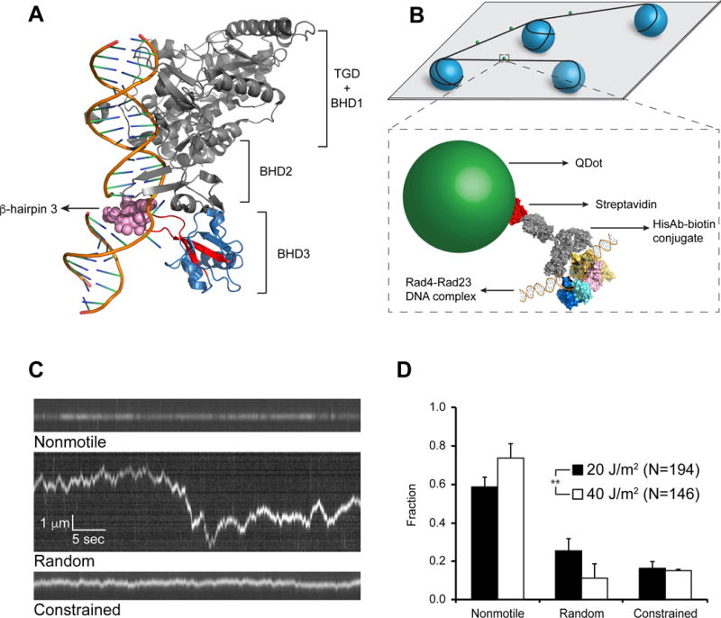
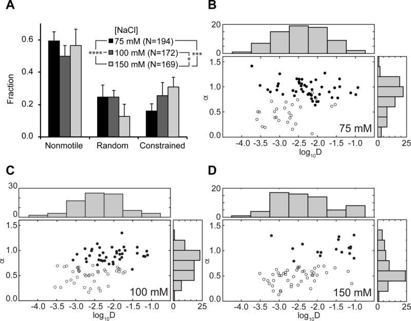
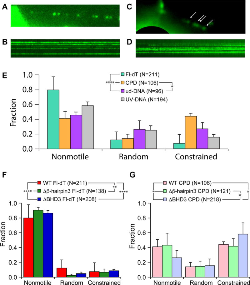
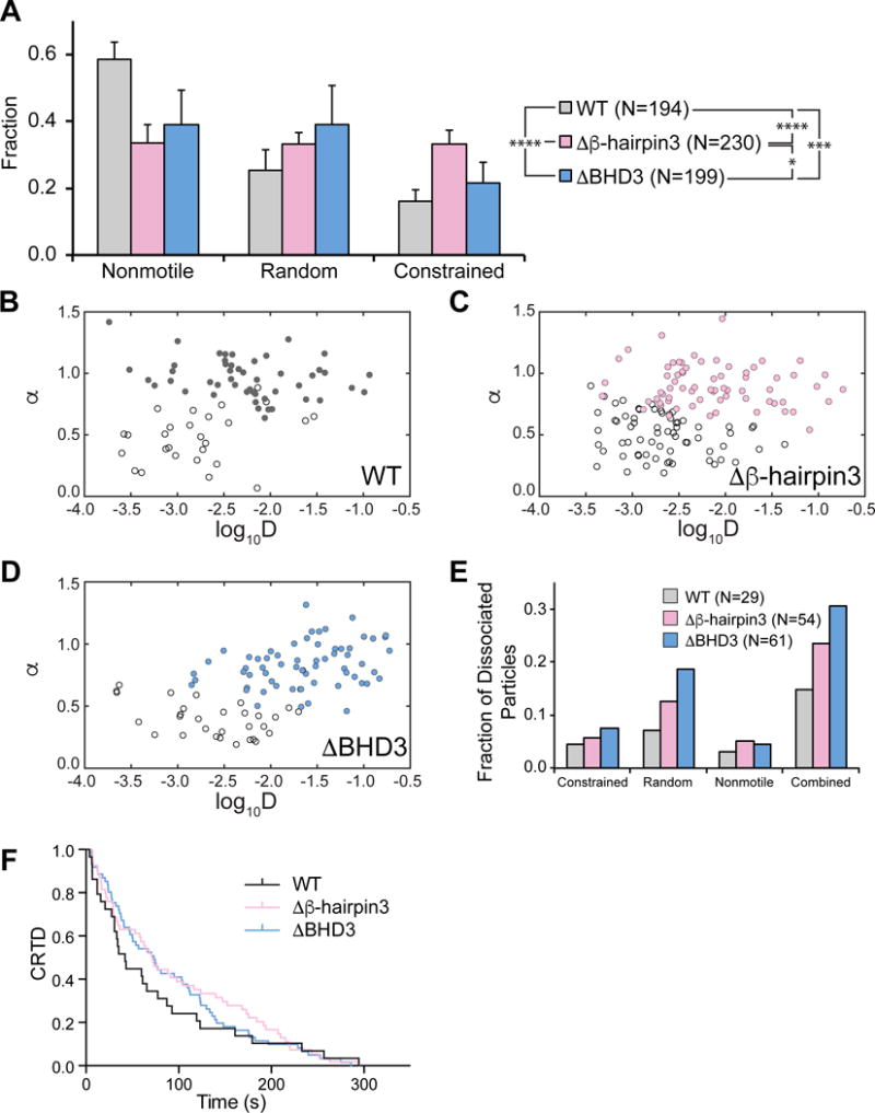
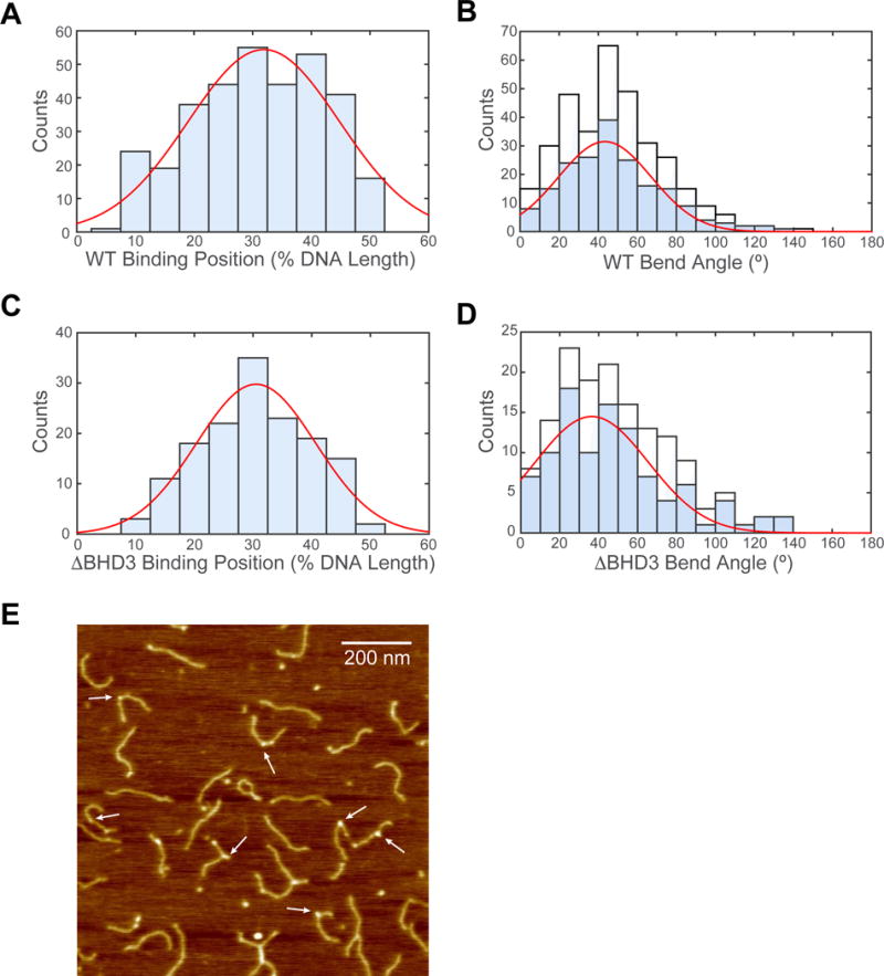
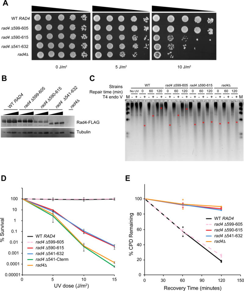
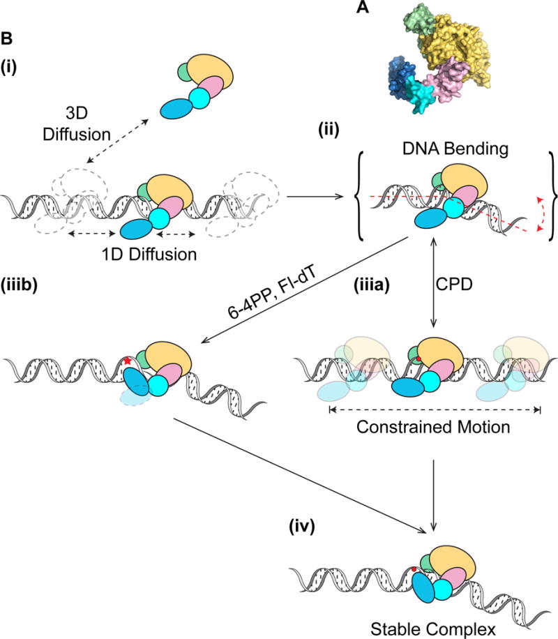
Nucleotide excision repair (NER) is an evolutionarily conserved mechanism that processes helix-destabilizing and/or -distorting DNA lesions, such as UV-induced photoproducts. Here, we investigate the dynamic protein-DNA interactions during the damage recognition step using single-molecule fluorescence microscopy. Quantum dot-labeled Rad4-Rad23 (yeast XPC-RAD23B ortholog) forms non-motile complexes or conducts a one-dimensional search via either random diffusion or constrained motion. Atomic force microcopy analysis of Rad4 with the β-hairpin domain 3 (BHD3) deleted reveals that this motif is non-essential for damage-specific binding and DNA bending. Furthermore, we find that deletion of seven residues in the tip of β-hairpin in BHD3 increases Rad4-Rad23 constrained motion at the expense of stable binding at sites of DNA lesions, without diminishing cellular UV resistance or photoproduct repair in vivo. These results suggest a distinct intermediate in the damage recognition process during NER, allowing dynamic DNA damage detection at a distance.

Keywords: DNA tightrope assay; Rad23; Rad4; XPC; dynamic DNA damage recognition; nucleotide excision repair; quantum dots; single particle tracking; xeroderma pigmentosum.


Figures

Figure 1
Figure 1. Rad4-Rad23 Crystal Structure, Experimental Schematics, and WT on UV-irradiated λ-DNA
(A) Co-crystal of Rad4-Rad23 with CPD-mismatch-containing DNA (PDB ID: 2QSG). TGD domain and β-hairpin domains 1 and 2 of Rad4, as well as Rad23, are shown in gray, β-hairpin domain 3 in blue, with β-hairpin 3 in red. Residues 599–605 are shown as pink spheres. (B) Schematics of flow cell and protein conjugation strategy. Top: 5 μm diameter poly-L-lysine coated silica beads (blue) are deposited on polyethylene glycol treated coverslip (gray). DNA (black) is elongated and strung up across beads by flow. Bottom: His-tagged Rad4-Rad23 (yellow, pink, cyan, and blue) is labeled with streptavidin (red)-coated quantum dot (SAQD, green) through a His-antibody (His-Ab)-biotin conjugate (gray). See also Figure S1. (C) Representative kymographs depicting top: nonmotile (see also Movie S1), middle: random diffusion (see also Movie S2), and bottom: constrained motion (see also Movie S3) particles. Scale bars in middle panel apply to all three kymographs. (D) Bar graph of fractions of each observed motion type on λ-DNA irradiated with 20 J/m2 (black bars) or 40J/m2 (white bars) UV light (p = 0.0026, χ2 test). All bar graph data in this study are represented as weighted means ± weighted standard deviations over four to five independent experimental days. (Statistical significance *: p ≤ 0.05, **: p ≤ 0.01, ***: p ≤ 0.001, ****: p ≤ 0.0001)
Figure 2
Figure 2. Behavior of WT Rad4-Rad23 on UV-irradiated λ-DNA at Different Salt Concentrations
(A) Distributions of observed motion types at different salt concentrations. Data at 75 mM NaCl reproduced from Figure 1D (p = 0.0005, χ2 test). (B – D) Anomalous diffusion exponent (α) vs. diffusion coefficient (log10D) plotted for random (filled circles) and constrained (empty circles) particles at (B) 75 mM, (C) 100 mM, and (D) 150 mM NaCl. Distributions of diffusion coefficients log10D and anomalous diffusion exponents α are plotted above and to the right of each scatter plot, respectively.
Figure 3
Figure 3. Lesion-Dependent Motions in Damage Recognition by Rad4-Rad23
(A) Single frame of quantum dot-labeled Rad4-Rad23 assembled in an array on Fl-dT-containing DNA. See also Movie S4. (B) Kymograph of Rad4-Rad23 particles assembled on Fl-dT array shown in (A). (C) Single frame of quantum dot-labeled Rad4-Rad23 assembled in an array on CPD-containing DNA. Rad4-Rad23 particles are indicated by white arrows. See also Movie S5. (D) Kymograph of Rad4-Rad23 particles assembled on CPD array shown in (C). (E) Distributions of motion types of WT Rad4-Rad23 observed on DNA damage arrays show lesion-dependent behavior (Fl-dT – green, CPD – orange, undamaged DNA – purple, UV-irradiated λ-DNA – gray). See also Figure S2 and S3. (F) and (G) Distributions of motion types of WT, Δβ-hairpin3, and ΔBHD3 observed on DNA damage arrays containing sites of Fl-dT (WT – red, Δβ-hairpin3 – green, ΔBHD3 – blue), and CPD (WT – pink, Δβ-hairpin3 – mint, ΔBHD3 – lavender), respectively. WT data reproduced from Figure 3E.
Figure 4
Figure 4. Motion and Dissociation Kinetics of Rad4 WT and β-hairpin 3 Mutants on UV-irradiated λ-DNA
(A) Distributions of observed motion types from Rad4 WT and mutants. (B – D) Anomalous diffusion exponent (α) vs. diffusion coefficient (log10D) plotted for random (filled circles) and constrained (empty circles) particles of WT (B), Δβ-hairpin3 (C), and ΔHD3 (D). See also Figure S4. (E) Dissociating particles as fractions of total particles observed increase with larger deletions in Rad4 BHD3 sequence. (F) Cumulative residence time distribution (CRTD) plot of lifetimes of Rad4 WT and mutants that dissociated during observation. See also Figure S5.
Figure 5
Figure 5. Specific Binding and DNA Bending by WT and ΔBHD3
(A) Histogram and Gaussian fitting (red curve) of WT binding positions (32 ± 13%, N = 335) on DNA fragment in terms of percentage of total contour length measured from one end. (B) Histogram of DNA bend angles at all internal WT binding sites (white, N = 335). Histogram (blue) and Gaussian fitting (red curve) of DNA bend angles (43 ± 24°, N = 189) at WT proteins specifically bound between 20% and 40%. See also Figure S6. (C) Histogram and Gaussian fitting (red curve) of ΔBHD3 binding positions (31 ± 10%, N = 148) on DNA fragment in terms of percentage of total contour length measured from one end. (D) Histogram of DNA bend angles at all internal ΔBHD3 binding sites (white). Histogram (blue) and Gaussian fitting (red curve) of DNA bend angles (37 ± 29°, N = 101) at ΔBHD3 specifically bound between 20% and 40%. (E) Representative AFM image of ΔBHD3 bound to Fl-dT-containing DNA fragments. White arrows highlight representative binding events scored in data analysis. See also Figure S6.
Figure 6
Figure 6. UV Survival and Rates of CPD Removal of Yeast Carrying Different Rad4 Variants
(A) Serial dilutions of yeast cells (BY4742) expressing different 3×FLAG-tagged Rad4 variants on YPD plates, 72 hours after UV irradiation. (B) Expression levels of 3×FLAG-tagged Rad4 variants detected with anti-FLAG antibody. (C) Genomic DNA of yeast cells after UV irradiation and recovery digested with T4 endo V, separated on alkaline agarose gel, and detected with SYBR Gold. Approximate positions of the ensemble average size of DNA in each lane are denoted with red asterisks (*). DNA marker (M, λ DNA-HindIII) was loaded in the left- and right-most lanes. (D) Quantitative UV-survival of yeast cells (BY4741) expressing different untagged Rad4 variants. WT RAD4 – black, rad4 Δ599–605 (Δβ-hairpin3) – pink dashed, rad4 Δ590–615 – red, rad4 Δ541–632 (ΔBHD3) – blue, rad4 Δ541-cterm – green, rad4Δ – orange. (E) Quantitative rates of CPD removal of yeast cells (BY4741) expressing different untagged Rad4 variants, determined by T4 endo V digestion. Color scheme same as in Figure 6D. See also Figure S7.
Figure 7
Figure 7. Working Model for Dynamic Lesion Recognition by Rad4-Rad23
(A) Domains of Rad4-Rad23 (PDB: 2QSF) color-coded as shown in (B): TGD – yellow, BHD1 – pink, BHD2 – cyan, BHD3 – blue, Rad23 – green. (B) Rad4-Rad23 scans DNA through 3D or 1D diffusion (i) and tests integrity of DNA via bending/twisting during 1D diffusion on DNA (ii). Depending on the type of damage encountered, Rad4-Rad23 can either undergo constrained motion around lesion due to lack of β-hairpin 3 insertion (iiia), or alternatively rapidly forms stable protein-DNA complex at site of lesion with β-hairpin 3 inserted for stabilization in a twist-open action (Velmurugu et al., 2016) (iiib). While it is possible that the DNA in (iiia) is bent, for simplicity this is not shown. Extra time spent probing the lesion, afforded by constrained motion of Rad4-Rad23, could also lead to stable binding of the protein at sites that require larger base opening/flipping energies (iv).

References

    1. Aboussekhra A, Biggerstaff M, Shivji MK, Vilpo JA, Moncollin V, Podust VN, Protic M, Hubscher U, Egly JM, Wood RD. Mammalian DNA nucleotide excision repair reconstituted with purified protein components. Cell. 1995;80:859–868. - PubMed
    1. Berg OG, Winter RB, von Hippel PH. Diffusion-driven mechanisms of protein translocation on nucleic acids. 1. Models and theory. Biochemistry. 1981;20:6929–6948. - PubMed
    1. Blainey PC, van Oijen AM, Banerjee A, Verdine GL, Xie XS. A base-excision DNA-repair protein finds intrahelical lesion bases by fast sliding in contact with DNA. Proceedings of the National Academy of Sciences of the United States of America. 2006;103:5752–5757. - PMC - PubMed
    1. Brown KL, Roginskaya M, Zou Y, Altamirano A, Basu AK, Stone MP. Binding of the human nucleotide excision repair proteins XPA and XPC/HR23B to the 5R-thymine glycol lesion and structure of the cis-(5R,6S) thymine glycol epimer in the 5′-GTgG-3′ sequence: destabilization of two base pairs at the lesion site. Nucleic acids research. 2010;38:428–440. - PMC - PubMed
    1. Camenisch U, Trautlein D, Clement FC, Fei J, Leitenstorfer A, Ferrando-May E, Naegeli H. Two-stage dynamic DNA quality check by xeroderma pigmentosum group C protein. The EMBO journal. 2009;28:2387–2399. - PMC - PubMed

MeSH terms

LinkOut - more resources